. 2020 Jan 6;52(1):38-52.e10.
doi: 10.1016/j.devcel.2019.11.007. Epub 2019 Dec 12.

GCNA Preserves Genome Integrity and Fertility Across Species

Affiliations

GCNA Preserves Genome Integrity and Fertility Across Species

Varsha Bhargava et al. Dev Cell. .

Abstract

The propagation of species depends on the ability of germ cells to protect their genome from numerous exogenous and endogenous threats. While these cells employ ubiquitous repair pathways, specialized mechanisms that ensure high-fidelity replication, chromosome segregation, and repair of germ cell genomes remain incompletely understood. We identified Germ Cell Nuclear Acidic Peptidase (GCNA) as a conserved regulator of genome stability in flies, worms, zebrafish, and human germ cell tumors. GCNA contains an acidic intrinsically disordered region (IDR) and a protease-like SprT domain. In addition to chromosomal instability and replication stress, Gcna mutants accumulate DNA-protein crosslinks (DPCs). GCNA acts in parallel with the SprT domain protein Spartan. Structural analysis reveals that while the SprT domain is needed to limit DNA damage, the IDR imparts significant function. This work shows that GCNA protects germ cells from various sources of damage, providing insights into conserved mechanisms that promote genome integrity across generations.

Keywords: ACRC; DNA repair; DNA-protein crosslink; SprT domain; acidic repeat containing; chromosome instability; chromosome segregation; germ cell; intrinsically disordered region; transgenerational inheritance.


Conflict of interest statement

DECLARATION OF INTERESTS

The authors declare no competing interests.

Figures

Figure 1.
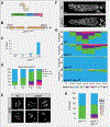
Figure 1.. Loss of Gcna results in chromosome instability in Drosophila.
(A) Domain organization of the Drosophila Gcna protein. (B) Drosophila Gcna gene locus showing the two isoforms and design of the GcnaKO allele with insertion of a dsRED cassette. UTRs in grey, exons in orange. (C) DAPI staining of control (wBerlin) and GcnaKO (maternal null) embryos reveals mitotic defects caused by loss of maternal Gcna. Scale bar represents 10μM. (D) Still images from movies of GcnaKO maternal-null embryos carrying a HistoneH2Av-mRFP transgene to visualize chromosomes. Yellow arrowheads indicate micronucleus formation (top) and nuclear fusion event (bottom). (E) Early Gcna maternal mutant Drosophila embryo stained for Lamin Dm0 (magenta) and DNA (green) to show that a nuclear envelope forms around a micronucleus (yellow arrow). Scale bar represents 5 μm. (F) Gynandromorph phenotypes in progeny of GcnaKO females. Left, note the line of dark and light pigmentation bisecting the thorax, reflecting the presence or absence of the yellow marker carried on one of the X chromosomes. Right, sex combs are seen on one forelimb (yellow arrow) but are missing from the other, reflecting adoption of male and female fates respectively. (G) Embryonic nuclei from control (wBerlin) and GcnaKO mutant females were labeled with FISH probes to the X (green) and second (red) chromosomes. The green and red numbers refer to number of X and second chromosome-specific foci observed in each half of the dividing nuclei. Controls show chromosomes dividing equally into daughter cells. GcnaKO cells have aberrant chromosome numbers and lagging chromosomes (green arrows). Scale bar represents 5 μm (H) Quantification and (I) corresponding images of Drosophila ovarian phenotypes. Hts marks the fusome; Vasa marks germ cells. Control (w1118) ovarioles contain the expected 16-cell cysts. GcnaKO/Df ovarioles have many cysts with abnormal numbers of cells, as well as tumors. The frequency of these phenotypes worsens with age. Scale bars represent 20 μm. (J) SIM images of wBerlin and GcnaKO mutant meiotic nuclei stained for synaptonemal complex marker C(3)G (red) and DNA damage marker γH2Av (green). (K) wBerlin, GcnaKO, mei-W860949, and GcnaKO; mei-W680949 ovarioles stained for C(3)G (red), γH2Av (green), and DNA (DAPI, blue). Scale bar represents 10 μm. (L) Average number of γH2Av labeled foci per nucleus in the indicated genotypes. 10 meiotic and non-meiotic nuclei were examined per genotype.
Figure 2.
Figure 2.. Loss of gcna-1 results in genomic instability in C. elegans.
(A) Domain structure of C. elegans GCNA-1 protein. (B) The C. elegans gcna-1 locus detailing the design of the gcna-1(ea43) allele. (C) The HIM phenotype increases over generations (n=12 parental worms for N2 (wild type control) and n= 21, 21, 34 for gcna-1 F1, F3, and F18, respectively) (D) gcna-1 mutant worms exhibit decreases in brood size with successive passaging (n=12 parental worms for wt, and 34 each for gcna-1 F1, F8, and F18). (E) DAPI staining of diakinesis nuclei from control (N2, wild type) and gcna-1 mutants. Controls showed the expected 6 DAPI+ bivalents; gcna-1 mutant nuclei contained mixtures of bivalents, univalents, and fused chromosomes. Scale bars represent 5 μm. (F) Mitotic germ cells from gcna-1 mutants exhibit chromosome bridges as seen by DAPI-stained DNA. Insets are examples of nuclei in anaphase. Scale bars represent 20 μm. (G) Quantification of meiotic (leptotene through onset of diplotene) RAD-51 foci in N2, gcna-1, spo-11, and gcna-1;spo-11 mutant C. elegans germ lines (n = 3 germ lines/genotype). (H) Quantification of DAPI positive bodies in diakinesis-stage, −1 oocytes of C. elegans (spo-11, n= 20; gcna-1;spo-11, n=42).
Figure 3.
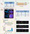
Figure 3.. GCNA mutant germ cells experience replication stress
(A) Frequency of microsatellite repeat length changes within the C. elegans vab-1 locus. (B-C) RPA foci (yellow arrows) accumulate in Drosophila GcnaKO germ cells. (B) Control and GcnaKO/Df germaria (left) and nurse cell nuclei (right) stained for DNA (blue), RPA (green), and Lamin C (red) as indicated. Scale bars represent 10 μm. (C) Quantification of RPA punctae per nucleus. (D) Quantification of γH2Av foci in region 1 of the Drosophila germarium in control and GcnaKO mutant germaria in response to HU treatment. (E) The dvc-1/Spartan locus of C. elegans showing the strategy for creating a complete null, dvc-1(ea65). (F) Brood analysis (total viable adult offspring) and embryonic lethality associated with N2, dvc-1 and gcna-1 single mutants, and dvc-1;gcna-1 double mutant worms. (G) Examples of DAPI-stained, diakinesis oocytes from dvc-1, gcna-1, and dvc-1; gcna-1 mutant germ cells show chromosome fragmentation in the double mutant. Scale bars represent 5 μm.
Figure 4.
Figure 4.. GCNA mutants exhibit increased DPC levels
(A) Schematic illustrating principle of the RADAR assay. (B) RADAR was performed on Drosophila ovaries and embryos. Samples were normalized to dsDNA, separated by SDS-PAGE, and silver stained. Red arrows mark examples of differentially enhanced bands in the Gcna mutant samples. * marks benzonase added to the prep. (C) Proteins enriched in DPCs from GcnaKO Drosophila embryos were analyzed by mass spectrometry and presented as enriched over wild-type controls. (D) List of proteins that co-immunoprecipate with Gcna expressed in Drosophila S2 cells. (E, F) Top2 is mislocalized in Gcna mutant flies and worms. (E) Early Drosophila embryos derived from control and GcnaKO females stained for DNA (blue), Top2 (green) and Tubulin (Tub, red). Grayscale shows Top2 alone. Scale bar represents 5 μm. (F) Control (N2) and gcna-1 mutant C. elegans gonads stained for DNA (green), TOP-2 (magenta), and staining control XND-1 (yellow). Scale bar represents 20 μm.
Figure 5.
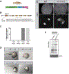
Figure 5.. Functional analysis of GCNA domain structure
(A, B) Expression of a UAS-GcnaWT transgene tagged at the C-terminus driven by nanos (nos)-gal4 shows prominent cytoplasmic staining. Germaria co-labeled for the Gcna-GFP transgene and (A) the nuage component, Aubergine (Red) or (B) DAPI (red) to visualize DNA. Yellow arrow points to a mitotic germ cell. Scale bars represent 20 μm. (C) Structure of the Drosophila GcnaWT and catalytic dead, GcnaHE>AA, mutant transgenes. (D) Comparison the GcnaWT and GcnaHE>AA proteins tagged at their N-termini. Note the abundant GcnaHE>AA punctae in the germarium and germ cells. DNA (red), Gcna (anti-HA, green). Scale bars represent 20 μm. (E-G) The catalytic dead transgene confers a separation-of-function phenotype and (E, F) cannot suppress the accumulation of γH2Av foci in (E) meiotic and (F) non-meiotic nuclei from wild-type, GcnaKO, GcnaKO; nos>GcnaWT, and GcnaKO; nos>GcnaHE>AA ovaries, but (G) can partially rescue the maternal-effect lethality conferred by loss of Gcna. Plotted are the percentage of eggs derived from females of the indicated genotypes that hatch into larvae. (H) Expression of C. elegans OLLAS::GCNA-1. Germ cells are labeled for DNA (blue), GCNA-1 (anti-OLLAS, yellow), and the nuclear pore antibody mAb414 (magenta). Scale bar represents 5 μm. (I) Structure of the C. elegans “IDR-only”, C-terminal truncation allele, gcna-1(ea76). (J) Comparison of brood sizes from N2 control, and homozygous, F3 and F8 gcna-1(ea43) null and gcna-1(ea76) truncation mutants. The IDR-only mutation does not exhibit the rapid transgenerational sterility seen in the null.
Figure 6.
Figure 6.. GCNA function is conserved in vertebrates
(A) Domain structure of the zebrafish GCNA protein. (B) Organization of the zebrafish gcna gene showing the lesions in the mut1 and mut2 alleles. (C, D) Zebrafish embryos derived from mutant gcna mutant females exhibit profound maternal-effect defects, regardless of the paternal genotype. (C) Quantification of defects and (D) Representative embryos from indicated crosses. (E) Fixed, DAPI-stained embryos derived from mutant gcna mutant females exhibit asynchronous divisions and extensive chromosome bridges during early development. Scale bar represents 10 μm. (F) RADAR was performed on zebrafish embryos, as described in Figure 4A, B. Samples were normalized to dsDNA input, separated by SDS-PAGE, and silver stained. Red arrows mark bands increased in gcna mutants. * marks benzonase.
Figure 7.
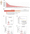
Figure 7.. GCNA is frequently altered in pediatric germ cell tumors (GCTs), and is associated with poor patient survival and genomic instability.
(A) GCNA genes display the most frequent copy-number loss and promoter hypermethylation in 94 childhood patients with GCTs. (B) Frequency of copy-number loss and promoter hypermethylation of GCNA gene in pediatric GCTs. (C) Low GCNA expression in pediatric GCTs is found in tumors with alterations of the GCNA locus. (D) Low GCNA expression is significantly associated with poor survival of GCT patients. (E,F) GCTs with copy number loss of GCNA exhibit elevated frequencies of both copy number (E) amplification and (F) loss relative to tumors with normal GCNA copy number.

Comment in

References

    1. Abdu U, Gonzalez-Reyes A, Ghabrial A, and Schupbach T (2003). The Drosophila spn-D gene encodes a RAD51C-like protein that is required exclusively during meiosis. Genetics 165, 197–204. - PMC - PubMed
    1. Arribere JA, Bell RT, Fu BX, Artiles KL, Hartman PS, and Fire AZ (2014). Efficient marker-free recovery of custom genetic modifications with CRISPR/Cas9 in Caenorhabditis elegans. Genetics 198, 837–846. - PMC - PubMed
    1. Banani SF, Lee HO, Hyman AA, and Rosen MK (2017). Biomolecular condensates: organizers of cellular biochemistry. Nat Rev Mol Cell Biol 18, 285–298. - PMC - PubMed
    1. Batool A, Karimi N, Wu XN, Chen SR, and Liu YX (2019). Testicular germ cell tumor: a comprehensive review. Cell Mol Life Sci 76, 1713–1727. - PMC - PubMed
    1. Borgermann N, Ackermann L, Schwertman P, Hendriks IA, Thijssen K, Liu JC, Lans H, Nielsen ML, and Mailand N (2019). SUMOylation promotes protective responses to DNA-protein crosslinks. EMBO J 38. - PMC - PubMed

Publication types

MeSH terms