. 2020 Jan 6;52(1):53-68.e6.
doi: 10.1016/j.devcel.2019.11.006. Epub 2019 Dec 12.

GCNA Interacts with Spartan and Topoisomerase II to Regulate Genome Stability

Affiliations

GCNA Interacts with Spartan and Topoisomerase II to Regulate Genome Stability

Gregoriy A Dokshin et al. Dev Cell. .

Abstract

GCNA proteins are expressed across eukarya in pluripotent cells and have conserved functions in fertility. GCNA homologs Spartan (DVC-1) and Wss1 resolve DNA-protein crosslinks (DPCs), including Topoisomerase-DNA adducts, during DNA replication. Here, we show that GCNA mutants in mouse and C. elegans display defects in genome maintenance including DNA damage, aberrant chromosome condensation, and crossover defects in mouse spermatocytes and spontaneous genomic rearrangements in C. elegans. We show that GCNA and topoisomerase II (TOP2) physically interact in both mice and worms and colocalize on condensed chromosomes during mitosis in C. elegans embryos. Moreover, C. elegans gcna-1 mutants are hypersensitive to TOP2 poison. Together, our findings support a model in which GCNA provides genome maintenance functions in the germline and may do so, in part, by promoting the resolution of TOP2 DPCs.

Keywords: DNA-protein crosslink (DPC) repair; DVC-1; GCNA; Spartan; SprT; Top1; Top2; germ cells; topoisomerase.


Conflict of interest statement

DECLARATION OF INTERESTS

The authors declare no competing interests.

Figures

Figure 1.
Figure 1.. gcna-1 Mutants Exhibit a Distinct Germline Phenotype Associated with Genomic Decline
(A) Brood size comparison between wild type, gcna-1(ne4356), dvc-1(ok260), and gcna-1(ne4356);dvc-1(ok260) double mutants (Error bars, SDM. ***p < 0.0001, *p < 0.05). (B) Brood sizes across twelve generations. Each symbol represents the number of progeny derived from a single hermaphrodite. p < 0.0001 at all generations. (Error bars, SDM.) See also Figure S1.
Figure 2.
Figure 2.. gcna-1 Is Required for Response to Replication Stress
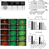
(A and B) Hatching rate of embryos after exposure of adults to UV (A) and hydroxyurea (B). Error bars, SDM. (C) Knockdown of chk-1 in gcna-1 mutants results in embryonic lethality. Values are normalized to the mean hatching rate of untreated controls. Box depicts 25th and 75th percentiles, and median. Whiskers represent the minimum and maximum values. ***p < 0.0001, *p < 0.05. See also Figure S2.
Figure 3.
Figure 3.. Absence of gcna-1 Causes a Potent Mutator Phenotype
(A) Schematic of unc-58(e665) mutator assay. (B) Frequencies of spontaneous mutation. Crosses denote >1 independent reversion event per plate, revealed by two distinct reverted phenotypes (n = 1 plate for gcna-1(ne4444) and n = 4 for dvc-1(ok260)). (C) Sequencing coverage surrounding unc-58 in gcna-1 (green) and dvc-1 (blue) mutant backgrounds. Deletions are indicated by absence of sequencing reads. Asterisks indicate increased copy number. Panel is modified from IGV. (D) Structural rearrangements at the unc-58 locus in revertant lines. See also Tables S2, S3, and S4 and Figures S1 and S3.
Figure 4.
Figure 4.. GCNA is Cell-Cycle Regulated and Localizes to Condensed Chromosomes during M Phase
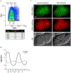
(A) Representative flow cytometry analysis of cell cycle and GCNA expression in mouse ESCs. (B) Cell-cycle regulated expression of S. pombe GCNA and wss1 transcripts (Gene Expression Viewer, Rustici et al., 2004). The timing of mitosis (M), S, and G2 phases are indicated. (C) Localization of GFP::GCNA-1 and DVC-1::mCherry in live C. elegans embryos during the second and third cell divisions. Nuclei in ABa and ABp cells are indicated by dashed circles. Scale bar, 5 μm. See also Figure S4; Video S1.
Figure 5.
Figure 5.. GCNA and TOP2 Physically Interact, Colocalize on Condensed Chromosomes, and Have a Functional Relationship
(A) Topoisomerase peptides recovered from anti-GCNA IP from mouse ESCs. Seven additional peptides are shared between TOP2 alpha and beta. No GCNA or topoisomerase peptides were recovered in an isotype control IP (Table S5). (B) Live-cell imaging of the first embryonic cell division in C. elegans showing colocalization of TOP-2:mCherry and GFP::GCNA-1. Scale bar, 5 μm. (C) Co-IP of TOP-2::mCherry with GFP::GCNA-1. Note: GFP::GCNA-1 is not abundant enough to be detected in input. (D) Hatching rate following treatment with TOP1 (camptothecin, CPT) and TOP2 (etoposide, ETP) poisons. n = 60, error bars, SDM. ***p < 0.0001. See also Figure S2; Video S2.
Figure 6.
Figure 6.. Gcna-Mutant Spermatocytes Exhibit DNA Damage, Crossover Defects, and Chromatin Condensation Abnormalities
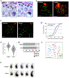
(A) Histology of Stage IX seminiferous tubules. Representative leptotene spermatocytes are indicated by arrowheads and detailed in insets. (B) Pachytene spermatocytes immunostained with SYCP3 (green) and γ-H2AX (red). (C) Analysis of crossovers in Gcna-mutant spermatocytes. For Ci, Cii, and Ciii, n = 300 nuclei per genotype. (1) Pachytene spermatocytes stained with SYCP3 (green) and MLH1 (red). Bivalents lacking MLH1 foci are indicated by white arrows and detailed in inset. X and Y chromosomes are labeled. (2) Quantification of MLH1 foci per nucleus. (3) Pachytene bivalents with 0, 1, 2, or 3 MLH1 foci. (4) Cumulative distribution curves of the distance between MLH1 foci normalized to the length of the synaptonemal complex (SC). n > 450 chromosomes. (5) Gcna-mutant diplotene spermatocyte stained with SYCP3 (black). Univalent chromosome pairs are indicated by colored highlights. (D) Morphology of DAPI-stained sperm heads. Scale bars, 5 μm. See also Figures S5 and S6.
Figure 7.
Figure 7.. Proposed Model for Germline-Specific DPC Processing by GCNA
(A) In S phase in both somatic and germline cells, ubiquitin signaling leads to Spartan-mediated proteolysis of DPCs. Remnants are bypassed by TLS machinery and repaired by NER. Spartan is not present in mitosis. (B) Outside of S, in germline and soma, DPC repair is mediated through SUMO signaling. In the soma (top), TDP2 hydrolyzes the bond between DPC and DNA and yields DNA ends predisposed to error prone NHEJ. NHEJ allows somatic cells to avoid LOH associated with HDR. In the germline (bottom), NHEJ is suppressed, and Spartan is complemented by GCNA, which recruits an MRE11-containing endonuclease complex to generate DNA ends that are compatible with HDR. Thus, germ cells can repair DPCs without risking NHEJ-associated mutations that would be detrimental to the heritable genome and incompatible with germline immortality.

Comment in

References

    1. Abyzov A, Urban AE, Snyder M, and Gerstein M (2011). CNVnator: an approach to discover, genotype, and characterize typical and atypical CNVs from family and population genome sequencing. Genome Res. 21, 974–984. - PMC - PubMed
    1. Adelman CA, and Petrini JH (2008). ZIP4H (TEX11)deficiency in the mouse impairs meiotic double strand break repair and the regulation of crossing over. PLoS Genet. 4, e1000042. - PMC - PubMed
    1. Ahmed EA, Philippens ME, Kal HB, de Rooij DG, and de Boer P (2010). Genetic probing of homologous recombination and non-homologous end joining during meiotic prophase in irradiated mouse spermatocytes. Mutat. Res. 688, 12–18. - PubMed
    1. Ahmed S, and Hodgkin J (2000). MRT-2 checkpoint protein is required for germline immortality and telomere replication in C. elegans. Nature 403, 159–164. - PubMed
    1. Akematsu T, Fukuda Y, Garg J, Fillingham JS, Pearlman RE, and Loidl J (2017). Post-meiotic DNA double-strand breaks occur in Tetrahymena, and require topoisomerase II and Spo11. Elife 6, e26176. - PMC - PubMed

Publication types

MeSH terms