

High-frequency ultrasound in the diagnosis of selected non-melanoma skin nodular lesions
- PMID: 31839774
- PMCID: PMC6906959
- DOI: 10.5114/ada.2019.89505
High-frequency ultrasound in the diagnosis of selected non-melanoma skin nodular lesions
Abstract
Introduction: Ultrasonographic examination is commonly used in an outpatient setting, possibly due to its low cost, low risk for patients and the possibility to obtain real time images. Typically used heads have the frequency ranging from 7.5 to 12 MHz. Higher frequencies ensure higher resolution, yet they are limited by the penetration depth - reaching from several to several tens of millimetres into the skin. High-frequency ultrasonography (HFUS) appears to be a promising method for the detection and differential diagnostics of selected nodular skin lesions.
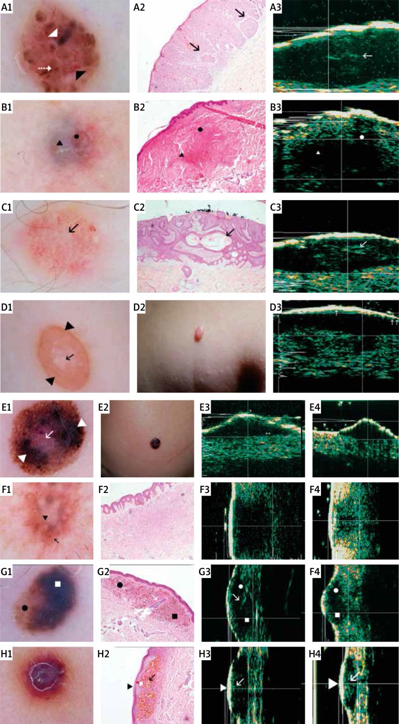
Aim: The study aimed at a comparison of the data obtained by using HFUS, histopathological and dermatoscopic images of selected skin lesions to determine their common features.
Material and methods: Nodular lesions classified as potentially malignant were subjected to clinical, dermatoscopic and high-frequency ultrasonographic examinations. Then the patients were referred for surgical removal with histopathological assessment.
Results: A total of 54 nodular lesions were examined, out of which 34 were diagnosed as non-melanoma. The most common lesions were melanocytic naevi dermatofibroma, nodular basal cell carcinoma and pyogenic granuloma. Other examined lesions included blue naevus, seborrheic wart, xanthogranuloma juvenile and Spits naevus. In all lesions except Spitz naevus, HFUS images corresponded at least with dermatoscopic or histopathology images.
Conclusions: HFUS can be used as a supporting diagnostic tool ensuring better pre-operative proceedings. HFUS is a non-invasive, easy and inexpensive screening method for the determination of different skin cancers as it provides valuable information allowing to determine the cutting margins and lesion shape.
Keywords: basal cell carcinoma; blue nevus; high-frequency ultrasonography; pyogenic granuloma; seborrheic wart; xanthogranuloma juvenile.
Copyright: © 2019 Termedia Sp. z o. o.
Conflict of interest statement
The authors declare no conflict of interest.
Figures
Similar articles
-
A Preliminary Study for Quantitative Assessment with HFUS (High- Frequency Ultrasound) of Nodular Skin Melanoma Breslow Thickness in Adults Before Surgery: Interdisciplinary Team Experience.Curr Radiopharm. 2020;13(1):48-55. doi: 10.2174/1874471012666191007121626. Curr Radiopharm. 2020. PMID: 31589132
-
Role of In Vivo Reflectance Confocal Microscopy in the Analysis of Melanocytic Lesions.Acta Dermatovenerol Croat. 2018 Apr;26(1):64-67. Acta Dermatovenerol Croat. 2018. PMID: 29782304 Review.
-
Assessment of tumor thickness in melanocytic skin lesions: comparison of optical coherence tomography, 20-MHz ultrasound and histopathology.Dermatology. 2011;223(2):161-8. doi: 10.1159/000332845. Epub 2011 Oct 20. Dermatology. 2011. PMID: 22024981
-
High frequency ultrasonography of the skin and its role as an auxillary tool in diagnosis of benign and malignant cutaneous tumors--a comparison of two clinical cases.Acta Dermatovenerol Croat. 2015;23(1):43-7. Acta Dermatovenerol Croat. 2015. PMID: 25969912
-
High-frequency ultrasound accuracy in preoperative cutaneous melanoma assessment: A meta-analysis.J Eur Acad Dermatol Venereol. 2025 Jan;39(1):86-96. doi: 10.1111/jdv.20179. Epub 2024 Jul 5. J Eur Acad Dermatol Venereol. 2025. PMID: 38967397 Free PMC article.
Cited by
-
Updated Role of High-frequency Ultrasound in Assessing Dermatological Manifestations in Autoimmune Skin Diseases.Acta Derm Venereol. 2022 Aug 24;102:adv00765. doi: 10.2340/actadv.v102.1969. Acta Derm Venereol. 2022. PMID: 36000997 Free PMC article. Review.
-
Diagnostic accuracy of high-frequency ultrasound for cutaneous neoplasms: a narrative review of the literature.Arch Dermatol Res. 2024 Jun 21;316(7):419. doi: 10.1007/s00403-024-03179-7. Arch Dermatol Res. 2024. PMID: 38904763 Free PMC article. Review.
-
Management of Non-Melanoma Skin Cancer: Radiologists Challenging and Risk Assessment.Diagnostics (Basel). 2023 Feb 20;13(4):793. doi: 10.3390/diagnostics13040793. Diagnostics (Basel). 2023. PMID: 36832281 Free PMC article. Review.
-
Significance of Dermoscopy in Association with Clinical Features in Differentiation of Basal Cell Carcinoma and Benign Trichoblastic Tumours.Cancers (Basel). 2022 Aug 17;14(16):3964. doi: 10.3390/cancers14163964. Cancers (Basel). 2022. PMID: 36010957 Free PMC article.
-
A Review of Non-Invasive Skin Imaging in Merkel Cell Carcinoma: Diagnostic Utility and Clinical Implications.Cancers (Basel). 2024 Oct 24;16(21):3586. doi: 10.3390/cancers16213586. Cancers (Basel). 2024. PMID: 39518026 Free PMC article. Review.
References
-
- Hayashi K, Koga H, Uhara H, Saida T. High-frequency 30-MHz sonography in preoperative assessment of tumor thickness of primary melanoma: usefulness in determination of surgical margin and indication for sentinel lymph node biopsy. Int J Clin Oncol. 2009;14:426–30. - PubMed
-
- Vilana R, Puig S, Sanchez M, et al. Preoperative assessment of cutaneous melanoma thickness using 10-MHz sonography. AJR Am J Roentgenol. 2009;193:639–43. - PubMed
-
- Gambichler T, Moussa G, Bahrenberg K, et al. Preoperative ultrasonic assessment of thin melanocytic skin lesions using a 100-MHz ultrasound transducer: a comparative study. Dermatol Surg. 2007;33:818–24. - PubMed
-
- Guitera P, Li LX, Crotty K, et al. Melanoma histological Breslow thickness predicted by 75-MHz ultrasonography. Br J Dermatol. 2008;159:364–69. - PubMed
LinkOut - more resources
Full Text Sources