

Epidemiology of dermatomycoses in southwest Poland, years 2011-2016
- PMID: 31839778
- PMCID: PMC6906960
- DOI: 10.5114/ada.2018.80615
Epidemiology of dermatomycoses in southwest Poland, years 2011-2016
Abstract
Introduction: Superficial mycosis is one of the most common diseases worldwide, however its epidemiology is changing over time.
Aim: To present epidemiological data of the skin fungal infections diagnosed in the years 2011-2016 in Lower Silesia.
Material and methods: A total of 11 004 patients with a clinically suspected superficial mycosis were investigated. Skin scrapings, nail clippings and plucked hair were examined with a direct microscopy, Wood's lamp and culture. Particular species were identified via polymerase chain reaction (PCR) examination. The lesions suspected for pityriasis versicolor were screened for Malassezia with Wood's lamp and direct microscopy.
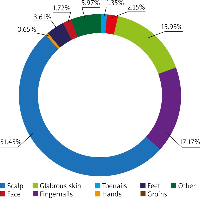
Results: Dermatomycosis was diagnosed in 1653 (15.00%) patients with 1795 fungi identified. 1858 specimens were indicative of fungal infection including dermatophytes, yeasts and moulds. Out of 924 cases of dermatophytic infections (51.48%), Trichophyton rubrum accounted for the majority (71.75%) and was followed by Trichophyton tonsurans (16.77%). Among the yeasts (716; 39.89%), Candida spp. was the most common agent identified (521; 67.66%). The sites affected most often were toenails (956; 51.45%) and fingernails (319; 17.17%). In paediatric population the most common diagnosis was tinea corporis (60, 41.10%).
Conclusions: Our study revealed that toenail onychomycosis remains the most common superficial mycosis and T. rubrum is the most common pathogen. However, in a longer period of observation, a decrease in the number of tinea capitis cases and an increase in infections caused by T. tonsurans were noticed. Observed changes indicate the need for continuing studies to detect the upcoming epidemiological trends.
Keywords: Poland; dermatomycoses; dermatophytes; epidemiology; moulds; yeasts.
Copyright: © 2018 Termedia Sp. z o. o.
Conflict of interest statement
The authors declare no conflict of interest.
Figures
References
-
- Zhan P, Liu W. The changing face of dermatophytic infections worldwide. Mycopathologia. 2017;182:77–86. - PubMed
-
- Jankowska-Konsur A, Dyląg M, Hryncewicz-Gwóźdź A, et al. A 5-year survey of dermatomycoses in southwest Poland, years 2003-2007. Mycoses. 2009;54:162–7. - PubMed
-
- Nenoff P, Krüger C, Ginter-Hanselmayer G, et al. Mycology – an update. Part 1: dermatomycoses: cusative agents, epidemiology and pathogenesis. J Dtsch Dermatol Ges. 2014;12:188–210. - PubMed
-
- Wlodek C, Trickey A, De Berker D, et al. Trends in laboratory-diagnosed onychomycosis between 2006 and 2014 in the South West of England. BJD. 2017;176:237–40. - PubMed
-
- Faergemann J, Baran R. Epidemiology, clinical presentation and diagnosis of onychomycosis. BJD. 2003;149(Suppl 65):1–4. - PubMed
LinkOut - more resources
Full Text Sources
Miscellaneous