Review
. 2020 Jan;598(2):227-247.
doi: 10.1113/JP278885.

Polyaromatic hydrocarbons in pollution: a heart-breaking matter

Affiliations
Review

Polyaromatic hydrocarbons in pollution: a heart-breaking matter

C R Marris et al. J Physiol. 2020 Jan.

Abstract

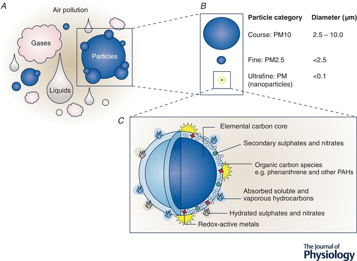
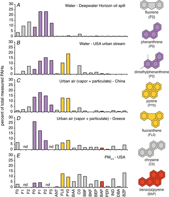
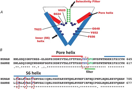
Air pollution is associated with detrimental effects on human health, including decreased cardiovascular function. However, the causative mechanisms behind these effects have yet to be fully elucidated. Here we review the current epidemiological, clinical and experimental evidence linking pollution with cardiovascular dysfunction. Our focus is on particulate matter (PM) and the associated low molecular weight polycyclic aromatic hydrocarbons (PAHs) as key mediators of cardiotoxicity. We begin by reviewing the growing epidemiological evidence linking air pollution to cardiovascular dysfunction in humans. We next address the pollution-based cardiotoxic mechanisms first identified in fish following the release of large quantities of PAHs into the marine environment from point oil spills (e.g. Deepwater Horizon). We finish by discussing the current state of mechanistic knowledge linking PM and PAH exposure to mammalian cardiovascular patho-physiologies such as atherosclerosis, cardiac hypertrophy, arrhythmias, contractile dysfunction and the underlying alterations in gene regulation. Our aim is to show conservation of toxicant pathways and cellular targets across vertebrate hearts to allow a broad framework of the global problem of cardiotoxic pollution to be established. AhR; Aryl hydrocarbon receptor. Dark lines indicate topics discussed in this review. Grey lines indicate topics reviewed elsewhere.

Keywords: PAH; PM; PM2.5; air pollution; cardiotoxicity; cardiovascular dysfunction; heart disease; oil spills; particulate matter; phenanthrene.


Figures

Figure 1
Figure 1. Constituents of air pollution
A, air pollution can be broadly characterized into gases, liquids and particles. PAHs can be found in the gaseous phase of air pollution as well as binding to the particle surface. B, particles can be further classified into size categories of particulate matter (PM). For perspective, if a human hair has a width of approximately 50 µm (0.05 mm) then PM10 encompasses particles with a diameter below a fifth of this size (<10 µm). Fine PM (<2.5 µm) would be a quarter of this size, and smaller still, ultrafine (nano) particles a hundredth of the size of PM10 (<0.1 µm). C, schematic diagram providing an example of the complex composition of a combustion derived ultrafine (nano) particle, such as a diesel exhaust particle (DEP), a common PM in urban air. The carbon core of DEP is coated with a diverse range of chemical species including reactive transition metals and polyaromatic hydrocarbons like phenanthrene. Detail of surface chemicals in C is not to scale. Figure reproduced from Environmental Health Perspectives with permission from the authors (Stone et al. 2017).
Figure 2
Figure 2. Comparison of PAH composition between water and air samples
Composition profiles represent the percentage of the indicated PAHs normalized to the sum total of the measured set, with ring number and molecular weight increasing to the right. Parent non‐alkylated compounds are denoted with a 0 (e.g. F0, P0), while alkylated compounds (e.g. those with 1 or 2 additional methyl groups) are indicated by F1, F2, P1, P2, etc. Selected individual structures are shown on the right, with phenanthrene and its alkylated homologues indicated in purple, the 4‐ringed pyrogenic compounds indicated in gold (pyrene and fluoranthene), and the archetypal carcinogenic 5‐ringed PAH benzo(a)pyrene indicated in red. Five sample sources are shown. A, water from the area of the Gulf of Mexico impacted by the 2010 Deepwater Horizon oil spill (Incardona et al. 2014); B, passive samplers placed in an urban stream in Seattle (USA) that receives large quantities of stormwater runoff from nearby roadways (Scholz et al. 2011; J. P. Incardona, unpublished); C, a total high volume air sample from Guangzhou (China) collected July 2001 by combined glass fibre filter (GFF) and polyurethane foam (PUF) plug (Bi et al. 2003); D, the mean of 16 total GFF/PUF samples collected between November 2000 and February 2002 in Heraklion (Greece) (Tsapakis & Stephanou, 2005); E, mean of quarterly samples collected over the year 2004 using a PM2.5 particle composition monitoring system at a heavily trafficked urban site in Atlanta, USA (Li et al. 2009). Abbreviations: F, fluorene; P, phenanthrene; ANT, anthracene; FL, fluoranthene; PY, pyrene; BAA, benz(a)anthracene; C, chrysene; BBF, benzo(b)fluoranthene; BKF, benzo(k)fluoranthene; BEP, benzo(e)pyrene; BAP, benzo(a)pyrene; PER, perylene; IND, indeno(123‐cd)pyrene; DBA, dibenzanthracene; BZP, benzo(ghi)perylene; nd, not determined.
Figure 3
Figure 3. The effects of Phe on excitation contraction coupling
The effect of Phe on the intracellular Ca2+ transient in sheep ventricular myocytes (25 µM) (A) and fish (bluefin tuna, 5 µM) ventricular myocytes (B) loaded with Fluo‐4AM and stimulated to contract at 0.5 Hz. The effect of Phe on the ventricular AP during whole‐cell current clamp in sheep (C) and bluefin tuna (D) ventricular myocytes. The red lines in both traces show data recorded during exposure to 25 µM Phe. The pink line shows the effect of 5 µM Phe exposure in tuna. No discernible effects were seen at 5 µM in sheep (not shown). Tuna data are from Brette et al. 2017, with permission from Scientific Reports, and sheep data are unpublished data of C.R.M., S.N.K. and H.A.S. in myocytes supplied by the members of the laboratories of Dr K. Dibb and Prof. A. Trafford at the University of Manchester.
Figure 4
Figure 4. PAH and EC coupling in cardiac myocytes
A, the top panel shows a simplified schematic of EC coupling in a cardiac cell, including the main ion channels, pumps, transporters, sarcoplasmic reticulum (SR), mitochondria and contractile proteins. EC coupling proceeds when an action potential (AP) opens L‐type Ca2+ channels (LTCC) in the cell membrane. This Ca2+ entry can directly activate the myofilament and can also initiate Ca2+‐induced Ca2+‐release from Ca2+ channels on SR membrane (ryanodine receptors, RyR). The global increase in the cytosolic [Ca2+]i transient activates contractile proteins leading to contraction. Repolarization of the AP occurs in large part by K+ efflux through ERG (ether‐à‐go‐go‐related gene) channel. Relaxation follows repolarization by removal of Ca2+ from the cytosol primarily via re‐uptake of Ca2+ into the SR (via the sarcoplasmic reticulum calcium ATPase, SERCA) and extrusion of Ca2+ out of the cell via the Na+‐Ca2+ exchanger (NCX). Exposure to PAHs, specifically Phe, impairs EC coupling as indicated by the red inhibition bars at various ionic flux pathways. Ion flux pathways where inhibition has only been measured indirectly have a red question mark beside them. Many pathways have not yet been investigated like direct effect on myofilaments, or the NCX. B, interactions between EC coupling and excitation‐transcription coupling, indicating points of PAH regulation which inhibit Ca2+ cycling and thus transcription. EC coupling is as in A. Excitation‐transcription coupling proceeds when puronergic G‐protein coupled receptors (P2Y) are activated by ATP (not shown), which activates phosphoinositide 3‐kinase (PI3K) via G protein. This causes phospholipase C (PLC) to be recruited to the membrane and produce inositol 1,4,5‐trisphosphate (IP3) and diacyl glycerol (DAG). IP3 receptor‐mediated Ca2+ signals cause Ca2+ to enter the nucleus and through kinases and/or phophatases (PKC, CamK, RhoA/ROK, CaN) activate transcription factors (CREB, NFAT, Myocd/SRF) ultimately leading to gene expression. Tricyclic PAHs disrupt Ca2+ cycling pathways in the cell, reducing Ca2+ stores, and affect Ca2+‐related gene expression pathways. CaN, calcineurin; CamK, calcium–calmodulin‐dependent protein kinase; PKC, protein kinase C; RhoA, Ras homolog gene family, member A; ROK, rho associated kinase; NFAT, nuclear factor of activated T‐cells; SRF, serum response factor; Myocd, myocardin; CREB, cAMP response element‐binding protein.
Figure 5
Figure 5. Homology of ERG channels
Schematic representation (A) and sequence homology (B) of pore‐helix and S6 helix regions between hERG and zERG channels. Sequence alignments made in UniProt (Q12809 and Q8JH78). The pore‐helix is highlighted in red and S6 helix in blue. The GFG selectivity sequence is highlighted in green. Residues known to contribute to the canonical drug binding site in hERG are highlighted by red dashed boxes. Note that residue numbering in A refers to amino acid position in hERG. hERG:zERG equivalents: T623 = T595; S624 = S596; V625 = V597; G648 = G620; Y652 = Y624; F656 = F628.

References

    1. Abdel‐Shafy HI & Mansour MSM (2016). A review on polycyclic aromatic hydrocarbons: Source, environmental impact, effect on human health and remediation. Egypt J Pet 25, 107–123.
    1. Abramochkin D V., Hassinen M & Vornanen M (2018). Transcripts of Kv7.1 and MinK channels and slow delayed rectifier K+ current (I Ks) are expressed in zebrafish (Danio rerio) heart. Pflugers Arch 470, 1753–1764. - PubMed
    1. European Environment Agency (2019). Air Quality in Europe — 2018 Report Available at: http://www.eea.europa.eu/publications/air-quality-in-europe-2012.
    1. Ainerua MO, Tinwell J, Kompella SN, Sørhus E, White KN, van Dongen BE & Shiels HA (2020). Understanding the cardiac toxicity of the anthropogenic pollutant phenanthrene on the freshwater indicator species (Salmo trutta): From whole heart to cardiomyocytes. Chemosphere 239, 124608. - PMC - PubMed
    1. Alday A, Alonso H, Gallego M, Urrutia J, Letamendia A, Callol C & Casis O (2014). Ionic channels underlying the ventricular action potential in zebrafish embryo. Pharmacol Res 84, 26–31. - PubMed

Publication types

MeSH terms

Substances