. 2020 Mar 15;201(6):650-660.
doi: 10.1164/rccm.201903-0563CI.

Etiology, Risk Factors, and Biomarkers in Systemic Sclerosis with Interstitial Lung Disease

Affiliations

Etiology, Risk Factors, and Biomarkers in Systemic Sclerosis with Interstitial Lung Disease

Dinesh Khanna et al. Am J Respir Crit Care Med. .

Abstract

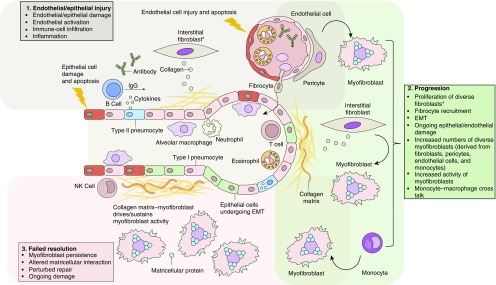
Systemic sclerosis (SSc) is a complex, multiorgan, autoimmune disease. Lung fibrosis occurs in ∼80% of patients with SSc; 25% to 30% develop progressive interstitial lung disease (ILD). The pathogenesis of fibrosis in SSc-associated ILD (SSc-ILD) involves cellular injury, activation/differentiation of mesenchymal cells, and morphological/biological changes in epithelial/endothelial cells. Risk factors for progressive SSc-ILD include older age, male sex, degree of lung involvement on baseline high-resolution computed tomography imaging, reduced DlCO, and reduced FVC. SSc-ILD does not share the genetic risk architecture observed in idiopathic pulmonary fibrosis (IPF), with key risk factors yet to be identified. Presence of anti-Scl-70 antibodies and absence of anti-centromere antibodies indicate increased likelihood of progressive ILD. Elevated levels of serum Krebs von den Lungen-6 and C-reactive protein are both associated with SSc-ILD severity and predict SSc-ILD progression. A promising prognostic indicator is serum chemokine (C-C motif) ligand 18. SSc-ILD shares similarities with IPF, although clear differences exist. Histologically, a nonspecific interstitial pneumonia pattern is commonly observed in SSc-ILD, whereas IPF is defined by usual interstitial pneumonia. The course of SSc-ILD is variable, ranging from minor, stable disease to a progressive course, whereas all patients with IPF experience progression of disease. Although appropriately treated patients with SSc-ILD have better chances of stabilization and survival, a relentlessly progressive course, akin to IPF, is seen in a minority. Better understanding of cellular and molecular pathogenesis, genetic risk, and distinctive features of SSc-ILD and identification of robust prognostic biomarkers are needed for optimal disease management.

Keywords: autoimmune diseases; biomarkers; interstitial lung diseases; risk factors; systemic sclerosis.


Figures

Figure 1.
Figure 1.
Cellular pathogenesis of fibrotic lung injury in systemic sclerosis. *Including SPINT2hi, MFAP5hi, and few WIF1hi fibroblasts. EMT = epithelial–mesenchymal transition; NK cell = natural killer T cell.
Figure 2.
Figure 2.
(AC) Limited disease (<20% extent) on high-resolution computed tomography (HRCT) imaging in a 72-year-old female nonsmoker. HRCT images at the level of (A) the aortic arch show no convincing interstitial lung disease (ILD), and (B and C) very limited subpleural ground-glass opacification. (DF) ILD of “indeterminate” extent on HRCT imaging in a 46-year-old female nonsmoker with systemic sclerosis. (AD) The upper zones show minor reticulation, (E) just below the level of the right hemidiaphragm, and (F) the costophrenic recesses demonstrating reticulation, ground-glass opacification, and traction bronchiectasis/bronchiolectasis. The morphologic features are in keeping with a fibrotic nonspecific interstitial pneumonia pattern. Disease extent on HRCT imaging with regard to the 20% threshold is difficult to gauge (i.e., “indeterminate” according to the Goh staging); FVC in this patient was 60% predicted, thereby indicating “extensive” ILD. Note the marked esophageal dilatation containing food residue.
Figure 3.
Figure 3.
High-resolution computed tomography images in a 58-year-old woman with systemic sclerosis who never smoked; DlCO 32% predicted and FVC 76% predicted. Axial images at (A) the level of the aortic arch, (B) the carina, and (C) the lower lobes demonstrate extensive disease (>20% extent by visual estimation) and (D) coronal reconstruction. There is marked honeycombing, particularly in the lower lobes, indicating a usual interstitial pneumonia pattern. The coronal image shows striking lower zone preponderance of disease.
Figure 4.
Figure 4.
Computed tomography imaging in a 52-year-old man, ex-smoker, with a DlCO of 22% and FVC 56% predicted. Axial images at (A) the level of the arch, (B) the pulmonary venous confluence, and (C) the costophrenic recesses show extensive (>20%) disease. There is predominant ground-glass opacification with fine reticulation, no honeycombing, but severe traction bronchiectasis. The computed tomography features are consistent with a fibrotic nonspecific interstitial pneumonia pattern. Note also the marked esophageal dilatation.
Figure 5.
Figure 5.
Histopathology of systemic sclerosis–associated interstitial lung disease (SSc-ILD) and idiopathic pulmonary fibrosis (13, 68). (A) SSc-ILD. (Ai) Nonspecific interstitial pneumonia; note the diffuse alveolar septal thickening throughout the lobule with lack of peripheral accentuation in the area of an interlobular septum on the left. (Aii) Usual interstitial pneumonia; note the peripheral involvement of a pulmonary lobule sparing the centrilobular area containing the bronchovascular bundle. Arrows indicate fibroblastic foci. (Aiii) Pulmonary arterial hypertension; note the hypertensive arterial changes with prominent intimal fibrosis. Arrow indicates separation of the media and intima by the internal elastic lamina. (Aiv) Pleural fibrosis; its presence supports the diagnosis of SSc-ILD in the appropriate clinical setting. Hematoxylin and eosin (H&E)-stained sections are shown in Ai, Aii, and Aiv; Verhoeff–van Gieson–stained sections in Aiii. Original magnification ×40 in i and ii; ×200 in iii; ×100 in iv. (B) Usual interstitial pneumonia. (Bi) At low magnification, the diagnostic key is the abrupt alternating of scarred and normal lung (patchwork pattern: scar-normal-scar-normal). In the scarred areas, the alveolar architecture is obliterated. (Bii) The fibrosis frequently prevails at the periphery of the lobule in the subpleural paraseptal regions (arrows), with relative sparing of the centrolobule. This is a useful diagnostic clue, particularly in early cases like this (H&E 20). (Biii) Honeycomb consists of enlarged airspaces lined by bronchiolar epithelium, frequently filled by mucus and surrounded by dense scars. Note the architectural distortion and the abrupt transition with residual normal lung seen in the right upper corner. (Biv) A fibroblastic focus consisting of a dome-shaped proliferation of myofibroblasts immersed in a myxoid matrix. Fibroblastic foci can be covered by bronchiolar epithelium, as here, or by hyperplasic pneumocytes. H&E-stained sections are shown in BiBiv. (A) Reprinted from Arthritis & Rheumatology, Vol. 66, Herzog EL, et al., Review: Interstitial Lung Disease Associated With Systemic Sclerosis and Idiopathic Pulmonary Fibrosis: How Similar and Distinct? 1967–1978, Copyright (2014), by permission from John Wiley & Sons. (B) Reprinted from Respiratory Medicine, Vol. 104, Cavazza A, et al., The role of histology in idiopathic pulmonary fibrosis: An update, S11–S22, Copyright (2010), by permission from Elsevier.

References

    1. Denton CP, Hughes M, Gak N, Vila J, Buch MH, Chakravarty K, et al. BSR and BHPR Standards, Guidelines and Audit Working Group. BSR and BHPR guideline for the treatment of systemic sclerosis. Rheumatology (Oxford) 2016;55:1906–1910. - PubMed
    1. Denton CP, Khanna D. Systemic sclerosis. Lancet. 2017;390:1685–1699. - PubMed
    1. Khanna D, Tashkin DP, Denton CP, Lubell MW, Vazquez-Mateo C, Wax S. Ongoing clinical trials and treatment options for patients with systemic sclerosis-associated interstitial lung disease. Rheumatology (Oxford) 2019;58:567–579. - PMC - PubMed
    1. Ranque B, Mouthon L. Geoepidemiology of systemic sclerosis. Autoimmun Rev. 2010;9:A311–A318. - PubMed
    1. van den Hoogen F, Khanna D, Fransen J, Johnson SR, Baron M, Tyndall A, et al. 2013 classification criteria for systemic sclerosis: an American College of Rheumatology/European League against Rheumatism collaborative initiative. Arthritis Rheum. 2013;65:2737–2747. - PMC - PubMed