. 2020 Feb 4;59(4):541-551.
doi: 10.1021/acs.biochem.9b00822. Epub 2019 Dec 31.

Protein Footprinting and X-ray Crystallography Reveal the Interaction of PD-L1 and a Macrocyclic Peptide

Affiliations

Protein Footprinting and X-ray Crystallography Reveal the Interaction of PD-L1 and a Macrocyclic Peptide

Ben Niu et al. Biochemistry. .

Abstract

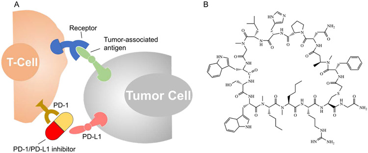
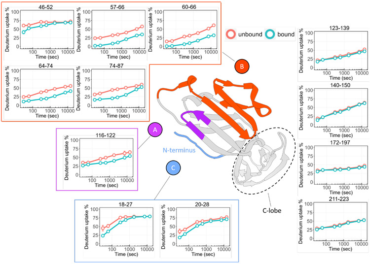
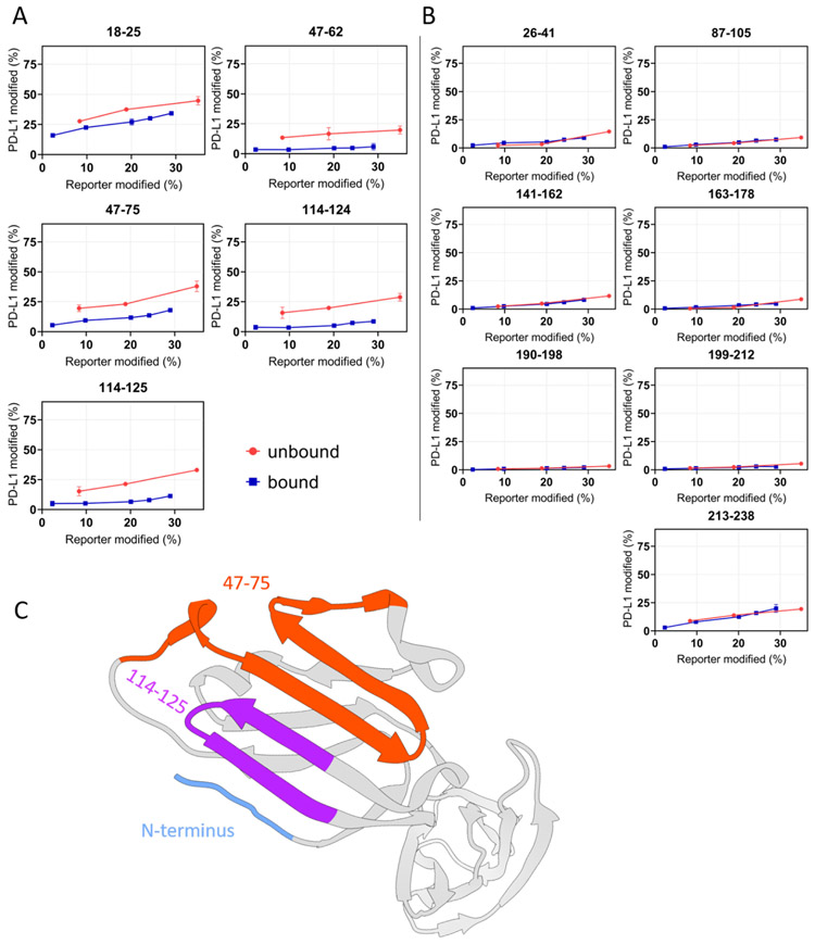
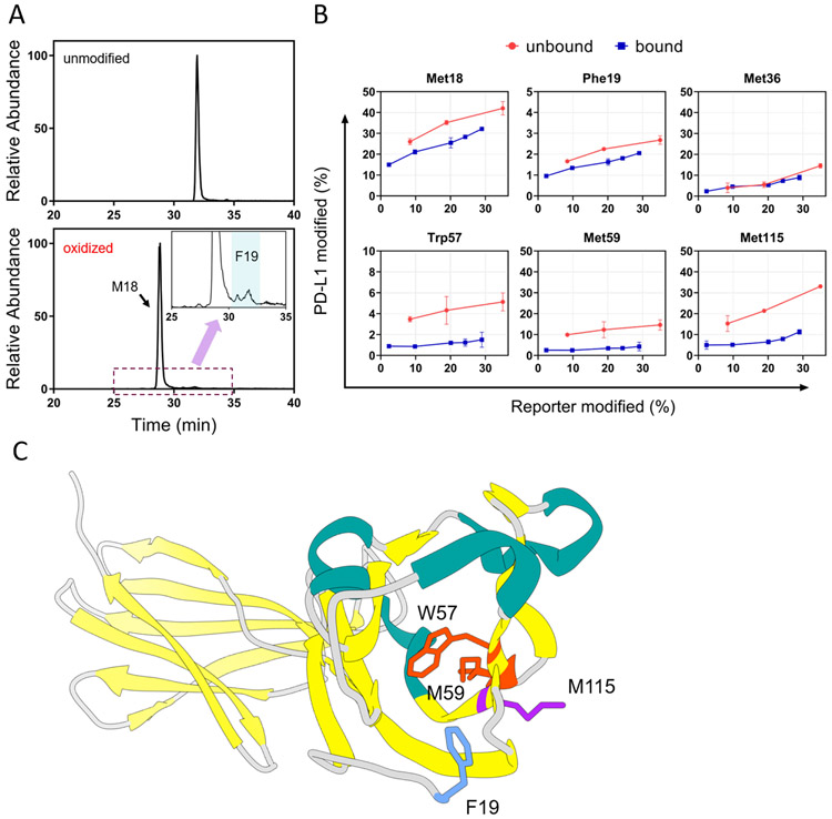
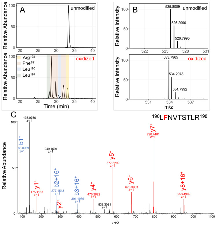
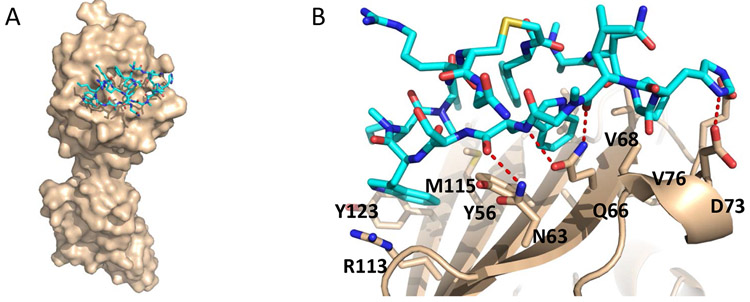
Blocking interactions between PD-1 and PD-L1 opens a new era of cancer treatment involving immunity modulation. Although most immunotherapies use monoclonal antibodies, small-molecule inhibitors offer advantages. To facilitate development of small-molecule therapeutics, we implemented a rapid approach to characterize the binding interfaces of small-molecule inhibitors with PD-L1. We determined its interaction with a synthetic macrocyclic peptide by using two mass spectrometry-based approaches, hydrogen-deuterium exchange and fast photochemical oxidation of proteins (FPOP), and corroborated the findings with our X-ray structure of the PD-L1/macrocycle complex. Although all three approaches show that the macrocycle binds directly to PD-L1 over the regions of residues 46-87 and 114-125, the two protein footprinting approaches show additional binding at the N-terminus of PD-L1, and FPOP reveals some critical binding residues. The outcomes not only show the binding regions but also demonstrate the utility of MS-based footprinting in probing protein/ligand inhibitory interactions in cancer immunotherapy.


Figures

Figure 1.
Figure 1.
(A) Cartoon showing inhibitors of programmed death-ligand 1 (PD-L1) or programmed cell death protein 1 (PD-1) can block the interaction between the two proteins, reactivating the immune response; (B) Structure of macrocyclic peptide (macrocycle), an inhibitor of PD-1/PD-L1 protein-protein interaction.
Figure 2.
Figure 2.. Peptide-level HDX kinetics analysis of PD-L1.
The comparison between macrocycle-bound (teal) and unbound (orange) states shows significant changes of HDX for mainly three regions, region A is represented by peptide 116-122 (denoted in purple), region B includes peptides 46-52, 57-66, 60-66, 64-74, 74-87 (denoted in orange), and region C that contains N-terminal peptides 18-27, 20-28 (denoted in light blue). The HDX results mapped onto the crystal structure of PD-L1 (PDB 4Z18) show all three regions are discontinuously located on the N-lobe. No deuterium uptake differences observed for peptides on C-lobe. Error bars correspond to ± SD from triplicate measurements and are nearly always smaller than the diameters of the open circles representing data points. For binding regions, the differences between the curves representing bound and unbound are at least 5 times the SD.
Figure 3.
Figure 3.. “Time-dependent” FPOP response curves of each peptide of PD-L1.
(A) Differential yields of FPOP observed between macrocycle-bound and unbound PD-L1 (represented by 5 tryptic peptides) suggest three discontinuous binding interfaces on PD-L1, including N-terminus, 47-75, and 114-125. (B) The overlapping of FPOP response curves indicate comparable surface solvent accessibility of these regions between bound and unbound states. These regions are not involved in the macrocycle binding. (C) FPOP results mapped onto the PD-L1 structure (PDB 4Z18) shows comparable outcome as that of HDX. The error bars correspond to ± SD from triplicate measurements and show that for binding sites, the differences between the accumulated differences for three points representing bound and unbound are at least 3 times the propagated errors (sq root of the sum of the squares of the SDs). This corresponds to 99.7% confidence in the assignment.
Figure 4.
Figure 4.
(A) XICs corresponding to modified and unmodified peptide 18-25 (amino acid sequence MFTVTVPK) showed Met18 is the major modification site, whereas modification on Phe19 is less abundant. The insert represents a zoomed-in region where peptides with Phe19 oxidation elutes. (B) Residue-level time-dependent curves of macrocycle-bound and unbound PD-L1. Residues identified that show significant differential FPOP yields between bound and unbound states of PD-L1 include M18, F19, W57, M59, and M115. Residues were identified by manual interpretation of the product-ion (MS/MS) spectra. Error bars correspond to ± SD from triplicate measurements. The error bars correspond to ± SD from triplicate measurements and show that for binding sites, the differences between the accumulated differences for three points representing bound and unbound are at least 3 times the propagated errors (sq root of the sum of the squares of the SDs). This corresponds to 99.7% confidence in the assignment. (C) Close-up view of these residues show they are spatially localized on the N-lobe of PD-L1.
Figure 5.
Figure 5.
Example of LC-MS/MS separation, detection, and identification of unmodified and oxidatively modified species of peptide 190-198. (A) XICs of unmodified and singly oxidized peptide 190-198; (B) mass spectra of precursor ions (+2 charge) show a 16 Da shift from unmodified to modified peptide 190-198; (C) the product-ion (MS/MS) spectra of the singly oxidized peptide 190-198 locates modification on Phe191.
Figure 6.
Figure 6.
(A) Overall structure of the macrocycle (cyan) bound to human PD-L1 (beige). The structure confirms only the N-terminal lobe is involved in binding; (B) Residue-specific binding interface between macrocycle and human PD-L1. The macrocycle carbon atoms are colored cyan while the PD-L1 carbon atoms are colored beige. Oxygen atoms (red), nitrogen atoms (blue), and sulfur atoms (yellow) are colored accordingly.

References

    1. Atkins MB; Sznol M, Cancer Immunotherapy: Past Progress and Future Directions. Semin Oncol 2015, 42 (4), 518–22. - PubMed
    1. Gubin MM; Schreiber RD, CANCER. The odds of immunotherapy success. Science 2015, 350 (6257), 158–9. - PubMed
    1. June CH; O'Connor RS; Kawalekar OU; Ghassemi S; Milone MC, CAR T cell immunotherapy for human cancer. Science 2018, 359 (6382), 1361–1365. - PubMed
    1. Page DB; Postow MA; Callahan MK; Allison JP; Wolchok JD, Immune modulation in cancer with antibodies. Annu Rev Med 2014, 65, 185–202. - PubMed
    1. Ribas A; Wolchok JD, Cancer immunotherapy using checkpoint blockade. Science 2018, 359 (6382), 1350–1355. - PMC - PubMed

Publication types

MeSH terms