. 2019 Dec 16;10(1):382.
doi: 10.1186/s13287-019-1481-9.

Relative safety of various spermatogenic stem cell purification methods for application in spermatogenic stem cell transplantation

Affiliations

Relative safety of various spermatogenic stem cell purification methods for application in spermatogenic stem cell transplantation

Jia Tian et al. Stem Cell Res Ther. .

Abstract

Background: Spermatogonial stem cell (SSC) transplantation technology as a promising option for male fertility preservation has received increasing attention, along with efficient SSC purification technology as a necessary technical support; however, the safety of such application in patients with tumors remains controversial.

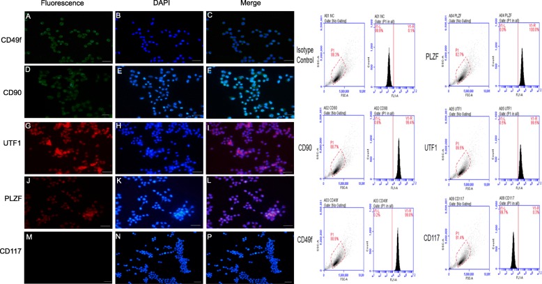
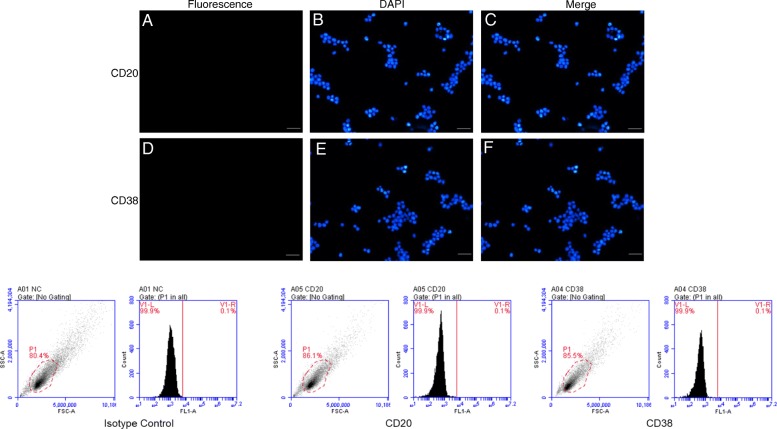
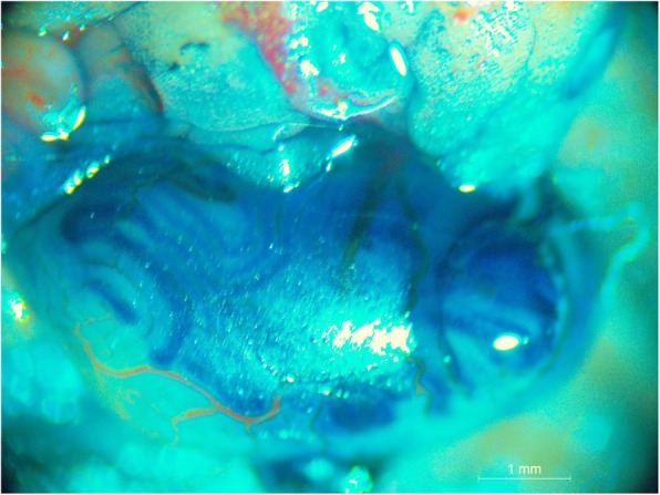
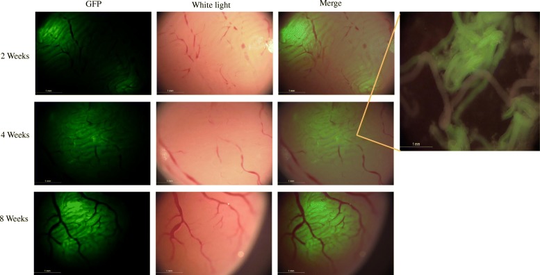
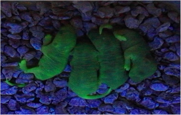
Methods: In this study, we used a green fluorescent protein mouse xenograft model of B cell acute lymphocytic leukemia. We isolated and purified SSCs from the testicular tissue of model mice using density gradient centrifugation, immune cell magnetic bead separation, and flow cytometry. The purified SSCs were transplanted into convoluted seminiferous tubules of the nude mice and C57BL/6 male mice subjected to busulfan. The development and proliferation of SSCs in the recipient testis were periodically tested, along with whether B cell acute lymphocytic leukemia was induced following SSC implantation. The genetic characteristics of the offspring obtained from natural mating were also observed.

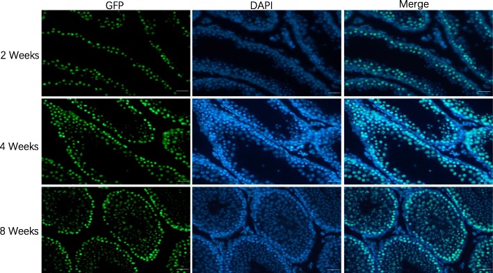
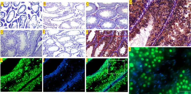
Results: In testicular leukemia model mice, a large number of BALL cells infiltrated into the seminiferous tubule, spermatogenic cells, and sperm cells in the testis tissue decreased. After spermatogonial stem cell transplantation, the transplanted SSCs purified by immunomagnetic beads and flow cytometry methods colonized and proliferated extensively in the basement of the seminiferous tubules of mice; a large number of spermatogenic cells and sperm were found in recipient testicular tissue after 12 weeks of SSC transplantation. In leukemia detection in nude mice after transplantation in the three SSC purification groups, a large number of BALL cells could be detected in the blood of recipient mice 2-3 weeks after transplantation in the density gradient centrifugation group, but not in the blood of the flow cytometry sorting group and the immunomagnetic bead group after 16 weeks of observation.

Conclusions: In this study, we confirmed that immunomagnetic beads and flow cytometry methods of purifying SSCs from the testicular tissue of the testicular leukemia mouse model could be safely applied to the SSC transplantation technology without concomitant tumor implantation. The results thus provide a theoretical basis for the application of tumor SSC cryopreservation for fertility preservation in patients with tumors.

Keywords: Density gradient centrifugation; Fertility preservation; Flow cytometry; Immunomagnetic bead separation; Spermatogonial stem cell; Tumor.


Conflict of interest statement

The authors declare that they have no competing interests.

Figures

Fig. 1
Fig. 1
Marker verification and validation of independent markers in BALL cells by immunofluorescence and flow cytometry. The expression of CD20 (ac) and CD38 (df), were positive on BALL cell membranes. CD90f (gi) and CD49f (j, k) were not expressed in BALL cells. DAPI indicates the cell nucleus. This finding was confirmed via the conducted flow cytometry analysis. Scale bar = 100 μm
Fig. 2
Fig. 2
Marker verification and validation of independent markers in SSCs by immunofluorescence and flow cytometry. Undifferentiated SSCs expressed CD49f (ac), CD90 (df), UTF1 (ef), and PLZF (jl), but not CD117 (mp). DAPI indicates the cell nucleus. This finding was confirmed via the conducted flow cytometry analysis. Scale bar = 100 μm
Fig. 3
Fig. 3
CD20 and CD38 verification and validation of independent markers in SSCs by immunofluorescence and flow cytometry. CD20 (ac) and CD38 (df) were not expressed in SSCs. DAPI indicates the cell nucleus. This finding was confirmed via the conducted flow cytometry analysis. Scale bar = 100 μm
Fig. 4
Fig. 4
Onset and survival of nude mice. Onset and survival time of leukemia after different numbers of BALL cells were injected into the tail vein of nude mice
Fig. 5
Fig. 5
Detection of testicular leukemia model mice. HE staining: a In normal C57BL/6 mice, a large number of spermatogenic cells and sperm cells could be seen in testis tissue. b In the model group, after 104 cells were injected to establish the model for 3 weeks, spermatogenic cells and sperm cells in the testis tissue decreased with the infiltration of BALL cells. Immunohistochemistry: c In normal C57BL/6 mice, the expression of CD20 protein in testicular tissue was negative. d In the model group, a large number of CD20-positive BALL cells infiltrated into the seminiferous tubule, spermatogenic cells and sperm cells in the testis tissue decreased. e Negative control. Scale bar = 100 μm
Fig. 6
Fig. 6
Transplantation of SSCs. Trypan blue staining was used to show the SSC transplantation procedure. A 3-mm incision was made at the dorsal tip of the testis tissue of the recipient mouse, and the seminiferous tubules were exposed. After the injection, the trypan blue solution gradually filled along the seminiferous tubules. One injection (40–50 μL) could fill one fourth to one third of the testicular tissue. Scale bar = 1 mm
Fig. 7
Fig. 7
Proliferation and differentiation of GFP mouse SSCs after transplantation. The GFP-positive volume in the testis of the recipient mice gradually expanded over time, and extensive GFP expression could be observed in the testis. Scale bar = 1 mm
Fig. 8
Fig. 8
Proliferation and differentiation of SSCs after transplantation. Paraffin sections were prepared from the transplanted mouse testes. Immunofluorescence was used to detect the colonization, proliferation, and differentiation of the transplanted SSCs in the seminiferous tubules of the mice. The transplanted SSCs colonized and proliferated extensively in the basement of the seminiferous tubules of mice and differentiated into spermatogenic cells at various levels. Scale bar = 100 μm
Fig. 9
Fig. 9
Morphological changes of testicular tissue before and after transplantation. a Testicle of SSC transplantation recipient; it can be seen that spermatogenic cells in testicular tissue disappeared. b After 12 weeks of natural recovery, the spermatogenic function was still not recovered. c A large number of spermatogenic cells and sperm were found in recipient testicular tissue after 12 weeks of SSC transplantation. dg Immunohistochemical staining for detection of GFP expression in testicular tissues. d Negative control. e Natural recovery of testicular tissue in mice after 12 weeks of treatment with busulfan. f, g Testicular tissue after 12 weeks of GFP+ SSC transplantation, a large number of GFP+ spermatogenic cells and spermatozoa in the convoluted tubule. Immunofluorescence assay staining for detection of GFP expression in testicular tissues after 12 weeks of SSC transplantation (hk). h GFP. i DAPI. j, k Meger. Magnification, scale bar = 100 μm
Fig. 10
Fig. 10
Fecundity of semen GFP sperm. Detection of epigenetic characteristics of the progeny of SSC transplantation mice: C57BL/6 mice after SSC transplantation and 4-week-old female C57BL/6 were bred at 1:2 to observe the epigenetic characteristics of the progeny as assessed by GFP expression. The offspring that were born by natural mating of the GFP-transplanted mice and the C57 mice expressed GFP protein, which emitted green fluorescence under UV excitation
Fig. 11
Fig. 11
Leukemia detection in nude mice after transplantation in the three SSC purification groups as assessed by blood flow cytometry and western blotting. A large number of BALL cells could be detected in the blood of recipient mice 2–3 weeks after transplantation in the density gradient centrifugation (DGU) group, but not in the blood of the flow cytometry sorting (FCMS) group and the immunomagnetic bead (MACS) group after 16 weeks of observation. CD20 detection in testicular tissue of nude mice after transplantation in the three groups by western blotting. High CD20 expression was detected in testicular tissue of nude mice in the density gradient centrifugation (DGU) group, but not in the flow cytometry sorting (FCMS) and immunomagnetic bead sorting (MACS) groups after 16 weeks of observation

References

    1. Siegel RL, Miller KD, Jemal A. Cancer statistics, 2018. CA Cancer J Clin. 2018;68:7–30. - PubMed
    1. Giudice MG, de Michele F, Poels J, Vermeulen M, Wyns C. Update on fertility restoration from prepubertal spermatogonial stem cells: how far are we from clinical practice? Stem Cell Res. 2017;21:171–177. doi: 10.1016/j.scr.2017.01.009. - DOI - PubMed
    1. Finney EL, Johnson EK. Challenges in fertility preservation among male adolescent and young adult cancer survivors. Pediatr Blood Cancer. 2018;65:e27042. doi: 10.1002/pbc.27042. - DOI - PubMed
    1. Lambertini M, Del Mastro L, Pescio MC, Andersen CY, Azim HA, Jr, et al. Cancer and fertility preservation: international recommendations from an expert meeting. BMC Med. 2016;14:1. doi: 10.1186/s12916-015-0545-7. - DOI - PMC - PubMed
    1. Williams DH. Fertility preservation in the male with cancer. Curr Urol Rep. 2013;14:315–326. doi: 10.1007/s11934-013-0345-6. - DOI - PubMed

Publication types

LinkOut - more resources