Review
. 2020 Apr 8;28(4):997-1015.
doi: 10.1016/j.ymthe.2019.11.006. Epub 2019 Nov 13.

A Molecular Revolution in the Treatment of Hemophilia

Affiliations
Review

A Molecular Revolution in the Treatment of Hemophilia

John S S Butterfield et al. Mol Ther. .

Abstract

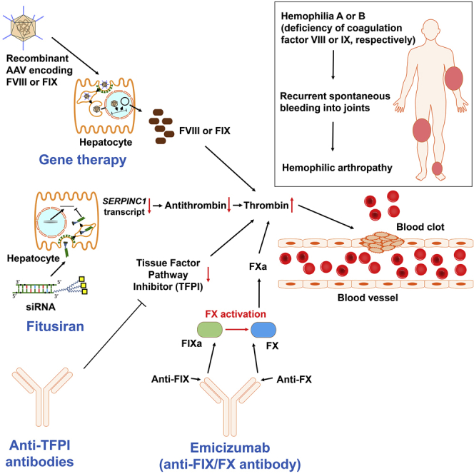
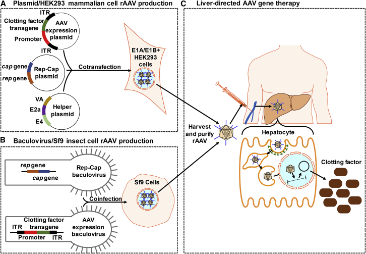
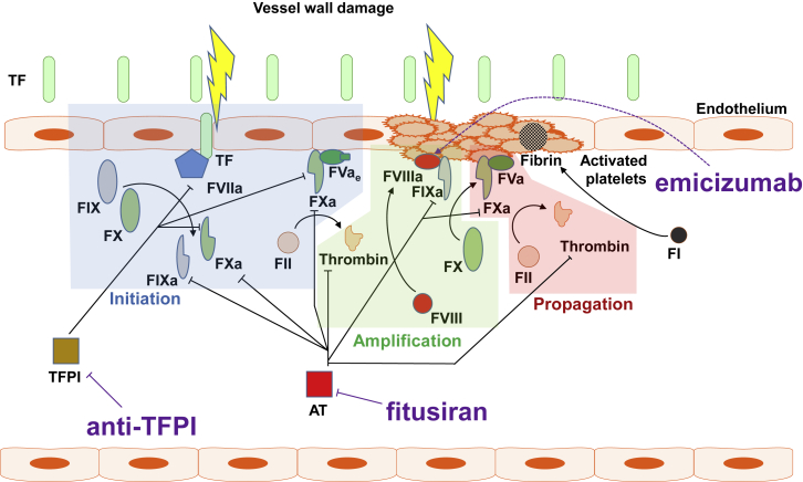
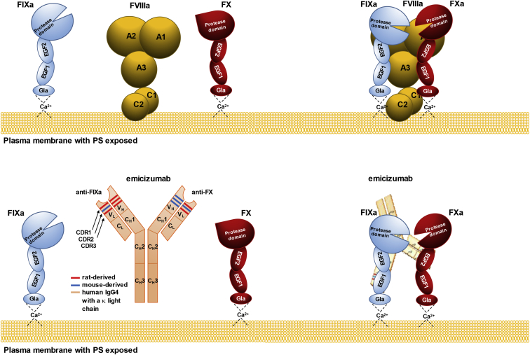
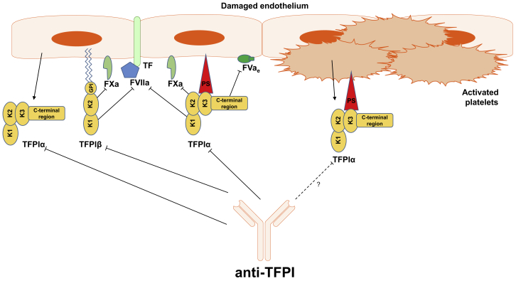
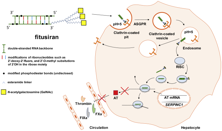
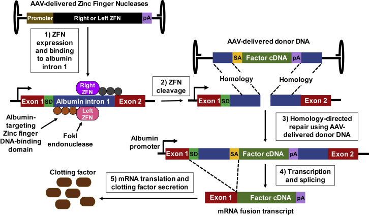
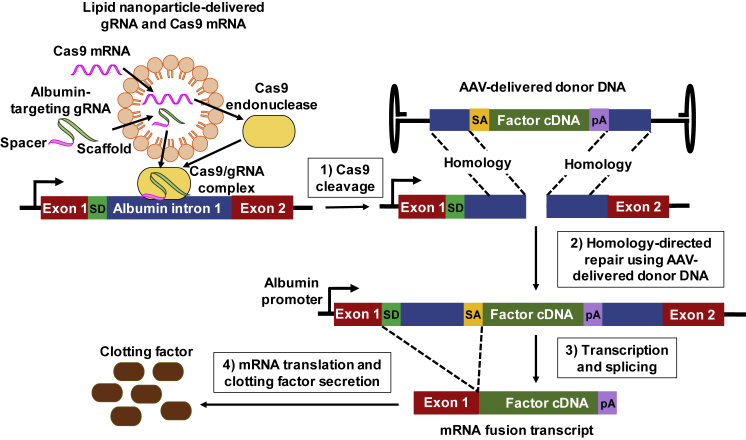
For decades, the monogenetic bleeding disorders hemophilia A and B (coagulation factor VIII and IX deficiency) have been treated with systemic protein replacement therapy. Now, diverse molecular medicines, ranging from antibody to gene to RNA therapy, are transforming treatment. Traditional replacement therapy requires twice to thrice weekly intravenous infusions of factor. While extended half-life products may reduce the frequency of injections, patients continue to face a lifelong burden of the therapy, suboptimal protection from bleeding and joint damage, and potential development of neutralizing anti-drug antibodies (inhibitors) that require less efficacious bypassing agents and further reduce quality of life. Novel non-replacement and gene therapies aim to address these remaining issues. A recently approved factor VIII-mimetic antibody accomplishes hemostatic correction in patients both with and without inhibitors. Antibodies against tissue factor pathway inhibitor (TFPI) and antithrombin-specific small interfering RNA (siRNA) target natural anticoagulant pathways to rebalance hemostasis. Adeno-associated virus (AAV) gene therapy provides lasting clotting factor replacement and can also be used to induce immune tolerance. Multiple gene-editing techniques are under clinical or preclinical investigation. Here, we provide a comprehensive overview of these approaches, explain how they differ from standard therapies, and predict how the hemophilia treatment landscape will be reshaped.


Figures

None
Graphical abstract
Figure 1
Figure 1
AAV Gene Therapy (A) Plasmid/HEK293 mammalian cell rAAV production: HEK293 cells are cotransfected with (1) AAV expression plasmid containing the clotting factor transgene and tissue-specific promoter, flanked by ITRs, (2) plasmid containing the rep and cap genes, and (3) helper plasmid containing adenovirus genes. (B) Baculovirus/Sf9 insect cell rAAV production: Sf9 cells are coinfected with (1) AAV expression baculovirus containing the clotting factor transgene and tissue-specific promoter, flanked by ITRs, and (2) baculovirus containing the rep and cap genes. (C) Liver-directed AAV gene therapy: rAAV is harvested by freeze-thawing transfected/infected cells, purified, and then injected intravenously. The transgene expression is targeted to hepatocytes using a liver-specific promoter and capsid with strong liver tropism. rAAV binds to a serotype-specific host cell receptor and is internalized by a clathrin-coated dynamin-dependent pathway into the endosomal compartment., , , The rAAV is quickly transported using microtubules to the perinuclear region and undergoes conformational changes that expose regions of its capsid to allow for escape from the endosome and nuclear localization signals for trafficking into the nucleus., , The clotting factor is expressed after AAV uncoats its capsid and converts to a circular double-stranded DNA episome by annealing of plus and minus strands delivered to the same cell or second-strand synthesis using host polymerase., rAAV, recombinant adeno-associated virus; ITR, inverted terminal repeat.
Figure 2
Figure 2
Key Events and Physiologic Inhibitors of Secondary Hemostasis Targeted by Non-replacement Hemophilia Therapies Upon vessel wall injury, normally subendothelial tissue factor (TF) is exposed to blood and binds to activated FVII (FVIIa), thus enhancing its catalytic activity. The TF-FVIIa complex generates small amounts of FIXa and FXa. FXa and early (partially activated) forms of FV (FVae) associate on negatively charged phospholipids (via calcium ions) exposed on damaged endothelial cells and activated platelets, where they form an early prothrombinase complex, which generates minute amounts of thrombin (initiation phase) by cleavage of FII (prothrombin). In the amplification phase, thrombin activates multiple coagulation factors, including cell surface-bound FVIII, FV, and FXI. FVIIIa enhances the catalytic activity of FIXa, which activates FX. FXa and thrombin-activated FV generated during the amplification phase now propel a thrombin burst on the surface of activated platelets (propagation phase), leading to cleavage of FI (fibrinogen) and formation of the fibrin clot.,, ,
Figure 3
Figure 3
Structure and Activity of FVIIIa and Emicizumab FVIIIa forms a complex with activated FIX (FIXa, a serine protease) and FX (the zymogen), so that FIXa can activate the latter. In this reaction, FVIIIa functions as a molecular scaffold for FIXa, whose protease activity toward FX is otherwise 105- to 106-fold lower and insufficient to drive the thrombin burst and, ultimately, optimal blood coagulation. Emicizumab is an asymmetric humanized bispecific anti-FIXa/anti-FX antibody with mouse and rat complementarity determining regions (CDRs) grafted on a human immunoglobulin (IgG4) with a kappa light chain. While FVIIIa binds multiple sites on FIXa and FX, emicizumab recognizes single epitopes within epidermal growth factor-like domain 1 (EGF1) of FIXa and EGF2 of FX. Unlike FVIIIa, emicizumab does not bind negatively charged phospholipids (e.g., PS [phosphatidylserine]), but their presence is necessary for its procoagulant activity, which suggests that bridging FIXa and FX in proper orientation requires that they sit on the cell surface.,
Figure 4
Figure 4
Key Isoforms of Tissue Factor Pathway Inhibitor and Their Activities Two predominant isoforms, tissue factor pathway inhibitor (TFPI)α and TFPIβ, emerge through alternative splicing and so differ in structure and localization. TFPIα is the full-length isoform consisting of three Kunitz-type domains, one of which (K3) shows no inhibitory function, but it binds protein S (PS), which localizes the circulating TFPIα to cell membrane surfaces upon blood vessel injury. TFPIβ is a truncated form of TFPIα, missing part of the C terminus including the K3 domain, but instead it contains a glycophosphatidylinositol (GPI) anchor, which moors it to the cell membrane. Both isoforms block FXa and the TF-FVIIa complex with K2 and K1 domains, respectively, but TFPIα displays one more mode of FXa inhibition, which is by blocking its association with early (partially activated) forms of FVa (FVae), and it operates in different mise-en-scènes. Endothelial cells and megakaryocytes are the main sites of TFPI expression. Megakaryocytes express TFPIα only, while endothelial cells produce both TFPIα and TFPIβ, so the GPI-anchored pool of TFPI resides on the endothelium. The plasma pool of TFPIα also comes from the endothelium, because the TFPIα synthesized by megakaryocytes is stored in platelets, which only release it upon activation. Although TFPIα is altogether the more abundant isoform, TFPIβ represents most of the TFPI that is readily available. Anti-TFPI antibodies currently evaluated in clinical trials block K2 or both K1 and K2 domains of TFPI.
Figure 5
Figure 5
Antithrombin Knockdown by Fitusiran Fitusiran is a double-stranded small interfering RNA (siRNA) with multiple chemical modifications, which protect it from degradation by nucleases and prevent innate immune sensing. The triantennary GalNAc moiety targets fitusiran to hepatocytes through asialoglycoprotein receptor (ASGPR, also known as Ashwell-Morell receptor) and clathrin-mediated endocytosis. Upon a drop in pH, the siRNA departs from ASGPR and exits the endosome. The cytosolic RNA-induced silencing complex (RISC) captures the molecule and ejects one strand, leaving the antisense strand to bind to antithrombin (AT) mRNA and induce its sequence-specific cleavage and degradation. This thwarts translation of the AT transcript, leading to a drop in blood AT levels.,,
Figure 6
Figure 6
Gene Editing by AAV-Delivered ZFN and Donor DNA Template The zinc finger nucleases (ZFNs) are designed to place the normal copy of the clotting factor gene within the albumin intron 1, under control of the endogenous albumin locus promoter. Three adeno-associated virus (AAV) vectors are delivered, each providing one of the three components: a right or left ZFN or clotting factor cDNA donor template.,, (1) The AAV-expressed ZFN fuses a DNA-cleavage domain (FokI endonuclease) to a zinc finger DNA-binding domain. (2) The ZFN binding domain targets a specific sequence and then the cleavage domain induces a double-strand break. (3) The double-strand break can be repaired by homologous recombination if a DNA donor template is present with flanking arms homologous to the DNA at the ZFN cleavage site. (4) Flanking the clotting factor cDNA is a poly(A) sequence (pA) and a splice acceptor signal (SA), which splices the transcribed clotting factor RNA to the splice donor (SD) of albumin exon 1 RNA to produce an mRNA fusion transcript. (5) The mRNA fusion transcript is translated into secreted clotting factor protein.
Figure 7
Figure 7
Gene Editing by Lipid Nanoparticle-Delivered CRISPR/Cas9 and AAV-Delivered Donor DNA The CRISPR/Cas9 system consists of guide RNA (gRNA), which is a combination of two single-stranded RNAs, CRISPR RNA (crRNA) and transactivating crRNA (tracrRNA), and a nuclease (Cas9) that form the Cas9-gRNA complex. The CRISPR/Cas9 components can be delivered by lipid nanoparticle in RNA form. (1) The gRNA scans the genome for a complementary sequence known as the protospacer adjacent motif (PAM) site, and cleavage by Cas9 occurs if the adjacent DNA sequence also matches the remaining gRNA. (2) The double-strand break can be repaired by homologous recombination if a DNA donor template, delivered by adeno-associated virus (AAV), is present with flanking arms homologous to the DNA at the Cas9 cleavage site. (3) Flanking the clotting factor cDNA is a poly(A) sequence (pA) and a splice acceptor signal (SA), which splices the transcribed clotting factor RNA to the splice donor (SD) of albumin exon 1 RNA to produce an mRNA fusion transcript. (4) The mRNA fusion transcript is translated into secreted clotting factor protein.

References

    1. World Federation of Hemophilia . World Federation of Hemophilia; 2017. Report of the Annual Global Survey 2016.http://www1.wfh.org/publications/files/pdf-1690.pdf
    1. Srivastava A., Brewer A.K., Mauser-Bunschoten E.P., Key N.S., Kitchen S., Llinas A., Ludlam C.A., Mahlangu J.N., Mulder K., Poon M.C., Street A., Treatment Guidelines Working Group on Behalf of the World Federation of Hemophilia Guidelines for the management of hemophilia. Haemophilia. 2013;19:e1–e47. - PubMed
    1. Mannucci P.M., Tuddenham E.G.D. The hemophilias—from royal genes to gene therapy. N. Engl. J. Med. 2001;344:1773–1779. - PubMed
    1. Stonebraker J.S., Bolton-Maggs P.H.B., Soucie J.M., Walker I., Brooker M. A study of variations in the reported haemophilia A prevalence around the world. Haemophilia. 2010;16:20–32. - PubMed
    1. Schrijvers L.H., Schuurmans M.J., Fischer K. Promoting self-management and adherence during prophylaxis: evidence-based recommendations for haemophilia professionals. Haemophilia. 2016;22:499–506. - PubMed

Publication types