. 2019 Dec 16;10(12):952.
doi: 10.1038/s41419-019-2184-y.

Primary cilia mediate mitochondrial stress responses to promote dopamine neuron survival in a Parkinson's disease model

Affiliations

Primary cilia mediate mitochondrial stress responses to promote dopamine neuron survival in a Parkinson's disease model

Ji-Eun Bae et al. Cell Death Dis. .

Abstract

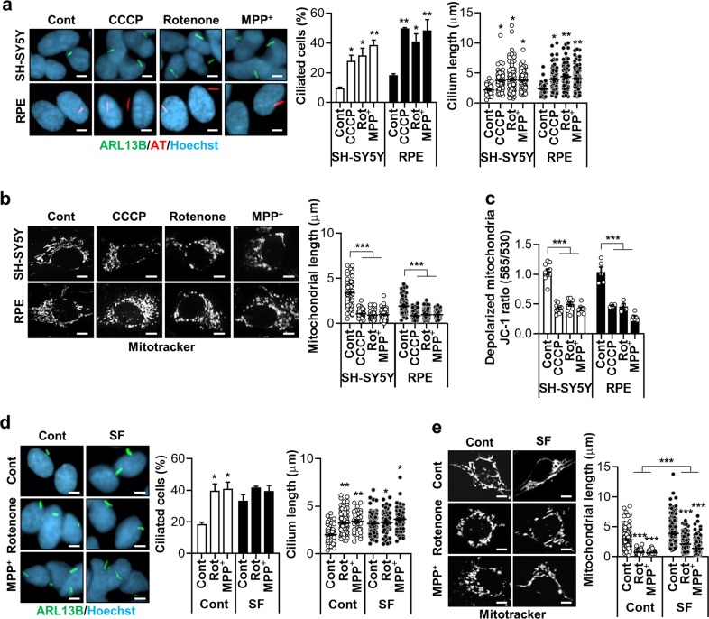
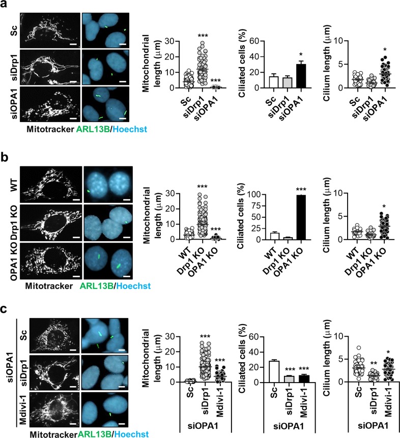
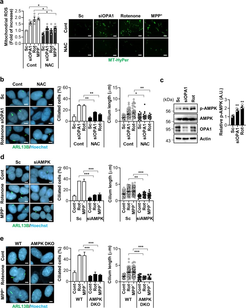
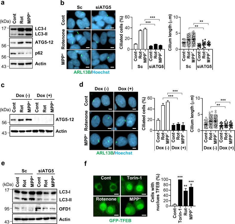
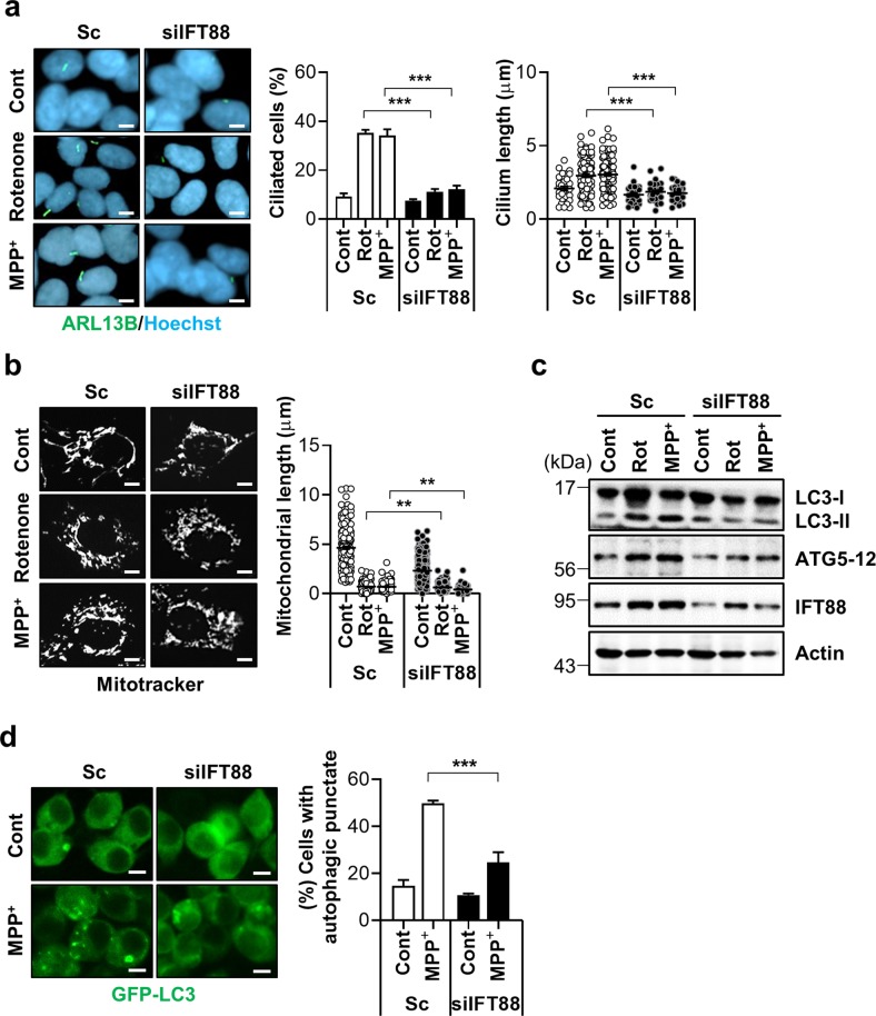
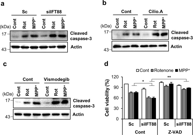
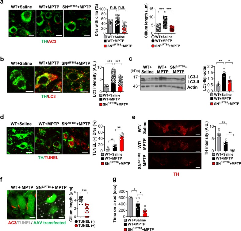
A primary cilium is an antenna-like structure on the cell surface that plays a crucial role in sensory perception and signal transduction. Mitochondria, the 'powerhouse' of the cell, control cell survival, and death. The cellular ability to remove dysfunctional mitochondria through mitophagy is important for cell survival. We show here that mitochondrial stress, caused by respiratory complex inhibitors and excessive fission, robustly stimulates ciliogenesis in different types of cells including neuronal cells. Mitochondrial stress-induced ciliogenesis is mediated by mitochondrial reactive oxygen species generation, subsequent activation of AMP-activated protein kinase and autophagy. Conversely, abrogation of ciliogenesis compromises mitochondrial stress-induced autophagy, leading to enhanced cell death. In mice, treatment with mitochondrial toxin, MPTP elicits ciliary elongation and autophagy in the substantia nigra dopamine neurons. Blockade of cilia formation in these neurons attenuates MPTP-induced autophagy but facilitates dopamine neuronal loss and motor disability. Our findings demonstrate the important role of primary cilia in cellular pro-survival responses during mitochondrial stress.


Conflict of interest statement

The authors declare that they have no conflict of interest.

Figures

Fig. 1
Fig. 1. Mitochondrial respiratory inhibitors stimulate ciliogenesis.
a Increased ciliogenesis occurs following treatment with the mitochondrial respiratory inhibitors CCCP (5 μM), rotenone (200 nM), and MPP+ (5 mM) in (upper) SH-SY5Y and (lower) RPE cells. Cells were cultured to almost 100% confluency and treated with these agents for 24 h. Primary cilia were stained with antibodies against ARL13B (green) or acetylated α-tubulin (AT) (red) and the nucleus (blue) was counterstained with Hoechst 33342 dye. b CCCP (5 μM), rotenone (200 nM), and MPP+ (5 mM) were applied to (upper) SH-SY5Y and (lower) RPE cells and mitochondria were stained with a MitoTracker (white) to measure mitochondrial length. c SH-SY5Y and RPE cells were treated with CCCP (5 μM), rotenone (200 nM), and MPP+ (5 mM). After 24 h, the alteration of mitochondrial membrane potential was measured with the MitoProbe JC-1 assay using the Attune NxT flow cytometer. d, e SH-SY5Y cells were treated with rotenone or MPP+ in the presence or absence of serum [normal (Cont) or serum-free (SF)]. Afterward, the ciliated cells and mitochondrial length were determined. Data are the mean ± SEM. *p < 0.05, **p < 0.01, ***p < 0.005 vs. untreated controls determined by ANOVA followed by a post hoc LSD test. Scale bar, 5 μm.
Fig. 2
Fig. 2. Mitochondrial fission induces ciliogenesis.
a Effects of mitochondrial fusion induced by Drp1 siRNA (siDrp1) treatment and fission by OPA1 siRNA (siOPA1) treatment. SH-SY5Y cells transfected with siDrp1 or siOPA1 were stained with a MitoTracker (white), ARL13B (green), and Hoechst 33342 dye (blue). b OPA1-knockout (KO) mouse embryonic fibroblasts (MEFs) and Drp1-KO MEFs were stained with a MitoTracker (white), ARL13B (green), and Hoechst 33342 dye (blue). c Effect of mitochondrial fusion on ciliogenesis in SH-SY5Y cells with siOPA1-enhanced ciliogenesis. Mitochondrial fusion was induced by siDrp1 transfection or Mdivi-1 (10 μM). Representative cilia images are presented. Cilia measurement data were obtained from about 200 cells per group and the experiments were repeated at least three times. Data are the mean ± SEM. *p < 0.05, **p < 0.01, ***p < 0.005 vs. scrambled non-targeting siRNA (Sc) treatment or wild type (WT) MEF cells determined by ANOVA followed by a post hoc LSD test. Scale bar, 5 μm.
Fig. 3
Fig. 3. Mitochondrial ROS and AMPK mediate mitochondrial stress-induced ciliogenesis.
a, b SH-SY5Y cells were transfected with scrambled control siRNA (Sc) or siRNA against OPA1 (siOPA1). After 2 days the cells were treated with NAC (1 mM) for 24 h. SH-SY5Y cells were treated with rotenone (200 nM) or MPP+ (5 mM) with or without NAC (1 mM) for 24 h. The enhanced mitochondrial ROS (mtROS) formation. The level of mitochondrial H2O2 was measured using the fluorescent intensity of the Mito-HyPer. Scale bar, 20 μm. b NAC (1 mM) treatment blocks the induction of ciliogenesis by siOPA1 or rotenone in SH-SY5Y cells. Primary cilia were immunostained with ARL13B antibody (green) and the nucleus was counterstained with Hoechst 33342 dye (blue). c SH-SY5Y cells transfected with siOPA1 for 3 days or treated with rotenone (200 nM) were analyzed by Western blotting with a phosphorylated-AMPK (p-AMPK) (T172) antibody. d SH-SY5Y cells transfected with Sc or siRNA for AMPK (siAMPK) were further tread with rotenone (200 nM) or MPP+ (5 mM). After 24 h, the cells were stained with ARL13B (green) or Hoechst 33342 dye (blue). Scale bar, 5 μm. e AMPK α1/α2 double-knockout (AMPK DKO) MEFs were treated with rotenone or MPP+. After 24 h, the cells were stained with ARL13B (green), Hoechst 33342 dye (blue). Scale bar, 5 μm. Experiments were repeated at least three times. Data are the mean ± SEM of about 200 cells per group. *p < 0.05, **p < 0.01, ***p < 0.005 between the indicated groups determined by ANOVA followed by a post hoc LSD test.
Fig. 4
Fig. 4. Inhibition of autophagy blocks mitochondrial stress-mediated ciliogenesis.
a Rotenone and MPP+ treatment enhance autophagy in SH-SY5Y cells. SH-SY5Y cells were treated with rotenone (200 nM) and MPP+ (5 mM) for 24 h, then the cells were assessed by Western blotting with indicated antibodies. b SH-SY5Y cells transfected with siATG5 were further treated with rotenone (200 nM) and MPP+ (5 mM) for 24 h, then primary cilia were immunostained with ARL13B antibody (green) and the nucleus was counterstained with Hoechst 33342 dye (blue). Scale bar, 5 μm. c, d Blunted rotenone- and MPP+-driven ciliogenesis in M5-7 cells. MEFs under doxycycline (Dox)-induced ATG5 depletion (M5-7 cells) were treated to rotenone (200 nM) and MPP+ (5 mM) for 24 h with or without Dox. Dox-induced ATG5 depletion was confirmed by Western blotting with ATG5 antibody (c). d Primary cilia were immunostained with ARL13B antibody (green) and the nucleus was counterstained with Hoechst 33342 dye (blue). Scale bar, 5 μm. e SH-SY5Y cells transfected with siATG5 were further treated with rotenone (200 nM) and MPP+ (5 mM) for 24 h, then the cells were analyzed by western blotting with indicated antibodies. f SY5Y/GFP-TFEB cells were treated with Torin-1 (1 μM for 1 h), rotenone (200 nM, 24 h) and MPP+ (5 mM, 24 h). Then, nuclear localization of GFP-TFEB was observed. Scale bar, 1 μm. Data were obtained from about 200 cells per group and experiments were repeated at least three times. Data are the mean ± SEM. **p < 0.01, ***p < 0.005 between indicated groups determined by ANOVA followed by a post hoc LSD test.
Fig. 5
Fig. 5. Ciliogenesis mediates mitochondrial stress-induced autophagy.
a, b SH-SY5Y cells transfected with siIFT88 were further treated with rotenone (200 nM) or MPP+ (5 mM) for 24 h. a Primary cilia were immunostained with ARL13B (green) and the nucleus was stained with Hoechst 33342 dye (blue). Cilia were measured in about 200 cells per group. b Mitochondria were stained with MitoTracker (white). Scale bar, 5 μm. c Inhibition of ciliogenesis with IFT88 siRNA (siIFT88) blocked rotenone- and MPP+-induced autophagy in SH-SY5Y cells. d SH-SY5Y/GFP-LC3 cells transfected with scrambled control siRNA (Sc) or siRNA against IFT88 (siIFT88) were further treated with rotenone (200 nM) and MPP+ (5 mM) for 24 h and then cells with autophagic punctate were counted under fluorescence microscopy. Scale bar, 10 μm. Data are the mean ± SEM. **p < 0.01, ***p < 0.005 between indicated groups determined by ANOVA followed by a post hoc LSD test.
Fig. 6
Fig. 6. Mitochondrial stress-induced ciliogenesis promotes cell survival.
ac Accelerated rotenone- and MPP+- induced cell death in SH-SY5Y cells with defective ciliogenesis. a SH-SY5Y cells transfected with Sc or siIFT88 were treated with rotenone (200 nM) and MPP+ (5 mM) for 24 h. Then the cells were further analyzed by Western blotting with cleaved caspase-3 antibody. b, c A blockade of ciliogenesis was induced by with (b) ciliobrevin A1 (Cilio.A, 10 μM) or (c) vismodegib (5 μM) treatment in SH-SY5Y cells and caspase-3 activation was examined by Western blotting with cleaved caspase-3 antibody. d SH-SY5Y cells were transfected with siRNA against IFT88 and further treated with rotenone (200 nM) or MPP+ (5 mM) for 24 h in the presence or absence of the pan caspase inhibitor Z-VAD (40 μM). Cell viability was measured by CCK-8 flow cytometric analysis. Data are the mean ± SEM (n = 3 per group). *p < 0.05, **p < 0.01 between the indicated groups determined by ANOVA followed by a post hoc LSD test.
Fig. 7
Fig. 7. Enhanced ciliogenesis in the substantia nigra dopamine neurons in the mice model of MPTP-induced Parkinson’s disease.
a Ciliary elongation in substantia nigra (SN) dopamine neurons (DNs) in the mouse after a single intraperitoneal administration of MPTP (30 mg/kg body weight) and blockade of cilia elongation with SN IFT88 knockdown. Two weeks before MPTP injection, the WT mice were injected with GFP-AAV and SN∆IFT88 mice were injected with IFT88-shRNA GFP-AAV into the bilateral SN. Primary cilia and DNs were stained using AC3 antibody and TH antibody. More than 100 TH-positive cells were analyzed in each animal. Scale bar, 10 μm. b, c Increased autophagy in SN DNs following MPTP treatment and blunted MPTP-induced autophagy in DNs with impaired ciliogenesis. Autophagy in DNs was evaluated by double staining with LC3 and TH antibodies and by LC3 immunoblotting. Scale bar, 10 μm. d Impaired ciliogenesis in the DNs enhances MPTP-induced apoptosis, as assessed by TUNEL staining. Scale bar, 10 μm. e MPTP treatment reduces the intensity of TH immunoreactivity in the SN pars compacta and this reduction is far greater in SNΔIFT88 mice. Scale bar, 500 μm. f Cilia elongation occurs before DN death. TUNEL-negative neurons of WT mice have long cilia but TUNEL-positive neurons in SNΔIFT88 mice have shorter or no cilia following MPTP treatment. Scale bar, 10 μm. g Motor function assessment using the rotarod test for 3 days after MPTP treatment in WT and SNΔIFT88 mice (n = 3~6 per group). Data are presented as the mean ± SEM. *p < 0.05, **p < 0.01, ***p < 0.005 between the indicated groups determined by ANOVA followed by a post hoc LSD test (n.s. = not significant).

References

    1. Singla V, Reiter JF. The primary cilium as the cell's antenna: signaling at a sensory organelle. Science. 2006;313:629–633. doi: 10.1126/science.1124534. - DOI - PubMed
    1. Ishikawa H, Marshall WF. Ciliogenesis: building the cell's antenna. Nat. Rev. Mol. Cell Biol. 2011;12:222–234. doi: 10.1038/nrm3085. - DOI - PubMed
    1. Cho DH, Nakamura T, Lipton SA. Mitochondrial dynamics in cell death and neurodegeneration. Cell. Mol. Life Sci. 2010;67:3435–3447. doi: 10.1007/s00018-010-0435-2. - DOI - PMC - PubMed
    1. Baek SH, et al. Inhibition of Drp1 Ameliorates Synaptic Depression, Abeta Deposition, and Cognitive Impairment in an Alzheimer's Disease Model. J. Neurosci. 2017;37:5099–5110. doi: 10.1523/JNEUROSCI.2385-16.2017. - DOI - PMC - PubMed
    1. Mizushima N. A brief history of autophagy from cell biology to physiology and disease. Nat. Cell Biol. 2018;20:521–527. doi: 10.1038/s41556-018-0092-5. - DOI - PubMed

Publication types

MeSH terms