. 2019 Dec 16;9(1):19214.
doi: 10.1038/s41598-019-55797-1.

A ligand-based system for receptor-specific delivery of proteins

Affiliations

A ligand-based system for receptor-specific delivery of proteins

Mariano Maffei et al. Sci Rep. .

Abstract

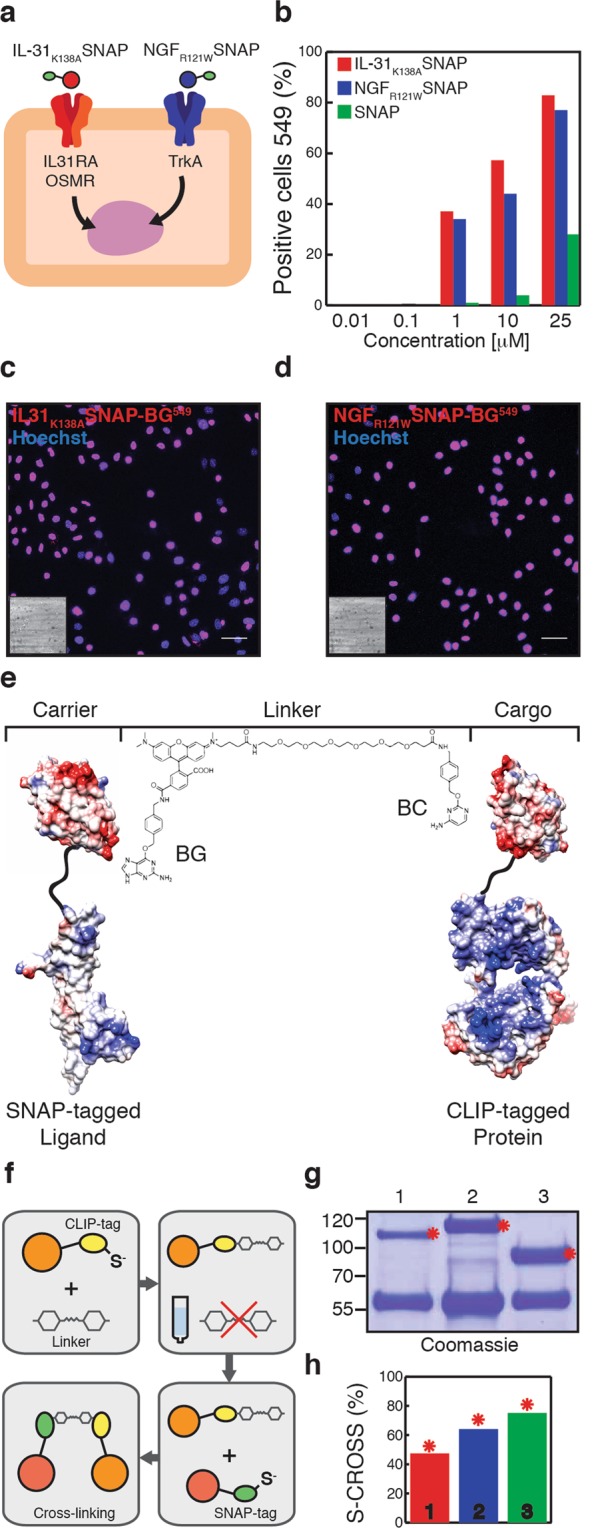
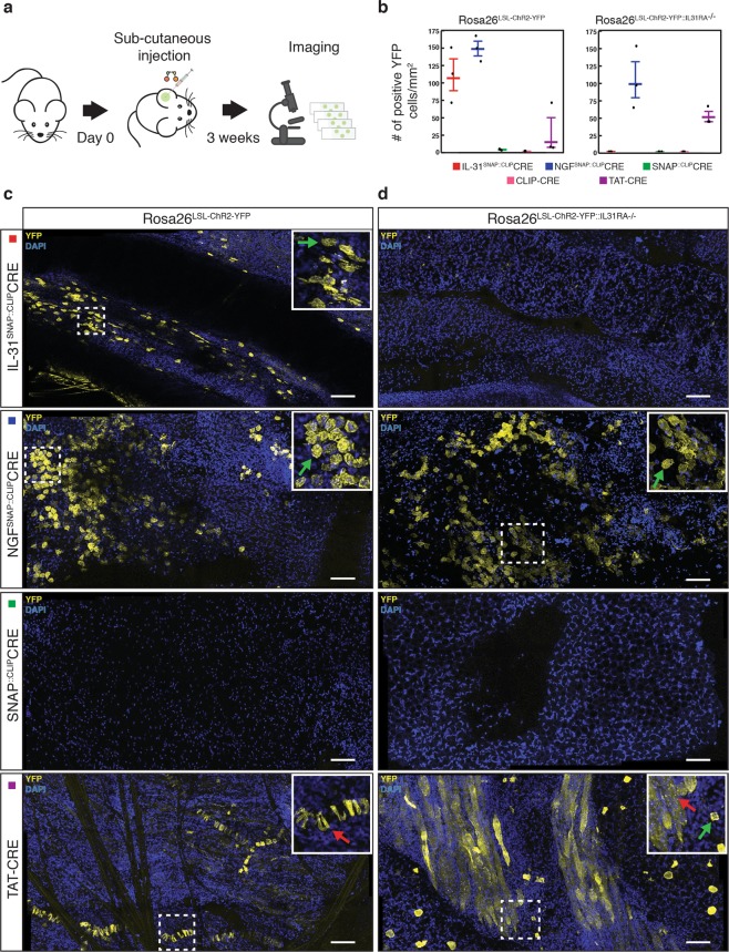
Gene delivery using vector or viral-based methods is often limited by technical and safety barriers. A promising alternative that circumvents these shortcomings is the direct delivery of proteins into cells. Here we introduce a non-viral, ligand-mediated protein delivery system capable of selectively targeting primary skin cells in-vivo. Using orthologous self-labelling tags and chemical cross-linkers, we conjugate large proteins to ligands that bind their natural receptors on the surface of keratinocytes. Targeted CRE-mediated recombination was achieved by delivery of ligand cross-linked CRE protein to the skin of transgenic reporter mice, but was absent in mice lacking the ligand's cell surface receptor. We further show that ligands mediate the intracellular delivery of Cas9 allowing for CRISPR-mediated gene editing in the skin more efficiently than adeno-associated viral gene delivery. Thus, a ligand-based system enables the effective and receptor-specific delivery of large proteins and may be applied to the treatment of skin-related genetic diseases.


Conflict of interest statement

The authors declare no competing interests.

Figures

Figure 1
Figure 1
Binding of SNAP-tagged ligands to keratinocytes and selective cross-linking to CLIP-tagged enzymes. (a) Schematic representation of one keratinocyte expressing the receptors of interest and the ligands used. (b) Quantification of labelled IL-31K138ASNAP-BG549 (c), NGFR121WSNAP-BG549 (d) and SNAP-BG549 (green bars) binding to primary keratinocytes. Nuclear localization was observed after 2 hours treatment. The nuclei were stained with Hoechst. Scale bars, 20 μm. The insets represent corresponding brightfield images. (e) 3D structures showing selective cross-linking of SNAP-tagged ligands (NGF-SNAP) and CLIP-tagged proteins (CLIP-Cre) through a BG-TMR-PEG-BC linker (PDB ID codes: 1BET, 1KBU, 3KZY). (f) Schematic representation of S-CROSS optimized chemical reaction. (g) Representative Coomassie gel showing cross-linking complexes (red asterisks). First lane (#1) is IL-31SNAP::CLIPCRE, second lane (#2) is NGFSNAP::CLIPCRE and third lane (#3) is SNAP::CLIP-CRE. (h) Quantification of cross-linking from Coomassie gel (g).
Figure 2
Figure 2
Ligand-mediated delivery of cross-linked CLIP-Cre. (a) Schematic of in-vitro keratinocytes treatment with cross-linked complexes. (b) Images and quantification of YFP positive primary keratinocytes (% of cells) from Rosa26LSL-ChR2-YFP mice 5 days after treatment with 2 μM of cross-linked complexes or CLIP-Cre alone. (c) Representative images and quantification of YFP positive primary keratinocytes from double transgenic Rosa26LSL-ChR2-YFP::IL31RA−/− mice 5 days after treatment with 2 μM of cross-linked complexes or CLIP-Cre alone. Scale bars, 20 μm. The insets represent corresponding brightfield images. The horizontal lines mark the geometric mean and the error bars mark the standard error. Representative data from n = 3 independent experiments.
Figure 3
Figure 3
Selective delivery of cross-linked CLIP-Cre in-vivo. (a) Schematic of in-vivo treatment with cross-linked complexes. (b) Quantification (number of cells per mm2) of YFP positive keratinocytes from Rosa26LSL-ChR2-YFP mice (c) and from double transgenic Rosa26LSL-ChR2-YFP::IL31RA−/− mice (d) 3 weeks after subcutaneous injection with 5 μM (0.85 mg/kg) of cross-linked complexes or TAT-Cre. The nuclei were stained with DAPI. Scale bars, 40 μm. The insets show the zoom of representative areas. Green arrows indicate YFP+ keratinocytes. Red arrows indicate non-selective YFP expression. The horizontal lines mark the geometric mean and the error bars mark the standard error. Data from n = 3 independent experiments.
Figure 4
Figure 4
CLIP-Cas9 activity and internalization in keratinocytes. (a) Schematic of CLIP-Cas9::sgRNA electroporation strategy. (b) Indel spectrum determined by TIDE of primary keratinocytes electroporated with CLIP-Cas9::sgRNA targeting the Atat1 gene. The inset show T7 endonuclease 1 assay performed on genomic DNA from electroporated keratinocytes. t.e. = total efficiency. (c) Quantification (% cells) and representative images (d) of TMR positive cells upon 2 hours treatment with 2 μM of ligand cross-linked Cas9 (#1 no sgRNA; #2 with sgRNA; #3 with sgRNA+ protamine; #4 with sgRNA + ppTG21). Nuclei were stained with Hoechst. Scale bars, 20 μm. The horizontal lines mark the geometric mean and the error bars mark the standard error.
Figure 5
Figure 5
Ligand-mediated delivery of cross-linked CLIP-Cas9 in cultured cells. (a) Schematic of plasmid electroporation strategy in primary keratinocytes. (b) % of indels detected from DNA sequencing of BFP-sorted keratinocytes expressing Atat1 U6-sgRNA and treated with: cross-linked complex IL-31SNAP::CLIPCas9 (3rd column from left), IL-31SNAP::CLIPCas9 + ppTG21 (4th column from left), NGFSNAP::CLIPCas9 (5th column from left) and NGFSNAP::CLIPCas9 + ppTG21 peptide (6th column from left). Untreated keratinocytes and untreated BFP-sorted keratinocytes are shown in column 1 and 2, respectively. (c) Graphical representation of AAV1/2::HDR and N2a cells experimental strategy. (d) Zoom of mEGFP-ß-Actin N2a positive cell. (e) Confocal images of AAV1/2::HDR transduced N2a WT cells overexpressing NGF receptors treated with NGFSNAP::CLIPCas9 alone (upper frames) or in presence of ppTG21 peptide (lower frames). First images on the left show control N2a cells infected only with AAV1/2::HDR. Scale bars, 40 μm. The insets represent corresponding brightfield images. (f) Quantification (% cells) of mEGFP positive cells from (e). The horizontal black lines mark the geometric mean and the error bars mark the standard error. Black triangles: cross-linking complexes carrying linker #5 (Table S1); green squares: cross-linking complexes carrying linker #3 (Table S1); red circles: cross-linking complexes carrying linker #6 (Table S1).
Figure 6
Figure 6
Ligand-mediated delivery of cross-linked CLIP-Cas9 in-vivo. (a) Schematic of subcutaneous injection of cross-linked CLIP-Cas9 and AAV1/2::HDR. (b) Confocal images showing control samples injected only with AAV1/2::HDR (left frame) or with dual AAV system AAV1/2::HDR + AAV1/2::Cas9 (right frame) (c) Confocal images of mEGFP positive keratinocytes 2–3 weeks after subcutaneous injection into the mouse ear, with AAV1/2::HDR and CLIP-Cas9 cross-linked to IL-31 (top) or NGF (bottom). The nuclei were stained with DAPI. Scale bars, 40 μm. The insets show enlarged representative areas with mEGFP-ß-Actin positive cells. Black triangles: cross-linking complexes carrying linker #5 (Table S1); green squares: cross-linking complexes carrying linker #3 (Table S1); red circles: cross-linking complexes carrying linker #6 (Table S1). (d) Mosaic confocal image (1.07 mm × 1.43 mm) of mEGFP-ß-Actin positive cells from WT mice 2–3 weeks after subcutaneous injection with AAV1/2::HDR and NGFSNAP::CLIPCas9 (linker #3). The nuclei were stained with DAPI. White dashed circles mark mEGFP positive targeted cell clusters. White arrows indicate mEGFP positive cells starting to express mEGFP. Scale bar, 100 μm.

Similar articles

Cited by

References

    1. Zuris JA, et al. Cationic lipid-mediated delivery of proteins enables efficient protein-based genome editing in vitro and in vivo. Nat Biotechnol. 2015;33:73–80. doi: 10.1038/nbt.3081. - DOI - PMC - PubMed
    1. Liu J, et al. Efficient delivery of nuclease proteins for genome editing in human stem cells and primary cells. Nat Protoc. 2015;10:1842–1859. doi: 10.1038/nprot.2015.117. - DOI - PubMed
    1. Dong Y, et al. A Dual Targeting Dendrimer-Mediated siRNA Delivery System for Effective Gene Silencing in Cancer Therapy. J Am Chem Soc. 2018;140:16264–16274. doi: 10.1021/jacs.8b10021. - DOI - PubMed
    1. Rosenberg SA, Restifo NP. Adoptive cell transfer as personalized immunotherapy for human cancer. Science. 2015;348:62–68. doi: 10.1126/science.aaa4967. - DOI - PMC - PubMed
    1. Owens B. Faster, deeper, smaller-the rise of antibody-like scaffolds. Nat Biotechnol. 2017;35:602–603. doi: 10.1038/nbt0717-602. - DOI - PubMed

Publication types