. 2020 Jan;52(1):40-47.
doi: 10.1038/s41588-019-0536-2. Epub 2019 Dec 16.

Loss of ADAMTS19 causes progressive non-syndromic heart valve disease

Collaborators, Affiliations

Loss of ADAMTS19 causes progressive non-syndromic heart valve disease

Florian Wünnemann et al. Nat Genet. 2020 Jan.

Abstract

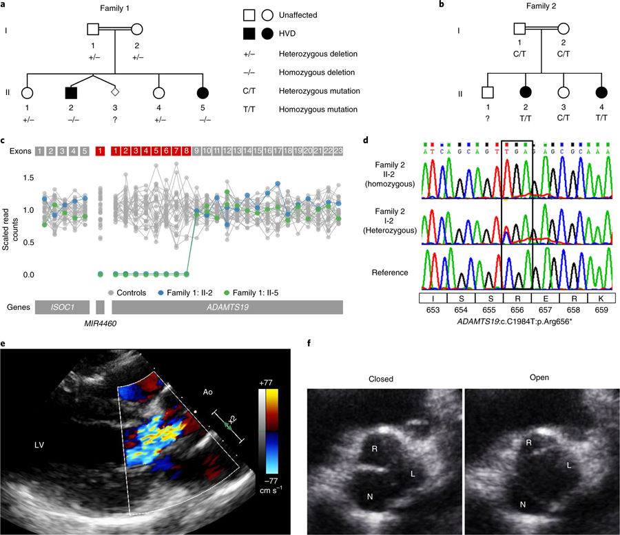
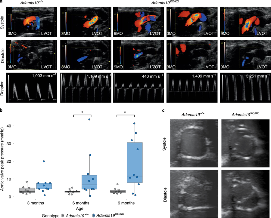
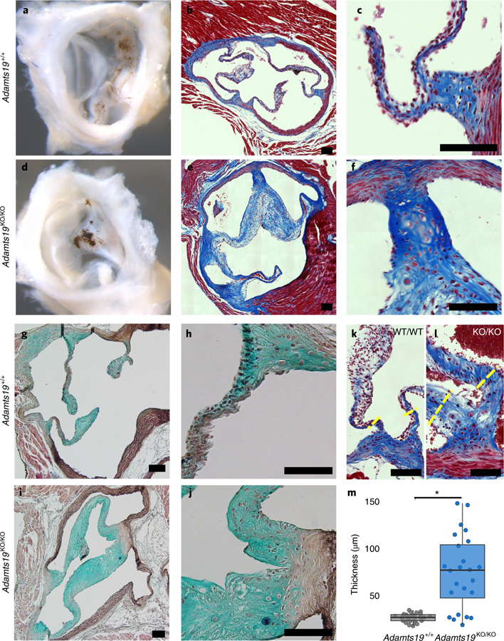
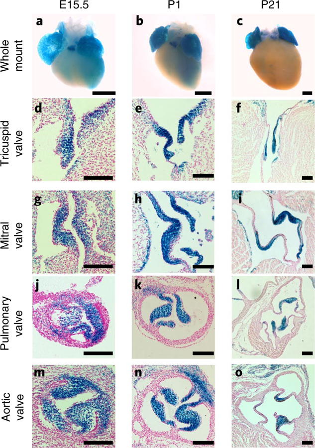
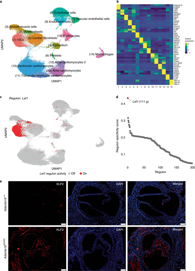
Valvular heart disease is observed in approximately 2% of the general population1. Although the initial observation is often localized (for example, to the aortic or mitral valve), disease manifestations are regularly observed in the other valves and patients frequently require surgery. Despite the high frequency of heart valve disease, only a handful of genes have so far been identified as the monogenic causes of disease2-7. Here we identify two consanguineous families, each with two affected family members presenting with progressive heart valve disease early in life. Whole-exome sequencing revealed homozygous, truncating nonsense alleles in ADAMTS19 in all four affected individuals. Homozygous knockout mice for Adamts19 show aortic valve dysfunction, recapitulating aspects of the human phenotype. Expression analysis using a lacZ reporter and single-cell RNA sequencing highlight Adamts19 as a novel marker for valvular interstitial cells; inference of gene regulatory networks in valvular interstitial cells positions Adamts19 in a highly discriminatory network driven by the transcription factor lymphoid enhancer-binding factor 1 downstream of the Wnt signaling pathway. Upregulation of endocardial Krüppel-like factor 2 in Adamts19 knockout mice precedes hemodynamic perturbation, showing that a tight balance in the Wnt-Adamts19-Klf2 axis is required for proper valve maturation and maintenance.


Conflict of interest statement

Competing interests

The authors declare no competing interests.

Figures

Fig. 1 |
Fig. 1 |. Homozygous loss of ADAMTS19 in human families causes progressive HVD.
a, Family 1 has a genomic deletion of ADAMTS19 coding exons 1–8. Individuals in family 1: II-2 and family 1: II-5 are homozygous for the deletion and have progressive HVD. Parents and siblings who are carriers do not show signs of HVD. b, In family 2, two offspring (family 2: II-2 and II-4) affected with progressive HVD are homozygous for a rare LOF mutation (ADAMTS19:c. C1984T:p.Arg656*). c, Scaled read counts of whole-exome sequencing for the deleted ADAMTS19 region in family 1. Exons missing both copies in affected individuals are marked in red. d, Representative Sanger sequencing of the nonsense allele in a reference individual, the heterozygous mother who is a carrier (family 2: I-2) and a homozygous patient (family 2: II-2). e, Color Doppler echocardiography of left ventricular outflow tract in diastole for individual family 2:II-4, highlighting aortic regurgitation in the patient. f, Echocardiographic view of aortic valve for individual family 2:II-4 at 15 years of age in opened (left) and closed (right) positions, showing thickened R-N commissure.
Fig. 2 |
Fig. 2 |. Homozygous Adamts19KO/KO mice develop aortic valve disease.
a, Representative modified ascending aortic view for one WT (Adamts19+/+, n = 9) and two homozygous Adamts19KO/KO mice. Aortic regurgitation in diastole was identified in 27% of Adamts19KO/KO mice. Color mode in systole shows abnormal blood flow and increased systolic aortic pressure in about 20% of Adamts19KO/KO mice. In total, 38% of mice were affected by some type aortic valve dysfunction (Supplementary Table 4). LVOT, left ventricular outflow tract. b, Aortic valve dysfunction is progressive in homozygous Adamts19KO/KO mice. Aortic peak pressures were significantly increased at 6 and 9 months in Adamts19KO/KO mice, while no increase in peak pressure was observed in Adamts19 WT mice (Adamts19+/+). The box plots show the interquartile range (IQR, 25th–75th percentile) with the 50th percentile as the solid line. Whiskers represent 1.5× IQR (n = 9 for WT, n = 10 for Adamts19KO/KO; the same mice were measured over time). c, Short axis view of aortic valves in B-mode. Adamts19+/+ valves show normal closing with three visible leaflets, whereas Adamts19KO/KO resemble a fused BAV in humans with only two leaflets visible and clear ‘fish mouth’ opening of the valve. Note that mice with this presentation have three formed leaflets and only functionally behave like BAV. Distribution of phenotypes across independent experiments is shown in Supplementary Table 4. LVOT, left ventricular outflow tract. *P < 0.05, two-sided Wilcoxon rank-sum test. P values: 6 months = 0.01721, 9 months = 0.03499.
Fig. 3 |
Fig. 3 |. Aortic valves from Adamts19KO/KO mice are thickened and have disorganized ECM at 9 months.
a,d, Whole mount of WT (Adamts19+/+) (a) and Adamts19KO/KO (d) aortic valve shows increased whitening of the valve tissue reflecting thickening at 9 months of age. b,c,e,f, Trichrome staining of aortic valve sections reveals substantial thickening of the valve leaflets of Adamts19KO/KO mice (e,f) compared to WT controls (b,c). gj, Pentachrome staining shows disorganization of the ECM in Adamts19KO/KO mice (i,j) versus WT control (g,h). Staining was performed on four mice per genotype. k,l, Examples of hinge regions in the aortic valves of normal WT (k) and thickened, homozygous knockout (l) mice. The yellow dashed lines represent regions of quantification for thickness. Black scale bars, 100 µM. m, Quantification of valve thickness reveals significant enlargement of the hinges of aortic valve leaflets in knockout mice (n = 4 mice for each genotype; each mouse has 6 valve hinge measurements). The box plots show the IQR (25th–75th percentile) with the 50th percentile as a solid line. The whiskers represent 1.5× IQR. *P = 3.336 × 10⁻⁶, two-sided, Student’s t-test.
Fig. 4 |
Fig. 4 |. Ultrastructural analysis of the ECM in WT and Adamts19KO/KO aortic valves.
a, Comparison of the three-layered stratification in the free part of the valve leaflets of Adamts19+/+ (top) versus Adamts19KO/KO animals (bottom). A trilaminar stratification (fibrosa, spongiosa, ventricularis) can be made out, but mutant leaflets are thickened and do not have the highly organized ECM architecture of the individual layers as seen in WT. b, Comparison between WT (left) and knockout (right) aortic valves at the level of the leaflet tip. Mutants have higher proteoglycan content in all layers, more cells with activated nuclei and more secretory granules, typical of a secretory VIC phenotype. c, At the hinge area of WT valves (left), fibroblasts and chondrocyte-like cells are surrounded by abundant collagen type I fibers. In knockout valves (right), fibroblasts and chondrocytes are found, with accumulation of cellular debris, vesicular structures and disorganized proteoglycan structure. d, The hinge area of WT valves contains bundles of typical collagen type I fibers (left). In mutants (right), these fibers are thinner with a less homogenous diameter. Ultrastructural findings were reproduced for three independent, healthy Adamts19+/+ and three independent Adamts19KO/KOmice with HVD.
Fig. 5 |
Fig. 5 |. Adamts19 is expressed in all four cardiac valves during valve remodeling and elongation.
ac, Whole-mount images of Adamts19KO/KO embryonic hearts show lacZ expression in both atria, the trabeculated myocardium as well as the pulmonary valve from E15.5 to P21. do, Histological sections from lacZ-stained hearts of Adamts19KO/KO animals corresponding to the stages shown in ac. Localized lacZ expression in the interstitial cell layer of the valves is observed in the tricuspid (df), mitral (gi), pulmonary (jl) and aortic valve (mo). Notably, the endothelial monolayer around the valve leaflets is lacZ-negative. Scale bars in whole-mount images, 500 µM. Scale bars in sections, 100 µM. LacZ staining was performed and the staining patterns consistently replicated in >10 independent mice.
Fig. 6 |
Fig. 6 |. Single-cell sequencing of E14.5 mouse hearts.
a, Joined UMAP-based clustering of a total of 55,152 single cells from Adamts19+/+ and Adamts19KO/KO mice. b, Z-scaled expression values for three marker genes for each respective cell cluster. Adamts19 strongly and specifically marks VICs. c, Binary regulon activity for the predicted Lef1 regulon, of which Adamts19 is part. d, Regulon specificity score distribution for the top 200 predicted regulons in VICs. Lef1 has the highest specificity score for VICs and represents the most distinct specific regulon for VICs compared to all other cell types (111 g = 111 genes predicted in the regulon). e, IHC for aortic valves of Adamts19+/+ and Adamts19KO/KO valves at 3 weeks show increased staining for KLF2 in sections from homozygous Adamts19 knockout mice. Scale bars, 100 μM. KLF2 staining was repeated for 11 independent mice per group; increased KLF2 was observed in 4 out of 11 (36%) Adamts19KO/KO mice.

References

    1. Nkomo VT et al. Burden of valvular heart diseases: a population-based study. Lancet 368, 1005–1011 (2006). - PubMed
    1. Garg V et al. Mutations in NOTCH1 cause aortic valve disease. Nature 437, 270–274 (2005). - PubMed
    1. Padang R, Bagnall RD, Richmond DR, Bannon PG & Semsarian C Rare non-synonymous variations in the transcriptional activation domains of GATA5 in bicuspid aortic valve disease. J. Mol. Cell. Cardiol 53, 277–281 (2012). - PubMed
    1. Tan HL et al. Nonsynonymous variants in the SMAD6 gene predispose to congenital cardiovascular malformation. Hum. Mutat 33, 720–727 (2012). - PMC - PubMed
    1. Durst R et al. Mutations in DCHS1 cause mitral valve prolapse. Nature 525, 109–113 (2015). - PMC - PubMed

Publication types