. 2020 Jan;52(1):29-34.
doi: 10.1038/s41588-019-0547-z. Epub 2019 Dec 16.

Extrachromosomal circular DNA drives oncogenic genome remodeling in neuroblastoma

Affiliations

Extrachromosomal circular DNA drives oncogenic genome remodeling in neuroblastoma

Richard P Koche et al. Nat Genet. 2020 Jan.

Erratum in

  • Publisher Correction: Extrachromosomal circular DNA drives oncogenic genome remodeling in neuroblastoma.
    Koche RP, Rodriguez-Fos E, Helmsauer K, Burkert M, MacArthur IC, Maag J, Chamorro R, Munoz-Perez N, Puiggròs M, Garcia HD, Bei Y, Röefzaad C, Bardinet V, Szymansky A, Winkler A, Thole T, Timme N, Kasack K, Fuchs S, Klironomos F, Thiessen N, Blanc E, Schmelz K, Künkele A, Hundsdörfer P, Rosswog C, Theissen J, Beule D, Deubzer H, Sauer S, Toedling J, Fischer M, Hertwig F, Schwarz RF, Eggert A, Torrents D, Schulte JH, Henssen AG. Koche RP, et al. Nat Genet. 2020 Apr;52(4):464. doi: 10.1038/s41588-020-0598-1. Nat Genet. 2020. PMID: 32107479

Abstract

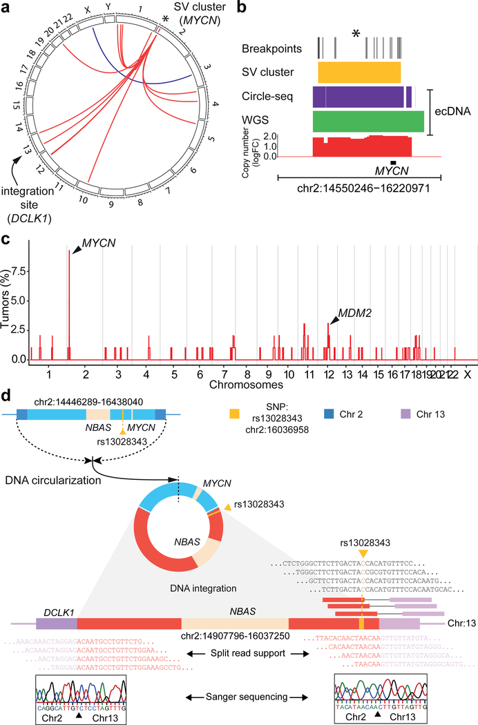
Extrachromosomal circularization of DNA is an important genomic feature in cancer. However, the structure, composition and genome-wide frequency of extrachromosomal circular DNA have not yet been profiled extensively. Here, we combine genomic and transcriptomic approaches to describe the landscape of extrachromosomal circular DNA in neuroblastoma, a tumor arising in childhood from primitive cells of the sympathetic nervous system. Our analysis identifies and characterizes a wide catalog of somatically acquired and undescribed extrachromosomal circular DNAs. Moreover, we find that extrachromosomal circular DNAs are an unanticipated major source of somatic rearrangements, contributing to oncogenic remodeling through chimeric circularization and reintegration of circular DNA into the linear genome. Cancer-causing lesions can emerge out of circle-derived rearrangements and are associated with adverse clinical outcome. It is highly probable that circle-derived rearrangements represent an ongoing mutagenic process. Thus, extrachromosomal circular DNAs represent a multihit mutagenic process, with important functional and clinical implications for the origins of genomic remodeling in cancer.


Figures

Fig. 1.
Fig. 1.. A genome-wide map of extrachromosomal circular DNAs in neuroblastoma.
a, Schematic representation of sequencing reads as predicted for circular genomic regions (bg = background/non-circular genome). b, Schematic representation of sequencing read positions on extrachromosomal circular DNA. c, Genome tracks comparing sequencing read densities on an ecDNA as detected via WGS (only circle-specific head-to-tail reads are depicted), Circle-seq followed by paired-end sequencing (ILM) and single molecule real-time sequencing (SMRT) in neuroblastoma cells. DNA digestion with an exonuclease and/or endonuclease is indicated (+/−). Dotted blue line indicates predicted circle junction. Interruption of read density profile is due to lack of read alignment. (y - axis: 0–30 reads). d, Chromosome ideogram with genome-wide somatic extrachromosomal circular DNA density as inferred from WGS (blue) compared to Circle-seq (red; M, circular mitochondrial DNA). e, Number of ecDNAs and eccDNAs per neuroblastoma (N=21 tumors, N=96,436 eccDNAs, N=14 ecDNAs). f, Size distribution of ecDNAs and eccDNAs identified using Circle-seq in neuroblastomas (N=21 tumors, N=96,436 eccDNAs, N=14 ecDNAs). g, Alternative-B-allele frequencies (BAF) in sequencing reads from Circle-Seq (N=21 tumors) and WGS (N=93 tumors). h, Density of extrachromosomal circular DNA detected using Circle-seq over genic compared to gene-surrounding regions in MYCN-amplified and non-amplified neuroblastomas (N=7 MYCN-amplified tumors, N=14 non-amplified tumors, lines represent mean signal and the shaded area represents the standard error of the mean). i, Fraction of genomic regions affected by eccDNA compared to ecDNA (N=21 tumors). TSS, transcription start site; TES, transcription end site.
Fig. 2.
Fig. 2.. Mono-allelic large extrachromosomal circular DNA are an origin of oncogene amplification and overexpression in neuroblastoma.
a, B-allele frequency of all extrachromosomal circular DNAs involving genes (both ecDNA and eccDNA) detected using Circle-seq (blue) compared to the corresponding genomic loci in whole-genome sequencing (red) and mRNA expressed from genes affected by DNA circularization measured using RNA sequencing (green, grey lines indicate corresponding measurements from Circle-seq, WGS and RNAseq, N=18 tumors). b, Genome track with phased reads from whole-genome sequencing of NB2013 (WGS), Circle-seq and RNA sequencing (RNA-seq) at the region of extrachromosomal circularization on chromosome 2 affecting MYCN. (BAF= B-allele frequency, blue and red colored dots represent reads from different haplotypes) c, Genes (rows) affected by circularization in neuroblastoma samples (columns) as detected using Circle-seq (N = 21 tumors). d, Relative mRNA expression (z-scores) of genes affected by extrachromosomal DNA circularization in the form of eccDNA (N=1,696) compared to ecDNA (N=24) as measured using total RNA sequencing (N=21 tumors). Normalized gene expression (mRNA) for MYCN proto-oncogene, bHLH transcription factor (MYCN, e) and neurotrophin 3 (NTF3, f) in neuroblastomas. The degree of gene circularization is indicated in red (see color scale).
Fig. 3.
Fig. 3.. The majority of structural rearrangements involve sites of extrachromosomal DNA circularization and form clustered rearrangement patterns in neuroblastoma.
a, Circos plot of inter-chromosomal rearrangements identified using five variant detection algorithms in one neuroblastoma genome (CB2013), shown exemplarily. Tree-shaped clustered rearrangement pattern (red), originating at a region of MYCN circularization (asterisk) is highlighted. b, Detailed view of genomic breakpoint localizations (black) at the base of the tree-shaped rearrangement cluster for the neuroblastoma shown in (a) define a region of clustered breakpoints (yellow) and overlaps with the region of extrachromosomal DNA circularization, as detected using Circle-seq (pink) and whole-genome sequencing (WGS, green). Copy number changes are highlighted in red. c, Genome-wide frequency of tree-shaped clusters of rearrangements in 93 primary neuroblastoma samples. The pattern is recurrently identified on chr2 (at the MYCN locus), chr11 and chr12 (at the MDM2 locus). d, Schematic representation of circle integration in one exemplary neuroblastoma (CB2013). Genomic region, including MYCN (blue), is circularized, and parts of the extrachromosomal circle are integrated (red) into chromosome 13 (pink) leading to a disruption of DCLK1. Sequencing reads supporting a circle-specific SNP as well as split reads supporting circle integration are shown below. Sanger sequencing of integration breakpoints are shown in boxes.
Fig. 4.
Fig. 4.. Rearrangement of extrachromosomal circular DNAs drives transcriptional deregulation and dismal prognosis in neuroblastoma.
a, Heatmap showing differential expression of up to 10 genes located both up- and downstream or a maximal distance of 2 Mb from each circle-derived rearrangement breakpoint (N=259 breakpoints, N=24 tumors). Modified z-scores for the expression of cancer-relevant genes DCLK1 (b) and TERT (c) affected by circle-derived rearrangements are shown for two representative genomic loci (in two neuroblastomas). d, Kaplan Meier analysis comparing neuroblastoma patient survival of patients with neuroblastomas affected by circle-derived clustered rearrangements (N=22 patients) to patients with tumors lacking such rearrangements (N=59 patients, P=0.00033 two-sided log-rank test). e, Kaplan Meier analysis comparing neuroblastoma patient survival with MYCN-amplified tumors affected by MYCN-circle-derived clustered rearrangements (N=10) to patients with tumors lacking such rearrangements (N=7, P=0.043 two-sided log-rank test). f, Schematic diagram of the proposed mechanism of circle-mediated genome remodeling.

Similar articles

Cited by

References

    1. Turner KM et al. Extrachromosomal oncogene amplification drives tumour evolution and genetic heterogeneity. Nature 543, 122–125, doi:10.1038/nature21356 (2017). - DOI - PMC - PubMed
    1. Moller HD et al. Circular DNA elements of chromosomal origin are common in healthy human somatic tissue. Nat Commun 9, 1069, doi:10.1038/s41467-018-03369-8 (2018). - DOI - PMC - PubMed
    1. Shibata Y. et al. Extrachromosomal microDNAs and chromosomal microdeletions in normal tissues. Science 336, 82–86, doi:10.1126/science.1213307 (2012). - DOI - PMC - PubMed
    1. Pennisi E. Circular DNA throws biologists for a loop. Science 356, 996, doi:10.1126/science.356.6342.996 (2017). - DOI - PubMed
    1. Verhaak RGW, Bafna V. & Mischel PS Extrachromosomal oncogene amplification in tumour pathogenesis and evolution. Nat Rev Cancer, doi:10.1038/s41568-019-0128-6 (2019). - DOI - PMC - PubMed

Publication types