. 2019 Nov 14;5(11):e02802.
doi: 10.1016/j.heliyon.2019.e02802. eCollection 2019 Nov.

Effect of an antimicrobial drug on lung microbiota in healthy dogs

Affiliations

Effect of an antimicrobial drug on lung microbiota in healthy dogs

Aline Fastrès et al. Heliyon. .

Abstract

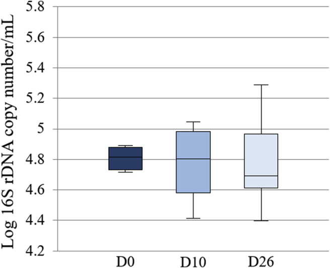
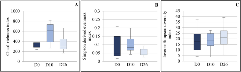
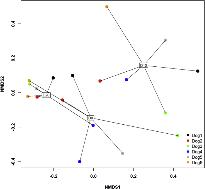
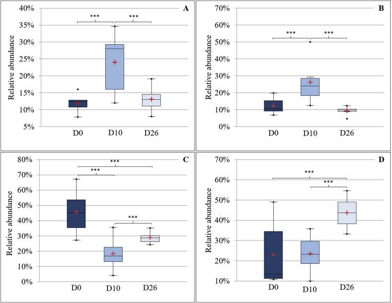
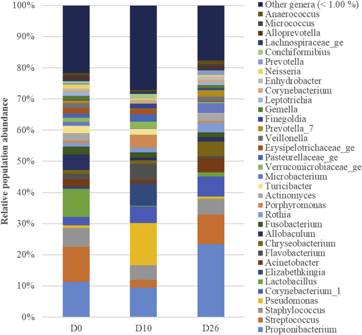
Alterations of the lung microbiota (LM) are associated with clinical features in chronic lung diseases (CLDs) with growing evidence that an altered LM contributes to the pathogenesis of such disorders. The common use of antimicrobial drugs in the management of CLDs likely represents a confounding factor in the study of the LM. The aim of the present study was to assess the effect of oral administration of amoxicillin/clavulanic acid (AC) on the LM in healthy dogs (n = 6) at short (immediately after stopping AC [D10]) and medium-term (16 days after stopping AC [D26]). Metagenetic analyses were performed on the V1-V3 hypervariable region of 16S rDNA after extraction of total bacterial DNA from samples of bronchoalveolar lavage fluid (BALF). AC did not induce significant changes in BALF cellular counts or in the bacterial load or microbial richness, evenness and α-diversity, while the β-diversity was clearly modified at D10 compared with D0 (before AC administration) and D26 (P < 0.01). The relative abundance of Bacteroidetes and Proteobacteria increased at D10 (P < 0.01) in comparison with D0 and D26 (P < 0.01). The relative abundance of Firmicutes decreased from D0 to D10 (P < 0.01) and increased from D10 to D26 (P < 0.01), but was still lower than at D0 (P < 0.01). The proportion of Actinobacteria increased at D26 compared with D0 and D10 (P < 0.01). Significant differences between timepoints at the level of family, genus or species were not found. In conclusion, in healthy dogs, oral administration of AC induces significant changes in LM at the phyla level and in the β-diversity. Most changes normalize within 2 weeks after discontinuation of AC.

Keywords: Antimicrobial; Antimicrobial drug; Bacteriology; Clinical research; Dog; Health sciences; Lung microbiota; Microbiology; Respiratory system; Veterinary medicine; Zoology.


Figures

Fig. 1
Fig. 1
Box plot representing the logarithm of the number of 16S rDNA copies per microliter (bacterial load) between timepoints. The medians are represented by the central horizontal bars. The lower and upper limits of the box are the first and third quartiles, respectively. There were no significant differences between timepoints.
Fig. 2
Fig. 2
Box plot graphs representing the bacterial richness (A), evenness (B) and alpha diversity (C) at the 3 timepoints. The medians are represented by the central horizontal bars. The lower and upper limits of the box are the first and third quartiles, respectively.
Fig. 3
Fig. 3
Two-dimensional non-parametric representation of the global bacterial composition at the species level between timepoints for each dog based on a Bray-Curtis matrix of dissimilarity. Lung communities are clustered by timepoints. D0: before antimicrobial administration; D10: just after antimicrobial discontinuation; D26: 16 days after antimicrobial discontinuation; NMDS: non-metric multidimensional scaling.
Fig. 4
Fig. 4
Phyla-level composition of bronchoalveolar lavage fluid (BALF) microbiota at the 3 timepoints. Bar charts showing relative abundance annotated to the taxonomic level of phylum for all taxa detected in BALF collected from 6 healthy adult beagle dogs, before (D0) and 10 days (D10) as well as 16 days after the discontinuation of the drug.
Fig. 5
Fig. 5
Box plot graphs representing Bacteroidetes (A), Proteobacteria (B), Firmicutes (C) and Actinobacteria (D) relative abundances between timepoints. The means and the medians are represented by the red crosses and the central horizontal bars respectively. The lower and upper limits of the box are the first and third quartiles, respectively. Points are considered as outliers. ***Statistically different (P < 0.001).
Fig. 6
Fig. 6
Genus-level composition of bronchoalveolar lavage fluid (BALF) microbiota at the 3 timepoints. Bar charts showing relative abundance annotated to the taxonomic level of genus of all taxa detected in BALF collected from 6 healthy adult beagle dogs, before (D0) and 10 days (D10) as well as 16 days after the discontinuation of the antimicrobial drug.

References

    1. Segal L.N., Rom W.N., Weiden M.D. Lung microbiome for clinicians: new discoveries about bugs in healthy and diseased lungs. Ann Am Thorac Soc. 2014;11(1):108–116. - PMC - PubMed
    1. Dickson R.P., Erb-Downward J.R., Martinez F.J., Huffnagle G.B. The microbiome and the respiratory tract. Annu. Rev. Physiol. 2016;78(1):481–504. - PMC - PubMed
    1. Dickson R.P., Erb-Downward J.R., Freeman C.M., McCloskey L., Beck J.M., Huffnagle G.B. Spatial variation in the healthy human lung microbiome and the adapted island model of lung biogeography. Ann Am Thorac Soc. 2015;12(6):821–830. - PMC - PubMed
    1. Costa A.N., Costa FM da, Campos S.V., Salles R.K., Athanazio R.A., Costa A.N. The pulmonary microbiome: challenges of a new paradigm. J. Bras. Pneumol. 2018;44(5):424–432. - PMC - PubMed
    1. Fastrès A., Felice F., Roels E., Moermans C., Corhay J.-L., Bureau F. The lung microbiome in idiopathic pulmonary fibrosis: a promising approach for targeted therapies. Int. J. Mol. Sci. 2017;18(12):2735. - PMC - PubMed

LinkOut - more resources