. 2019 Oct 19;4(12):577-583.
doi: 10.1016/j.vgie.2019.08.012. eCollection 2019 Dec.

Usefulness of a multibending endoscope in gastric endoscopic submucosal dissection

Affiliations

Usefulness of a multibending endoscope in gastric endoscopic submucosal dissection

Koichi Hamada et al. VideoGIE. .

Abstract

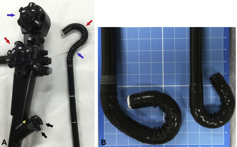
Background and aims: Intraoperative perforation is a major adverse event of endoscopic submucosal dissection (ESD). To avoid perforation, it is important for the endoscope to approach the portion to be resected carefully and to ensure that the knife can approach the submucosa at an angle parallel to the muscle layer. The multibending endoscope has 2 bends at its tip and may facilitate the ESD procedure. To the best of our knowledge, very few studies have reported the use of the multibending endoscope during gastric ESD. The aim of this study was, therefore, to introduce the usefulness of the multibending endoscope for gastric ESD.

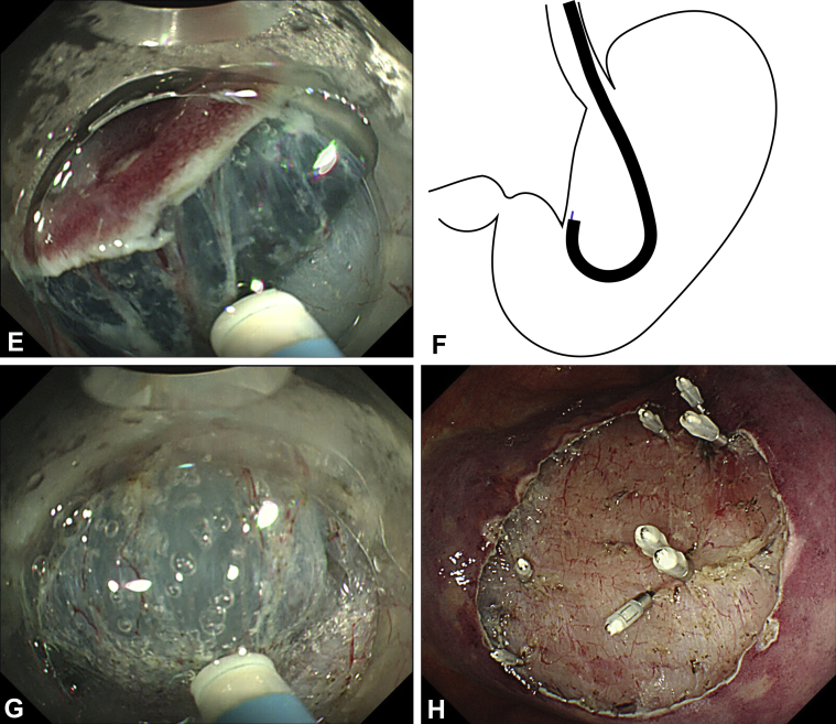
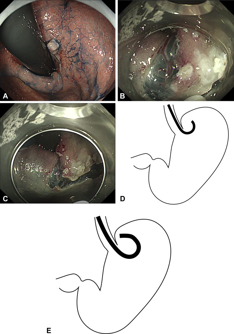
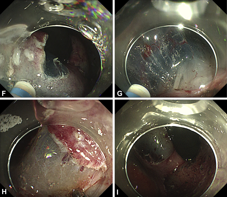
Methods: We report 2 cases of early gastric cancer in which ESD was performed using a multibending endoscope.

Results: Unlike conventional single-bending endoscopes that have only 1 moveable part, the multibending endoscope allowed difficult areas to be approached more easily. Small adjustments could be made to the upward or downward angle of both the first and the second bending sections of the endoscope. This ensured that the knife would approach the submucosa at an angle parallel to the muscle layer. In patient 1, initially the conventional endoscope was used, but it became more difficult to approach the site, and paradoxic movement occurred. When the conventional endoscope was changed to the multibending endoscope, the ESD procedure became safer and more efficient. Another ESD using the multibending endoscope was performed successfully without any adverse events.

Conclusions: The use of a multibending endoscope for ESD will enable safer and faster treatment of patients.

Keywords: ESD, endoscopic submucosal dissection.


Figures

Figure 1
Figure 1
Multibending endoscope (GIF-2TQ260M). A, This multibending endoscope has 2 bending sections and 2 channels (black arrows). The first bending section angulates up (red arrow), whereas the second angulates down (blue arrow). B, Comparison of the multibending endoscope (left) and the conventional endoscope (right). In the multibending endoscope, both bending sections angulate up.
Figure 2
Figure 2
Patient 1. A, Type 0-IIc lesion at the lesser curvature of the lower body of the stomach. B, During use of a conventional endoscope, the knife-edge points toward the muscle layer. C, Approach to the lesser curvature of the lower body of the stomach by use of a conventional endoscope. The knife is angled toward the muscle layer. D, Pushing a conventional endoscope in the forward direction causes it to move away from the lesion (paradoxical movement). E, Using a multibending endoscope, we approached the resection site and adjusted the knife-edge angle to be parallel with the muscle layer. F, Approach to the lesser curvature of the lower body of the stomach by use of the multibending endoscope. The knife-edge approaches the dissection site and is angled so that it is parallel with the muscle layer. G, Performing dissection while maintaining a parallel angle between the knife-edge and muscle layer. H, Ulcer after endoscopic submucosal dissection. We used a hemostatic clip on large blood vessels to prevent secondary bleeding.
Figure 2
Figure 2
Patient 1. A, Type 0-IIc lesion at the lesser curvature of the lower body of the stomach. B, During use of a conventional endoscope, the knife-edge points toward the muscle layer. C, Approach to the lesser curvature of the lower body of the stomach by use of a conventional endoscope. The knife is angled toward the muscle layer. D, Pushing a conventional endoscope in the forward direction causes it to move away from the lesion (paradoxical movement). E, Using a multibending endoscope, we approached the resection site and adjusted the knife-edge angle to be parallel with the muscle layer. F, Approach to the lesser curvature of the lower body of the stomach by use of the multibending endoscope. The knife-edge approaches the dissection site and is angled so that it is parallel with the muscle layer. G, Performing dissection while maintaining a parallel angle between the knife-edge and muscle layer. H, Ulcer after endoscopic submucosal dissection. We used a hemostatic clip on large blood vessels to prevent secondary bleeding.
Figure 3
Figure 3
Patient 2. A, Type 0-IIc lesion at the anterior wall to posterior wall of the upper body of the stomach. B, Incision on the fornix side, the most difficult area to reach with the endoscope. C, Incision on the fornix side. D, Approach to the fornix by use of a conventional endoscope. The endoscope cannot reach the dissection site. E, Approach to the fornix by use of the multibending endoscope. The multibend functionality of the endoscope enables us to efficiently approach and treat the site. F, Extension of the knife from the left channel while incision was made on the posterior wall expanded the right visual field. G, In dissection, when performing a cut toward the right, extending the knife from the left channel secured the visual field in the direction of the cut. H, Close approach to the site enabled us to visually confirm location of the blood vessels, and these vessels can be precisely targeted and grasped with hemostatic forceps. I, Ulcer after endoscopic submucosal dissection.
Figure 3
Figure 3
Patient 2. A, Type 0-IIc lesion at the anterior wall to posterior wall of the upper body of the stomach. B, Incision on the fornix side, the most difficult area to reach with the endoscope. C, Incision on the fornix side. D, Approach to the fornix by use of a conventional endoscope. The endoscope cannot reach the dissection site. E, Approach to the fornix by use of the multibending endoscope. The multibend functionality of the endoscope enables us to efficiently approach and treat the site. F, Extension of the knife from the left channel while incision was made on the posterior wall expanded the right visual field. G, In dissection, when performing a cut toward the right, extending the knife from the left channel secured the visual field in the direction of the cut. H, Close approach to the site enabled us to visually confirm location of the blood vessels, and these vessels can be precisely targeted and grasped with hemostatic forceps. I, Ulcer after endoscopic submucosal dissection.

References

    1. Yamamoto Y., Kikuchi D., Nagami Y. Management of adverse events related to endoscopic resection of upper gastrointestinal neoplasms: review of the literature and recommendations from experts. Dig Endosc. 2019;31:4–20. - PubMed
    1. Daegu-Kyungpook Gastrointestinal Study Group (DGSG) Kim M., Jeon S.W., Cho K.B. Predictive risk factors of perforation in gastric endoscopic submucosal dissection for early gastric cancer: a large, multicenter study. Surg Endosc. 2013;27:1372–1378. - PubMed
    1. Imagawa A., Okada H., Kawahara Y. Endoscopic submucosal dissection for early gastric cancer: results and degrees of technical difficulty as well as success. Endoscopy. 2006;38:987–990. - PubMed
    1. Kawamura M., Sekine H., Kikuchi T. Endoscopic submucosal dissection for gastric neoplasms by using a novel attachment device-a one-sided, expandable balloon. Gastrointest Endosc. 2011;74:415–418. - PubMed
    1. Isshi K., Tajiri H., Fujisaki J. The effectiveness of a new multibending scope for endoscopic mucosal resection. Endoscopy. 2004;36:294–297. - PubMed

LinkOut - more resources