Comment
. 2019 Dec 17:8:e45120.
doi: 10.7554/eLife.45120.

Replication Study: Biomechanical remodeling of the microenvironment by stromal caveolin-1 favors tumor invasion and metastasis

Collaborators, Affiliations
Comment

Replication Study: Biomechanical remodeling of the microenvironment by stromal caveolin-1 favors tumor invasion and metastasis

Mee Rie Sheen et al. Elife. .

Abstract

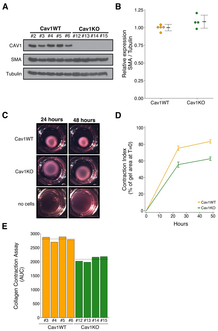
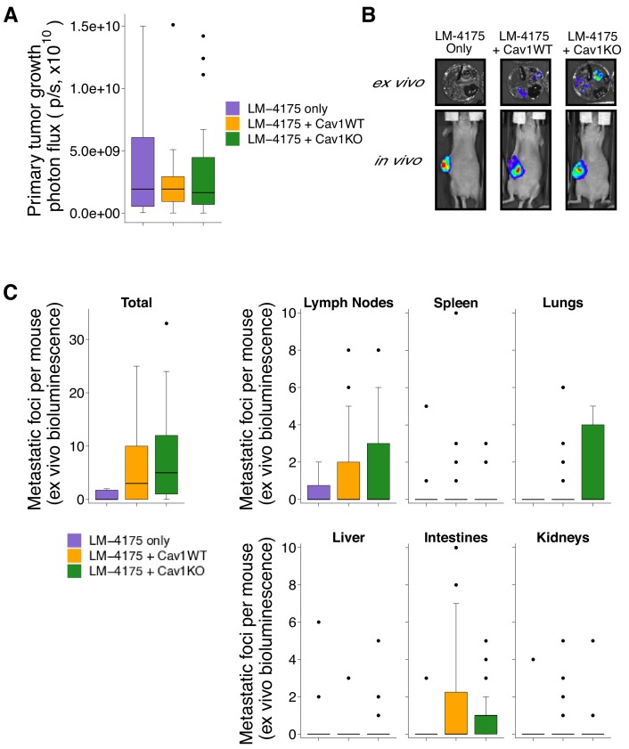
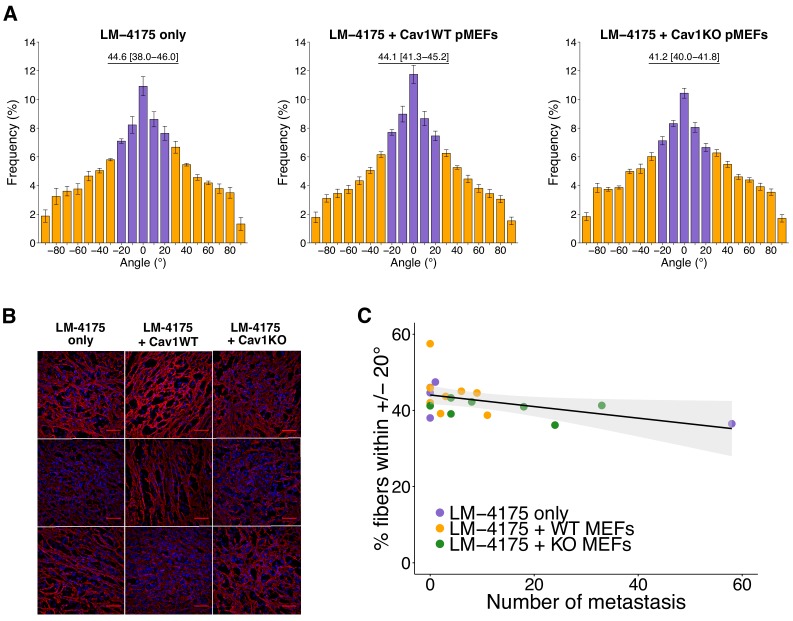
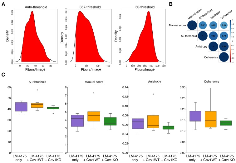
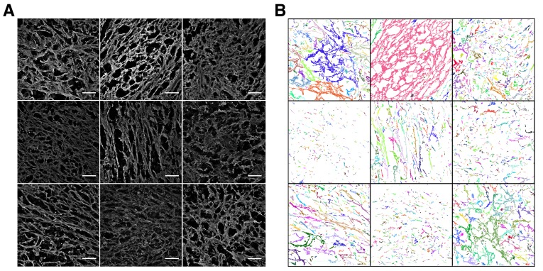
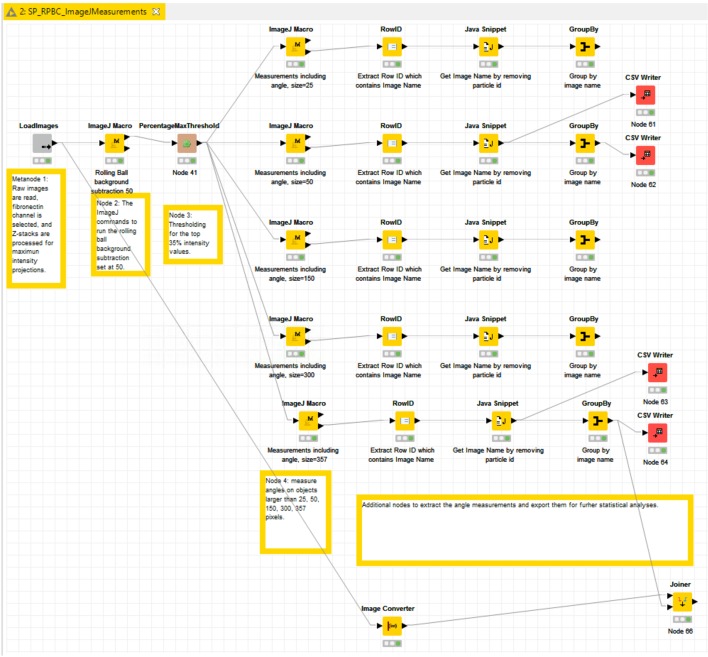
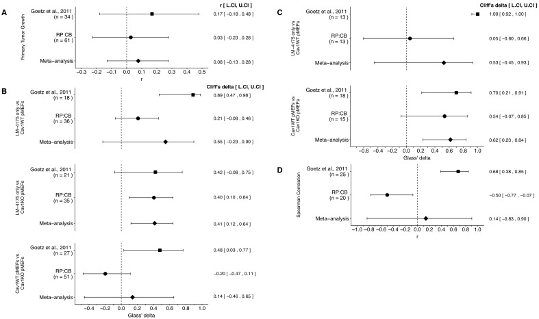
As part of the Reproducibility Project: Cancer Biology we published a Registered Report (Fiering et al., 2015) that described how we intended to replicate selected experiments from the paper 'Biomechanical remodeling of the microenvironment by stromal caveolin-1 favors tumor invasion and metastasis' (Goetz et al., 2011). Here we report the results. Primary mouse embryonic fibroblasts (pMEFs) expressing caveolin 1 (Cav1WT) demonstrated increased extracellular matrix remodeling in vitro compared to Cav1 deficient (Cav1KO) pMEFs, similar to the original study (Goetz et al., 2011). In vivo, we found higher levels of intratumoral stroma remodeling, determined by fibronectin fiber orientation, in tumors from cancer cells co-injected with Cav1WT pMEFs compared to cancer cells only or cancer cells plus Cav1KO pMEFs, which were in the same direction as the original study (Supplemental Figure S7C; Goetz et al., 2011), but not statistically significant. Primary tumor growth was similar between conditions, like the original study (Supplemental Figure S7Ca; Goetz et al., 2011). We found metastatic burden was similar between Cav1WT and Cav1KO pMEFs, while the original study found increased metastases with Cav1WT (Figure 7C; Goetz et al., 2011); however, the duration of our in vivo experiments (45 days) were much shorter than in the study by Goetz et al. (2011) (75 days). This makes it difficult to interpret the difference between the studies as it is possible that the cells required more time to manifest the difference between treatments observed by Goetz et al. We also found a statistically significant negative correlation of intratumoral remodeling with metastatic burden, while the original study found a statistically significant positive correlation (Figure 7Cd; Goetz et al., 2011), but again there were differences between the studies in terms of the duration of the metastasis studies and the imaging approaches that could have impacted the outcomes. Finally, we report meta-analyses for each result.

Keywords: Reproducibility Project: Cancer Biology; biochemical remodeling; cancer biology; human; metascience; mouse; replication; reproducibility; tumor microenvironment.


Conflict of interest statement

MS, JF, SF Transgenics and Genetic Constructs Shared Resource Center, Geisel School of Medicine at Dartmouth is a Science Exchange associated lab, BN, JL Cellavie Inc is a Science Exchange associated lab, LA Confocal Imaging Core Facility, Beth Israel Deaconess Medical Center was a Science Exchange associated lab, EI, RT, NP: Employed by and hold shares in Science Exchange Inc.

Figures

Figure 1.
Figure 1.. Characterization of Cav1 wild-type and Cav1 knockout pMEFs.
Primary MEFs (pMEFs) from wild-type (WT) or knockout (KO) embryos were examined for increased fibroblast activation and extracellular matrix (ECM) remodeling capabilities in vitro. (A) Western blots of the indicated pMEFs probed with antibodies against caveolin-1 (CAV1), alpha-smooth muscle actin (SMA), and gamma-Tubulin. Numbers indicate individual pMEF clones. (B) Western blot bands were quantified, SMA levels were normalized to Tubulin, and protein expression are presented relative to Cav1WT. Dot plot with means reported as crossbars and error bars represent SD. Number of individual clones per group: Cav1WT = 5, Cav1KO = 4. Exploratory analysis: Student’s two-tailed t-test; t(7) = 1.791, p=0.116; Cohen’s d = 1.20, 95% CI [−0.52, 2.92]. (C) Representative images from collagen contraction assay of the indicated conditions at 24 or 48 hr after plating. (D) Line graph of contraction index, measured as the change in percent of gel area at time of plating, of Cav1WT and Cav1KO pMEFs at the indicated times after plating. Means reported and error bars represent SD. Number of individual clones per group: Cav1WT = 4, Cav1KO = 4. (E) The contraction index was used to calculate the area under the curve (AUC) for each clone. Bar plots for each individual clone tested (numbers indicate same clone number as in A). Dashed lines indicate means of each group. Exploratory analysis: Student’s two-tailed t-test; t(6) = 10.80, p=3.72×10⁻⁵; Cohen’s d = 7.64, 95% CI [2.66, 12.62]. Additional details for this experiment can be found at https://osf.io/na5h2/.
Figure 2.
Figure 2.. Primary tumor growth and metastatic burden from subcutaneous tumorigenicity assay.
Female nude mice were subcutaneously injected with 1 × 10⁶ LM-4175 cells mixed with or without 1 × 10⁶ Cav1WT or Cav1KO pMEFs and monitored for 45 days. (A) At the end of the experiment primary tumors were imaged in vivo. Box and whisker plot of primary tumor photon flux with median represented as the line through the box and whiskers representing values within 1.5 IQR of the first and third quartile. Number of primary mice per group: LM-4175 only (control group)=10, LM-4175 plus Cav1WT pMEFs = 26, LM-4175 plus Cav1KO pMEFs = 25. Kruskal-Wallis test on all three groups: H(2) = 0.0439, p=0.978. (B) Representative images of primary tumors in vivo and extracted organs ex vivo. (C) The indicated organs were dissected, imaged ex vivo, and individual metastatic foci were blindly quantified. Box and whisker plots of metastatic foci counts for each organ and total metastatic counts with median represented as the line through the box and whiskers representing values within 1.5 IQR of the first and third quartile (dots represent outliers). Note: the y-axes have been truncated for visualization purposes and excludes two outliers from Total (LM-4175 only), one from Lymph Nodes (LM-4175 plus Cav1WT), seven from Lung (two from LM-4175 only; five from LM-4175 plus Cav1KO), and one from Intestines (LM-4175 only). The excluded outliers were included in the statistical analysis below. Number of mice per group: LM-4175 only = 10, LM-4175 plus Cav1WT pMEFs = 26, LM-4175 plus Cav1KO pMEFs = 25. Planned Wilcoxon-Mann-Whitney comparison on total metastatic counts between LM-4175 only and LM-4175 plus Cav1WT pMEFs: U = 103, uncorrected p=0.318 with a priori alpha level of 0.0167, Bonferroni corrected p=0.954, Cliff’s delta = 0.21, 95% CI [−0.08, 0.46]. Planned Wilcoxon-Mann-Whitney comparison on total metastatic counts between LM-4175 only and LM-4175 plus Cav1KO pMEFs: U = 175.5, uncorrected p=0.062, Bonferroni corrected p=0.185, Cliff’s delta = 0.40, 95% CI [0.10, 0.64]. Planned Wilcoxon-Mann-Whitney comparison on total metastatic counts between LM-4175 plus Cav1WT pMEFs and LM-4175 plus Cav1KO pMEFs: U = 389.5, uncorrected p=0.219, Bonferroni corrected p=0.657, Cliff’s delta = −0.20, 95% CI [−0.47, 0.11]. Additional details for this experiment can be found at https://osf.io/bq54u/.
Figure 3.
Figure 3.. Intratumoral fibronectin fiber orientation and correlation to metastasis.
A random subset of the primary tumors from the subcutaneous tumorigenicity assay (20 of 61 mice) were stained for fibronectin and analyzed to determine the average percentage of fibers oriented within 20° of the modal angle. (A) Bar graphs of the frequency of fibronectin fiber angle plotted relative to the modal angle (set at 0°). Means reported and error bars represent s.e.m. Number of mice, and thus tumors, per group: LM-4175 only = 5, LM-4175 plus Cav1WT pMEFs = 8, LM-4175 plus Cav1KO pMEFs = 7. Values reported above bar graphs indicate the median and interquartile range (IQR) of percent of fibers oriented within 20° of the modal angle (−20° to 20°, represented as purple bars) for each tumor. Planned Wilcoxon-Mann-Whitney comparison on percent of fibers oriented within 20° of the modal angle between LM-4175 only and LM-4175 plus Cav1WT pMEFs: U = 19, uncorrected p=0.884 with a priori alpha level of 0.025, Bonferroni corrected p>0.99. Planned Wilcoxon-Mann-Whitney comparison on percent of fibers oriented within 20° of the modal angle between LM-4175 plus Cav1WT pMEFs and LM-4175 plus Cav1KO pMEFs: U = 13, uncorrected p=0.0826, Bonferroni corrected p=0.165. (B) Different fields of views (fov) of the immunostained primary tumors for fibronectin and Hoechst. Three independent tumors derived from LM4175 only (first column, top to bottom: tumor 2 fov5, tumor 4 fov9, tumor 34 fov3), LM-4175 plus Cav1WT pMEFs (second column, top to bottom: tumor 16 fov7, tumor 51 fov 7, tumor 61 fov2), and LM-4175 plus Cav1KO pMEFs (third column, top to bottom: tumor 20 fov4, tumor 30 fov1, tumor 47 fov6). Scale bar: 50 µm. Fibronectin signal is pseudo-colored in red (microscope emission peak wavelength: 614 nm) and Hoechst signal is pseudo-colored in blue (microscope peak emission wavelength: 454 nm). Images are maximum intensity projections of the Z-stacks, corrected for background (as described in Materials and methods - Fibronectin fiber analysis) and displayed in the same range of grey levels. (C) Scatter plot of percentage of fibers within 20° of the modal angle and total number of metastatic counts for 20 tumors analyzed for fibronectin orientation. Line represents spearman rank correlation and light gray region represents 95% CI. Spearman rank-order correlation analysis: rs(18) = −0.50, p=0.025. Additional details for this experiment can be found at https://osf.io/bq54u/.
Figure 3—figure supplement 1.
Figure 3—figure supplement 1.. Immunostaining of primary tumors.
Primary tumors from the subcutaneous tumorigenicity assay were sectioned, fixed, and stained for fibronectin and SMA. (A) Representative images of tumor sections stained for fibronectin, no primary antibody control, or isotype control antibody. Tumor derived from LM-4175 plus Cav1WT pMEFs (tumor 8). (B) Representative images of tumor sections stained for SMA, no primary antibody control, or isotype control antibody. Tumor derived from LM-4175 plus Cav1WT pMEFs (tumor 8). (C) Representative images of tumor sections stained for SMA, no primary antibody control, or isotype control antibody after performing mouse-on-mouse (MOM) blocking Tumor derived from LM-4175 plus Cav1WT pMEFs (tumor 16). Images are maximum intensity projections of the original Z-stacks and each row are displayed in the same grey level range. Scale bar: 50 µm. Additional details for this experiment can be found at https://osf.io/bq54u/.
Figure 3—figure supplement 2.
Figure 3—figure supplement 2.. Additional measures of fibronectin fiber analysis.
Image thresholding parameters tested and exploratory analysis approaches. (A) Kernel density estimations of number of fibers per image for each thresholding parameter tested with range of number of fibers per image observed colored as red. Auto-threshold used object sizes greater than 50 and KNIME automatic thresholding, 357-threshold used object sizes greater than 357 and global threshold of 35% of max signal, and 50-threshold used object sizes greater than 50 and global threshold of 35% of max signal. Of note, 50-threshold parameter was used for results presented in Figure 3 of this study and used to compare results to the original study. (B) Correlation matrix of additional methods to assess fibronectin fiber orientation. Positive correlations are displayed in blue with color intensity and circle size proportional to the correlation coefficients, which are displayed in each circle. Exploratory spearman rank-order correlation analyses. Manual score vs 50-threshold: rs(198) = 0.67, p=1.04×10⁻²⁷; Manual score vs Anisotropy: rs(198) = 0.66, p=2.33×10⁻²⁶; Manual score vs Coherency: rs(198) = 0.56, p=6.29×10⁻¹⁸; 50-threshold vs Anisotropy: rs(198) = 0.82, p=1.02×10−50; 50-threshold vs Coherency: rs(198) = 0.72, p=5.85×10⁻³³; Anisotropy vs Coherency: rs(198) = 0.94, p=5.68×10−96. (C) Box and whisker plots of average percent of fibers oriented within 20° of the modal angle using 50-threshold (same parameter presented in Figure 3A), manual score, anisotropy score, or coherency score for each tumor with median represented as the line through the box and whiskers representing values within 1.5 IQR of the first and third quartile (dots represent outliers). Number of tumors per group: LM-4175 only = 5, LM-4175 plus Cav1WT pMEFs = 8, LM-4175 plus Cav1KO pMEFs = 7. Confirmatory analysis with 50-threshold reported in Figure 3 figure legend. Exploratory Wilcoxon-Mann-Whitney analyses for each additional method. Manual score: LM-4175 vs LM-4175 plus Cav1WT pMEFs: U = 13, p=0.306, Cliff’s delta = 0.35, 95% CI [−0.27, 0.76]; LM-4175 plus Cav1WT pMEFs vs LM-4175 plus Cav1KO pMEFs: U = 17, p=0.203, Cliff’s delta = 0.39, 95% CI [−0.18, 0.77]. Anisotropy score: LM-4175 vs LM-4175 plus Cav1WT pMEFs: U = 18, p=0.770, Cliff’s delta = 0.10, 95% CI [−0.52, 0.65]; LM-4175 plus Cav1WT pMEFs vs LM-4175 plus Cav1KO pMEFs: U = 19, p=0.298, Cliff’s delta = 0.32, 95% CI [−0.26, 0.73]. Coherency score: LM-4175 vs LM-4175 plus Cav1WT pMEFs: U = 21, p=0.884, Cliff’s delta = −0.05, 95% CI [−0.61, 0.55]; LM-4175 plus Cav1WT pMEFs vs LM-4175 plus Cav1KO pMEFs: U = 22, p=0.487, Cliff’s delta = 0.21, 95% CI [−0.39, 0.69]. Additional details for this experiment can be found at https://osf.io/bq54u/.
Figure 3—figure supplement 3.
Figure 3—figure supplement 3.. Fields of view from fibronectin fiber analysis.
These are the same fields of views of immunostated primary tumors for fibronectin displayed in Figure 3B. (A) Monochrome display presented in grey levels for fibronectin only (microscope emission peak wavelength: 614 nm). Scale bar: 50 µm. (B) Display of the corresponding segmented objects obtained with the described workflow, the angles of which were measured.
Figure 3—figure supplement 4.
Figure 3—figure supplement 4.. KNIME workflow.
Screenshot of the processing/analysis steps performed using the KNIME analytics platform.
Figure 4.
Figure 4.. Meta-analyses of each effect.
Effect size and 95% confidence interval are presented for Goetz et al. (2011), this replication attempt (RP:CB), and a random effects meta-analysis to combine the two effects. The effect size r is a standardized measure of the correlation (strength and direction) of the association between two variables and Cliff’s delta is a standardized measure of how often a value in one group is larger than the values from another group. Sample sizes used in Goetz et al. (2011) and this replication attempt are reported under the study name. (A) Primary tumor growth between mice injected with LM-4175 cells with or without Cav1WT or Cav1KO pMEFs (meta-analysis p=0.467). (B) Total metastasis counts between mice injected with LM-4175 cells and LM-4175 plus Cav1WT pMEFs (meta-analysis p=0.107), mice injected with LM-4175 cells and LM-4175 plus Cav1KO pMEFs (meta-analysis p=0.0027), and mice injected with LM-4175 plus Cav1WT pMEFs and LM-4175 plus Cav1KO pMEFs (meta-analysis p=0.680). (C) Fibronectin fiber orientation (average percentage of fibers oriented within 20° of the mode angle) between tumors from mice injected with injected with LM-4175 cells and LM-4175 plus Cav1WT pMEFs (meta-analysis p=0.269) and mice injected with LM-4175 plus Cav1WT pMEFs and LM-4175 plus Cav1KO pMEFs (meta-analysis p=1.11×10⁻⁴). (D) Rank-order correlation between fibronectin fiber orientation and total metastasis counts (meta-analysis p=0.837). Additional details for these meta-analyses can be found at https://osf.io/rvf57/.

Comment on

References

    1. Amatangelo MD, Bassi DE, Klein-Szanto AJ, Cukierman E. Stroma-derived three-dimensional matrices are necessary and sufficient to promote desmoplastic differentiation of normal fibroblasts. The American Journal of Pathology. 2005;167:475–488. doi: 10.1016/S0002-9440(10)62991-4. - DOI - PMC - PubMed
    1. Antonello ZA, Nucera C. Orthotopic mouse models for the preclinical and translational study of targeted therapies against metastatic human thyroid carcinoma with BRAF(V600E) or wild-type BRAF. Oncogene. 2014;33:5397–5404. doi: 10.1038/onc.2013.544. - DOI - PMC - PubMed
    1. Attieh Y, Clark AG, Grass C, Richon S, Pocard M, Mariani P, Elkhatib N, Betz T, Gurchenkov B, Vignjevic DM. Cancer-associated fibroblasts lead tumor invasion through integrin-β3-dependent fibronectin assembly. The Journal of Cell Biology. 2017;216:3509–3520. doi: 10.1083/jcb.201702033. - DOI - PMC - PubMed
    1. Baba S, Cho SY, Ye Z, Cheng L, Engles JM, Wahl RL. How reproducible is bioluminescent imaging of tumor cell growth? single time point versus the dynamic measurement approach. Molecular Imaging. 2007;6:7290.2007.00031–7290.2007.07290. doi: 10.2310/7290.2007.00031. - DOI - PubMed
    1. Bailoo JD, Reichlin TS, Würbel H. Refinement of experimental design and conduct in laboratory animal research. ILAR Journal. 2014;55:383–391. doi: 10.1093/ilar/ilu037. - DOI - PubMed

Publication types