Review
. 2020 Jan;76(1):52-63.
doi: 10.1111/his.14015.

Incorporation of molecular characteristics into endometrial cancer management

Affiliations
Review

Incorporation of molecular characteristics into endometrial cancer management

Lisa Vermij et al. Histopathology. 2020 Jan.

Abstract

Histopathological evaluation including subtyping and grading is the current cornerstone for endometrial cancer (EC) classification. This provides clinicians with prognostic information and input for further treatment recommendations. Nonetheless, patients with histologically similar ECs may have very different outcomes, notably in patients with high-grade endometrial carcinomas. For endometrial cancer, four molecular subgroups have undergone extensive studies in recent years: POLE ultramutated (POLEmut), mismatch repair-deficient (MMRd), p53 mutant (p53abn) and those EC lacking any of these alterations, referred to as NSMP (non-specific molecular profile). Several large studies confirm the prognostic relevance of these molecular subgroups. However, this 'histomolecular' approach has so far not been implemented in clinical routine. The ongoing PORTEC4a trial is the first clinical setting in which the added value of integrating molecular parameters in adjuvant treatment decisions will be determined. For diagnostics, the incorporation of the molecular parameters in EC classification will add a level of objectivity which will yield biologically more homogeneous subclasses. Here we illustrate how the management of individual EC patients may be impacted when applying the molecular EC classification. We describe our current approach to the integrated diagnoses of EC with a focus on scenarios with conflicting morphological and molecular findings. We also address several pitfalls accompanying the diagnostic implementation of molecular EC classification and give practical suggestions for diagnostic scenarios.

Keywords: POLE; adjuvant treatment; endometrial carcinoma; lymphovascular space invasion; mismatch repair; molecular classification; p53; risk stratification.


Conflict of interest statement

All authors declare no conflicts of interest.

Figures

Figure 1
Figure 1
Diagnostic algorithm for a ‘histomolecular’ endometrial cancer classification. 1Pathogenic polymerase‐epsilon (POLE) variants include: P286R, V411L, S297F, A456P and S459F. 2Mismatch repair protein (MMR) deficiency is defined by the loss of one or more MMR‐proteins (MLH1, PMS2, MSH2 and MSH6). 3p53 immunohistochemistry (IHC) is an acceptable surrogate marker for TP53 mutational status in MMR‐proficient, POLE wild‐type endometrial cancer (EC).50
Figure 2
Figure 2
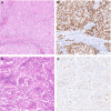
Two examples of polymerase‐epsilon mutated endometrial cancer (EC) with a pathogenic polymerase‐epsilon ultramutated (POLEmut) EC. A,B, Haematoxylin and eosin (H&E) stain of an EC diagnosed as endometrioid EC (FIGO Grade 3), based on solid growth, with aberrant mutant‐like p53 immunostaining. Molecular profiling showed the presence of a POLE P286R variant and a TP53 mutation. C,D, H&E of an EC case originally diagnosed as mixed endometroid and clear cell EC with scattered nuclear p53 immunostaining interpreted as wild‐type p53. Molecular profiling showed a POLE V411L variant and no mutations in TP53.
Figure 3
Figure 3
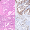
Example of a mismatch repair‐deficient (MMRd) endometrioid endometrial cancer (EEC) with lymphovascular space invasion (LVSI). A, Haematoxylin and eosin (H&E) stain of an endometrioid EC (FIGO grade 1), with prominent peritumoural lymphocytes. B, Representative image of the invasive front with foci of LVSI; the tumour contained >5 foci and was thereby reported as EC with substantial LVSI. C,D, Absence of nuclear immunoreactivity of PMS2 (C) and retained nuclear staining of MSH6 (D).
Figure 4
Figure 4
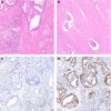
Two examples of low‐grade p53 mutation endometrioid endometrial cancer (EC). A, Representative haematoxylin and eosin (H&E) stain of an EC diagnosed as FIGO grade 2 (based on nuclear atypia) endometrioid EC. B, This case showed diffuse nuclear overexpression of p53 by IHC, interpreted as mutant‐like expression. Next‐generation sequencing (NGS) confirmed the presence of a TP53 mutation. C, Another example of an EC diagnosed as FIGO grade 1 endometrioid EC with aberrant p53 staining. NGS confirmed the presence of a pathogenic TP53 mutation. Both cases were mismatch repair protein (MMR)‐proficient and did not carry a polymerase‐epsilon (POLE) mutation.
Figure 5
Figure 5
Example of human epidermal growth factor receptor 2 (HER2) overexpressing serous endometrial cancer (EC). A, Representative haematoxylin and eosin (H&E) stain of a serous EC with profound nuclear atypia and presence of a rugged luminal surface. B, Complete strong membranous HER2 immunohistochemical staining. In this case the ERBB2 amplification was confirmed with in‐situ hybridisation (see inset).

References

    1. Kurman RJ, Carcangiu ML, Herrington S, Young RH. WHO classification of tumours of female reproductive organs. 4th ed Lyon: WHO Press, 2014.
    1. de Boer SM, Wortman BG, Bosse T et al Clinical consequences of upfront pathology review in the randomised portec‐3 trial for high‐risk endometrial cancer. Ann. Oncol. 2018; 29; 424–430. - PMC - PubMed
    1. Gilks CB, Oliva E, Soslow RA. Poor interobserver reproducibility in the diagnosis of high‐grade endometrial carcinoma. Am. J. Surg. Pathol. 2013; 37; 874–881. - PubMed
    1. Cancer Genome Atlas Research N , Kandoth C, Schultz N et al Integrated genomic characterization of endometrial carcinoma. Nature 2013; 497; 67–73. - PMC - PubMed
    1. Stelloo E, Nout RA, Osse EM et al Improved risk assessment by integrating molecular and clinicopathological factors in early‐stage endometrial cancer‐combined analysis of the portec cohorts. Clin. Cancer Res. 2016; 22; 4215–4224. - PubMed

Substances