. 2019 Dec 13;8(12):599.
doi: 10.3390/plants8120599.

The Striking Flower-in-Flower Phenotype of Arabidopsis thaliana Nossen (No-0) is Caused by a Novel LEAFY Allele

Affiliations

The Striking Flower-in-Flower Phenotype of Arabidopsis thaliana Nossen (No-0) is Caused by a Novel LEAFY Allele

Anne Mohrholz et al. Plants (Basel). .

Abstract

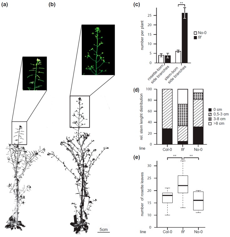
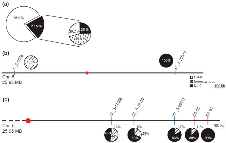
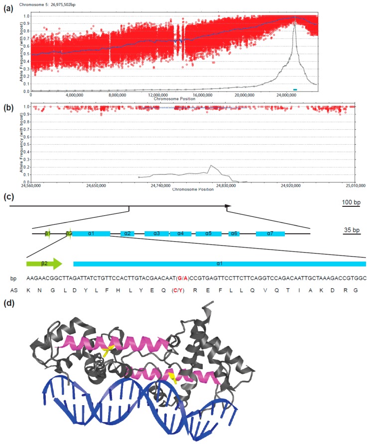
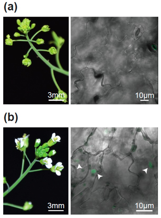
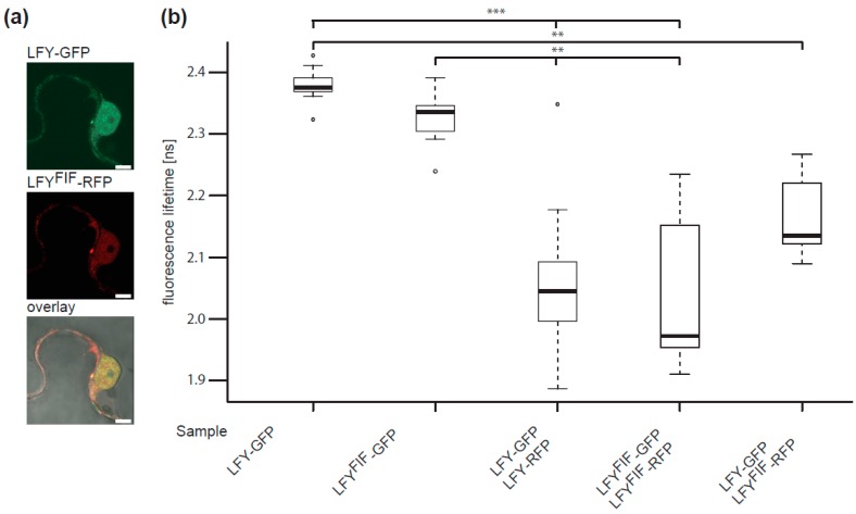
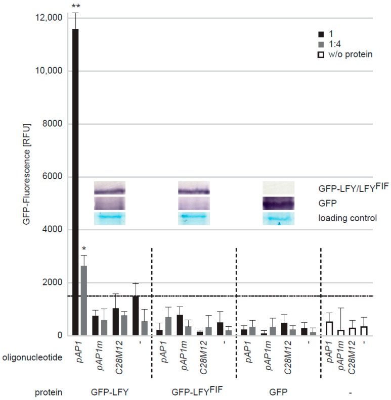
The transition to reproduction is a crucial step in the life cycle of any organism. In Arabidopsis thaliana the establishment of reproductive growth can be divided into two phases: Firstly, cauline leaves with axillary meristems are formed and internode elongation begins. Secondly, lateral meristems develop into flowers with defined organs. Floral shoots are usually determinate and suppress the development of lateral shoots. Here, we describe a transposon insertion mutant in the Nossen accession with defects in floral development and growth. Most strikingly is the outgrowth of stems from the axillary bracts of the primary flower carrying secondary flowers. Therefore, we named this mutant flower-in-flower (fif). However, the transposon insertion in the annotated gene is not the cause for the fif phenotype. By means of classical and genome sequencing-based mapping, the mutation responsible for the fif phenotype was found to be in the LEAFY gene. The mutation, a G-to-A exchange in the second exon of LEAFY, creates a novel lfy allele and results in a cysteine-to-tyrosine exchange in the α1-helix of LEAFY's DNA-binding domain. This exchange abolishes target DNA-binding, whereas subcellular localization and homomerization are not affected. To explain the strong fif phenotype against these molecular findings, several hypotheses are discussed.

Keywords: Arabidopsis thaliana; DNA-binding; Ds transposon; LEAFY; classical/sequencing-based mapping; floral development; flower morphology.


Conflict of interest statement

The authors declare no conflict of interest. The funders had no role in the design of the study; in the collection, analyses, or interpretation of data; in the writing of the manuscript, or in the decision to publish the results.

Figures

Figure 1
Figure 1
Flower phenotype of the Arabidopsis thaliana (No-0) flower-in-flower (fif) mutant. (a) Overview over a representative fif mutant “inflorescence” displaying different flower types 1 to 4. (b) Floral organs of the primary fif flower (1) and different secondary fif flowers (2–4). (c) Flower of the wild type No-0 accession. (d) Primary flowers of the fif mutant with stems that outgrow from axillary bract meristems (red arrow heads) and carry secondary flowers. Size bar: 1 mm.
Figure 2
Figure 2
Growth habitus and flowering time of wild type No-0, wild type Col-0 and fif mutant plants. Overview over the growth habitus and magnification of the inflorescence of 6.5-weeks old wild type No-0 (a) and fif plants (b), grown side-by-side in the greenhouse. Size bar: 5.0 cm. (c) Number of rosette-born side branches and stem-born side branches of wild type No-0 (white bars) and fif (black bars) plants. Error bars indicate the standard deviation of the mean. The statistical significance (nNo-0 = 33, nfif = 25) was testet by two-sited t-test (***: p = 2 × 10⁻²³). (d) Relative distribution of stem length in wild type No-0, wild type Col-0 and fif mutant plants 31 days after sowing (nCol-0 = 7, nNo-0 = 16, nfif = 89). (e) Number of rosette leaves at the onset of senescence for wild type No-0, wild type Col-0 and fif mutant plants. The data are presented in Box-and-Whisker plots including the median (thick line), the upper and lower quartile (+/− 25%, white boxes) and the maximum and minimum (dottet line). The statistical significance (nNo-0 = 4, nCol-0 = 9, nfif = 38) was tested with ANOVA followed by a Tukey honest significant difference post-hoc test (*: p < 0.05; **: p < 0.01; ***: p < 0.001).
Figure 3
Figure 3
Segregation analysis and mapping of the mutant locus causal for the fif mutant phenotype. (a) Segregation of the floral phenotype and the Ds transposon insertion within the combined F2 population of (♀fif × ♂No-0) and (♀No-0 × ♂fif) backcrosses showing either the wild type (78.4%) or the fif floral phenotype (21.6%) (left) and distribution of the transposon insertions within the plants of the F2 population that displayed the fif floral phenotype (right); white circle outcut: no transposon insertion (29.2%), striped outcut: heterozygous for the Ds transposon insertion (45.8%), black outcut: homozygous for the Ds transposon insertion (25.0%). (b,c) Insertion and DELetion INDEL marker- and single nucleotide polymorphism (SNP)-based derived cleaved amplified polymorphic sequences (dCAP) marker-associated containment of the fif locus using a mapping population generated by a cross of the fif mutant (No-0) with wild type Col-0. Schematic representation of the Arabidopsis thaliana chromosome 5 (sizes in MB) and the localization of the chromosome-specific INDEL markers initially used for mapping (codes above blue lines) (b). Schematic representation of the q-arm of chromosome 5 and the localization of INDEL (codes above the blue lines) and SNP-based dCAP markers (codes above red lines) used for fine mapping (c). The pie charts show the distribution of the No-0 and Col-0 genotypes for chromosome 5 (b) and the q-arm of chromosome 5 (c). White circular outcut: homozygous for Col-0, striped outcut: heterozygous for Col-0/No-0, black outcut: homozygous for No-0; red dot: localization of the centromere.
Figure 4
Figure 4
Identification of the fif-related SNP in the second exon of the LEAFY (LFY) locus on chromosome 5 by genome sequencing of a mapping population generated by a cross of the fif mutant (No-0) with wild type Col-0. (a) Allele frequency analysis of the No-0 genotype within chromosome 5 of the recombinant mutant pool. Each red circle refers to a SNP marker distinguishing the No-0 and Col-0 genotypes. The blue line refers to a 200 kb sliding window analysis of the allele frequencies. The brown line and blue box highlight the estimated mapping intervals (x-axis: genomic location; y-axis: Nos allele frequency). (b) Like (a), but only showing the 300 kb mapping interval. (c) Sequence of the LFY gene showing the fif SNP (G to A exchange, red) and the resulting amino acid exchange (C to Y, red) within the DNA-binding domain of the LFY protein. Green boxes: β-sheets; blue boxes: α-helices. (d) Structural representation of the wild type LFY dimer bound to DNA according to Hames and colleagues (2008). The α1 helix in the monomers is shown in magenta and the cysteine (C) in yellow that are mutated to tyrosine (Y) in the fif mutant.
Figure 5
Figure 5
Complementation of the fif mutant phenotype by the UBI10 promoter-driven expression of wild type LFY-GFP. (a) Inflorescence (left) and confocal image of epidermal cells (right) of a representative non-transformed fif mutant plant. (b) As in (a), but for a representative LFY-GFP transgenic plant in the fif mutant background. The fluorescent nuclei are highlighted by arrowheads. Three independent transgenic lines were obtained displaying nuclear localization of LFY-GFP and in parallel the complementation of the fif phenotype.
Figure 6
Figure 6
Comparative analysis of the intracellular localization and homomerization capacity of LFY and LFYFIF. (a) Confocal fluorescence images of transiently transformed Nicotiana benthamina epidermal leaf cells expressing LFY-GFP and LFYFIF-RFP in the same cell. Size bar: 5 µm. (b) FRET-FLIM analysis of the homo- and heterotypic interaction of LFY and LFYFIF. LFY-GFP or LFYFIF-GFP were expressed either alone or together with the indicated RFP fusions and the fluorescence lifetime of the GFP fusions measured in the nuclei. A reduction of the GFP fluorescence lifetime indicates interaction. The data are presented in Box-and-Whisker plots including the median (thick line), the upper and lower quartile (+/− 25%, white boxes), the maximum and minimum (dottet line) and outlier points (n > 20, each). The variance was analyzed by a Levene test and statistical significance was determined with an all-pair, two-sided Kruskal–Wallis test followed by an all-pair Steel-Dwass test (**: p < 0.01; ***: p < 0.001).
Figure 7
Figure 7
Comparative analysis of the in vitro DNA-binding capacity of LFY and LFYFIF using a GFP-fluorescence-based DPI-ELISA approach. GFP-LFY, GFP-LFYFIF and GFP were expressed in E. coli. After extraction, crude extracts containing either no recombinant protein (w/o protein) or, based on GFP fluorescence, equal amounts of GFP or the GFP fusion proteins were added to ELISA plates covered with either the double-stranded (ds) DNA oligonucleotide pAP1, which contains a LFY binding site, an altered version of pAP1 (pAP1m), in which the binding site was mutated, a dsDNA oligonucleotide unrelated to the pAP1 and pAP1m sequences (C28M12) or without any DNA-oligonucleotide. The amount of DNA-bound fusion protein was detected by reading out the GFP fluorescence. The crude extract was either used undiluted (black bars) or in a 1:4 dilution (grey bars). Error bars indicate the standard deviation of the mean (n = 3) and asterisk statistically significant differences to the background fluorescence (dotted horizontal line), determined by two-sided t-test (*: p < 0.05; **: p < 0.01). The inlet shows a Western-blot of the crude extracts using a GFP polyclonal antiserum for detection of GFP, GFP-LFY and GFP-LFYFIF as well as a Coomassie stain as loading control.

Similar articles

Cited by

References

    1. Pineiro M., Coupland G. The control of flowering time and floral identity in Arabidopsis. Plant Physiol. 1998;117:1–8. doi: 10.1104/pp.117.1.1. - DOI - PMC - PubMed
    1. Ma H. To be, or not to be, a flower–control of floral meristem identity. Trends Genet. 1998;14:26–32. doi: 10.1016/S0168-9525(97)01309-7. - DOI - PubMed
    1. Pidkowich M.S., Klenz J.E., Haughn G.W. The making of a flower: Control of floral meristem identity in Arabidopsis. Trends Plant Sci. 1999;4:64–70. doi: 10.1016/S1360-1385(98)01369-7. - DOI - PubMed
    1. Denay G., Chahtane H., Tichtinsky G., Parcy F. A flower is born: An update on Arabidopsis floral meristem formation. Curr. Opin. Plant Biol. 2017;35:15–22. doi: 10.1016/j.pbi.2016.09.003. - DOI - PubMed
    1. O’Maoileidigh D.S., Graciet E., Wellmer F. Gene networks controlling Arabidopsis thaliana flower development. New Phytol. 2014;201:16–30. doi: 10.1111/nph.12444. - DOI - PubMed

LinkOut - more resources