. 2019 Dec 13;20(24):6292.
doi: 10.3390/ijms20246292.

Detection of ALDH3B2 in Human Placenta

Affiliations

Detection of ALDH3B2 in Human Placenta

Sylwia Michorowska et al. Int J Mol Sci. .

Abstract

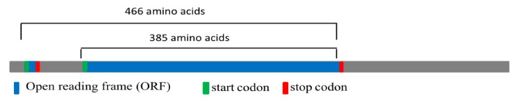
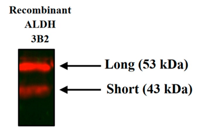
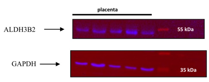
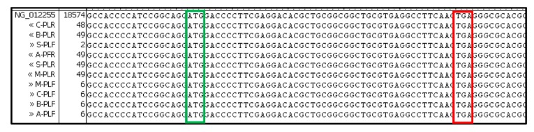
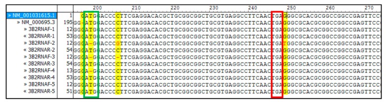
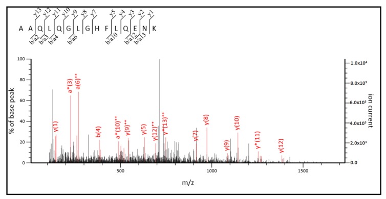
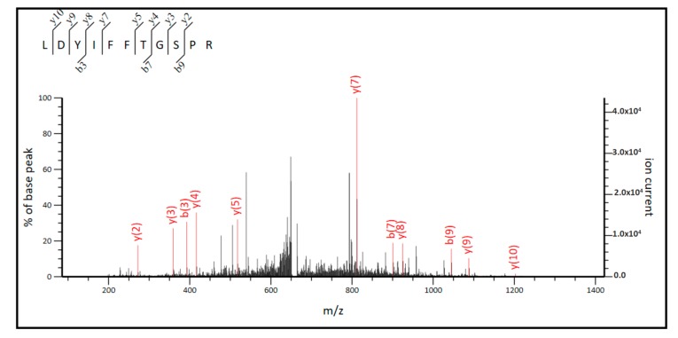
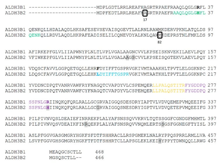
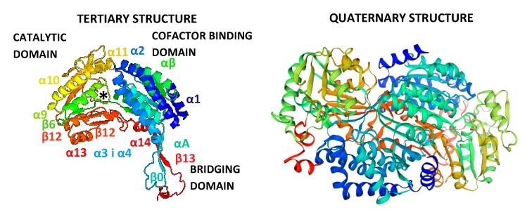
Aldehyde dehydrogenase 3B2 (ALDH3B2) gene contains a premature termination codon, which can be skipped or suppressed resulting in full-length protein expression. Alternatively, the longest putative open reading frame starting with the second in-frame start codon would encode short isoform. No unequivocal evidence of ALDH3B2 expression in healthy human tissues is available. The aim of this study was to confirm its expression in human placenta characterized by the highest ALDH3B2 mRNA abundance. ALDH3B2 DNA and mRNA were sequenced. The expression was investigated using western blot. The identity of the protein was confirmed using mass spectrometry (MS). The predicted tertiary and quaternary structures, subcellular localization, and phosphorylation sites were assessed using bioinformatic analyses. All DNA and mRNA isolates contained the premature stop codon. In western blot analyses, bands corresponding to the mass of full-length protein were detected. MS analysis led to the identification of two unique peptides, one of which is encoded by the nucleotide sequence located upstream the second start codon. Bioinformatic analyses suggest cytoplasmic localization and several phosphorylation sites. Despite premature stop codon in DNA and mRNA sequences, full-length ALDH3B2 was found. It can be formed as a result of premature stop codon readthrough, complex phenomenon enabling stop codon circumvention.

Keywords: ALDH3B2; placenta; premature stop codon; readthrough.


Conflict of interest statement

The authors declare no conflict of interest.

Figures

Figure 1
Figure 1
Structure of ALDH3B2 gene. Blue represents open reading frames (ORF), green represents the start codon, and red represents the stop codon. Black lines show lengths of two ALDH3B2 isoforms: the short one with 385 amino acids and the long one with 466 amino acids.
Figure 2
Figure 2
Western blot analysis using anti-ALDH3B2 antibody confirmed the expression of both short and long isoforms of recombinant ALDH3B2 protein in E. coli. Purified recombinant proteins were applied onto the polyacrylamide gel. After separation, they were transferred onto the PVDF membrane. Their immunodetection was performed using anti-ALDH3B2 antibody. The image was taken using ChemiDoc XRS+ (Bio-Rad).
Figure 3
Figure 3
Western blot analysis of placenta homogenates using anti-ALDH3B2 and anti-GAPDH antibodies revealed bands corresponding to the molecular weight of 53 kDa (long isoform of ALDH3B2) and bands corresponding to molecular weight of 37 kDa (GAPDH). Proteins in homogenates were applied onto the polyacrylamide gel. After separation, they were transferred onto the PVDF membrane. Immunodetection of ALDH3B2 protein was performed using anti-ALDH3B2 antibody. After membrane stripping, the immunodetection was repeated with anti-GAPDH antibody. GAPDH detection was used as a loading control, as GAPDH gene is constitutively expressed at high levels in many tissues. The image was taken using ChemiDoc XRS+ (Bio-Rad). Additionally, the image of the whole membrane was added into the Supplementary Materials (Supplementary Figure S1).
Figure 4
Figure 4
All sequences of ALDH3B2 DNA isolated from placenta samples contained premature stop codon. Sequence alignment was performed using Vector NTI AdvanceTM 11.0 software. As a reference ALDH3B2 sequence from NCBI (NG_012255.1) was used. Green frame shows first start codon and red one premature stop codon.
Figure 5
Figure 5
All sequences of ALDH3B2 cDNA synthesized in RT-PCR reaction using mRNA isolated from placenta samples contained premature stop codon. Sequence alignment was performed using Vector NTI AdvanceTM 11.0 software. Isolated RNA was treated with DNase to remove contaminating DNA and then subjected to RT-PCR. The obtained cDNA was sequenced. As a reference, ALDH3B2 cDNA sequences from NCBI (NM_001031615.1 and NM_000695.3) were used. The green frame shows first start codon and red frame shows the premature stop codon. Yellow indicates differences in the aligned sequences.
Figure 6
Figure 6
MS/MS spectrum for the tryptic peptide, AAQLQGLGHFLQENK, found in the sequence of long ALDH3B2 isoform. The upper left corner indicates the source of the fragment ions identified as either b, a, or y ions. ++ designates doubly charged fragment ions, * designates fragment ions with neutral loss.
Figure 7
Figure 7
MS/MS spectrum for the tryptic peptide, LDYIFFTGSPR, found in the sequence of long and short ALDH3B2 isoform. The upper left corner indicates the source of the fragment ions identified as either b or y ions.
Figure 8
Figure 8
Peptides identified in MS/MS analysis of placenta homogenates. Peptide sequences identified in the MS/MS analysis were mapped to the long ALDH3B2 isoform sequence (translation product of mRNA transcript number U37519.1 from NCBI obtained using ExPASy server [14] with alanine in the position encoded by premature stop codon) and compared with the sequences of the corresponding peptides of ALDH3B1 (NCBI: NP_001154945.1). Colors indicate sequences of identified peptides: yellow and violet—sequences common to ALDH3B1 as well as short and long ALDH3B2; blue—sequence common to both, short and long ALDH3B2 isoforms; and green—sequence unique to long ALDH3B2 isoform. Residues in bold represent amino acids different for ALDH3B1 and ALDH3B2 within sequences of identified peptides. Grey shading indicates positions of different amino acids present in different ALDH3B2 natural variants that can be found in UniProt database [15]: residue 50: A/T, residue 52: S/N, residue 203:H/R, residue 220: G/S, residues 276: R/W, residue 302: S/R, residue 361: H/R. Two black frames in the sequence of ALDH3B2 indicate: the alanine in the position encoded by the premature stop codon (17th aa) and methionine encoded by the first in-frame start codon (82nd aa), respectively.
Figure 9
Figure 9
Comparison of ALDH3 isoenzymes’ primary structures. The alignment of ALDH3 sequences was performed using Clustal Omega server [16] and the following sequences: NCBI sequence NP_001128640.1 of ALDH3A1, NCBI sequence NP_000373.1 of ALDH3A2, NCBI sequence NP_001154945.1 of ALDH3B1 and long ALDH3B2 isoform sequence, the product of translation of mRNA transcript number U37519.1 from NCBI obtained using ExPASy server [14], with alanine in the position encoded by premature stop codon. Orange indicates residues involved in substrate binding, red residues involved in cofactor binding, blue three proline residues contributing to the tertiary structure, violet residue 236, gray shading residues highly conserved: Cys-244 acting as a nucleophile; Glu-334 activating thiol group of Cys-244; Gly-241 positioning Cys-244; Glu-210 activating water molecule that hydrolyzes ester group, residues responsible for cofactor binding: Gly-188 and Gly-193 forming Rossmann fold; Lys-138, Glu-334, and Phe-336 responsible for nicotinamide ring positioning and hydrogen bond formation to the NAD(P)+ adenine ribose; Ile-335 and Lys-214 ensuring appropriate geometry of cofactor binding domain; Asn-115 crucial for catalytic activity, transiently stabilizing oxygen of carboxyl group of tetrahedral intermediate; Asp-248 conserved in ALDHs belonging to the third subfamily, responsible for appropriate geometry of active site; Arg-26, Gly-106, Pro-117, Gly-132, Pro-338, Gly-384, Asn-389, and Gly-404 [1,17].
Figure 10
Figure 10
Predicted tertiary and quaternary structure of human long ALDH3B2 isoform, with alanine in the position encoded by the premature stop codon. The tertiary structure model was prepared using Phyre2 server [18] and the quaternary structure model was prepared using Swiss Model server [19]. ALDH3B2 sequence used was the product of translation of mRNA transcript number U37519.1 from NCBI obtained using ExPASy server [14], with alanine in the position encoded by premature stop codon. In both cases, the rat class 3 aldehyde dehydrogenase template at 2.6 Å resolution was used (PDB ID: 1AD3). The confidence was 100%, coverage 95% (444 residues: 4-447) with 50% sequence identity. The rainbow color code describes the predicted 3D structures from N- (blue) to C-termini (red). The designation of the three domains as well as that of alpha helices and beta strands was based on the rat ALDH3A1 structure (PDB ID: 1AD3) [20]. The asterisk indicates the catalytic Cys-244.
Figure 11
Figure 11
Nucleotide sequence surrounding the premature stop codon in ALDH3B2 mRNA.

References

    1. Holmes R.S., Hempel J. Comparative studies of vertebrate aldehyde dehydrogenase 3: Sequences, structures, phylogeny and evolution. Evidence for a mammalian origin for the ALDH3A1 gene. Chem. Biol. Interact. 2011;191:113–121. doi: 10.1016/j.cbi.2011.01.014. - DOI - PubMed
    1. Marchitti S.A., Brocker C., Stagos D., Vasiliou V. Non-P450 aldehyde oxidizing enzymes: The aldehyde dehydrogenase superfamily. Expert Opin. Drug Metab. Toxicol. 2008;4:697–720. doi: 10.1517/17425255.4.6.697. - DOI - PMC - PubMed
    1. Giebułtowicz J., Wroczyński P., Samolczyk-Wanyura D. Can lower aldehyde dehydrogenase activity in saliva be a risk factor for oral cavity cancer? Oral Dis. 2013;19:763–766. doi: 10.1111/odi.12064. - DOI - PubMed
    1. Hsu L.C., Chang W.C., Yoshida A. Human aldehyde dehydrogenase genes, ALDH7 and ALDH8: Genomic organization and gene structure comparison. Gene. 1997;189:89–94. doi: 10.1016/S0378-1119(96)00839-6. - DOI - PubMed
    1. Hsu L.C., Chang W.-C. Sequencing and expression of the human ALDH8 encoding a new member of the aldehyde dehydrogenase family. Gene. 1996;174:319–322. doi: 10.1016/0378-1119(96)00087-X. - DOI - PubMed

LinkOut - more resources