

Four-Dimensional Flow Magnetic Resonance Imaging for Assessment of Velocity Magnitudes and Flow Patterns in The Human Carotid Artery Bifurcation: Comparison with Computational Fluid Dynamics
- PMID: 31847224
- PMCID: PMC6963916
- DOI: 10.3390/diagnostics9040223
Four-Dimensional Flow Magnetic Resonance Imaging for Assessment of Velocity Magnitudes and Flow Patterns in The Human Carotid Artery Bifurcation: Comparison with Computational Fluid Dynamics
Abstract
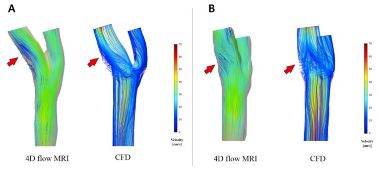
Purpose: Knowledge of the hemodynamics in the vascular system is important to understand and treat vascular pathology. The present study aimed to evaluate the hemodynamics in the human carotid artery bifurcation measured by four-dimensional (4D) flow magnetic resonance imaging (MRI) as compared to computational fluid dynamics (CFD).
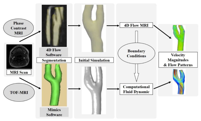
Methods: This protocol used MRI data of 12 healthy volunteers for the 3D vascular models and 4D flow MRI measurements for the boundary conditions in CFD simulation. We compared the velocities measured at the carotid bifurcation and the 3D velocity streamlines of the carotid arteries obtained by these two methods.
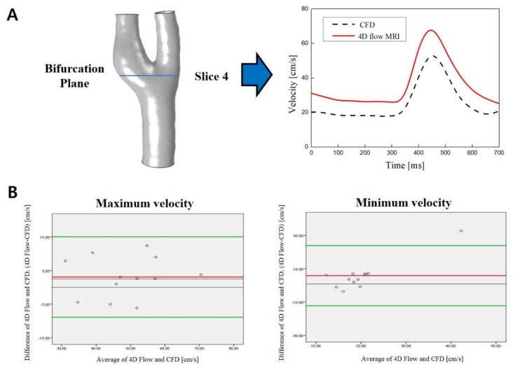
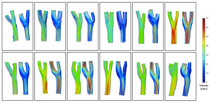
Results: There was a good agreement for both maximum and minimum velocity values between the 2 methods for velocity magnitude at the bifurcation plane. However, on the 3D blood flow visualization, secondary flows, and recirculation regions are of poorer quality when visualized through the 4D flow MRI.
Conclusion: 4D flow MRI and CFD show reasonable agreement in demonstrated velocity magnitudes at the carotid artery bifurcation. However, the visualization of blood flow at the recirculation regions and the assessment of secondary flow characteristics should be enhanced for the use of 4D flow MRI in clinical situations.
Keywords: carotid artery; carotid bifurcation; computational fluid dynamics (CFD); flow patterns; four-dimension flow magnetic resonance imaging (4D flow MRI); velocity magnitudes.
Conflict of interest statement
The authors declare no conflict of interest.
Figures

References
-
- Harloff A., Strecker C., Reinhard M., Kollum M., Handke M., Olschewski M., Weiller C., Hetzel A. Combined Measurement of Carotid Stiffness and Intima-Media Thickness Improves Prediction of Complex Aortic Plaques in Patients With Ischemic Stroke. Stroke. 2006;37:2708–2712. doi: 10.1161/01.STR.0000244763.19013.dc. - DOI - PubMed
-
- Harloff A., Albrecht F., Spreer J., Stalder A.F., Bock J., Frydrychowicz A., Schöllhorn J., Hetzel A., Schumacher M., Hennig J., et al. 3D blood flow characteristics in the carotid artery bifurcation assessed by flow-sensitive 4D MRI at 3T. Magn. Reson. Med. 2009;61:65–74. doi: 10.1002/mrm.21774. - DOI - PubMed
Grants and funding
LinkOut - more resources
Full Text Sources
Miscellaneous
