

Applicability of Anti-Human Estrogen Receptor β Antibody PPZ0506 for the Immunodetection of Rodent Estrogen Receptor β Proteins
- PMID: 31847265
- PMCID: PMC6941125
- DOI: 10.3390/ijms20246312
Applicability of Anti-Human Estrogen Receptor β Antibody PPZ0506 for the Immunodetection of Rodent Estrogen Receptor β Proteins
Abstract
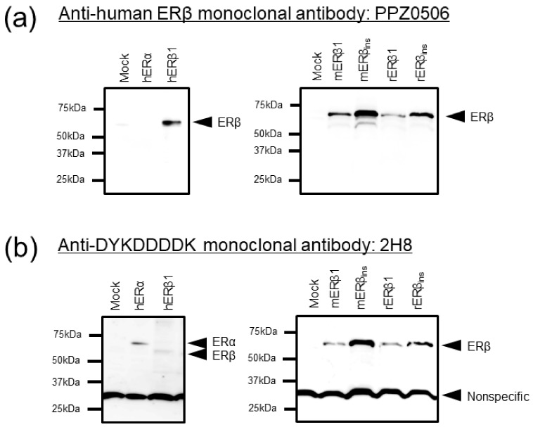
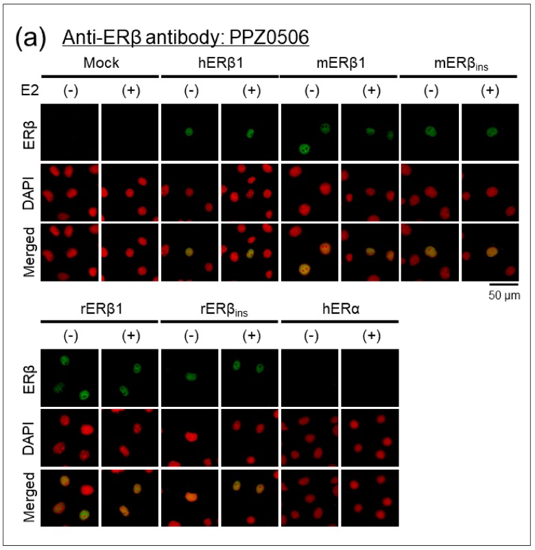
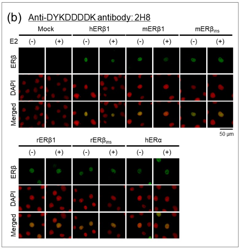
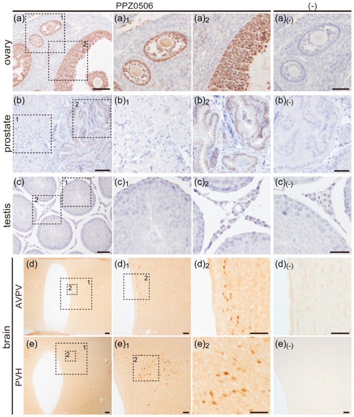
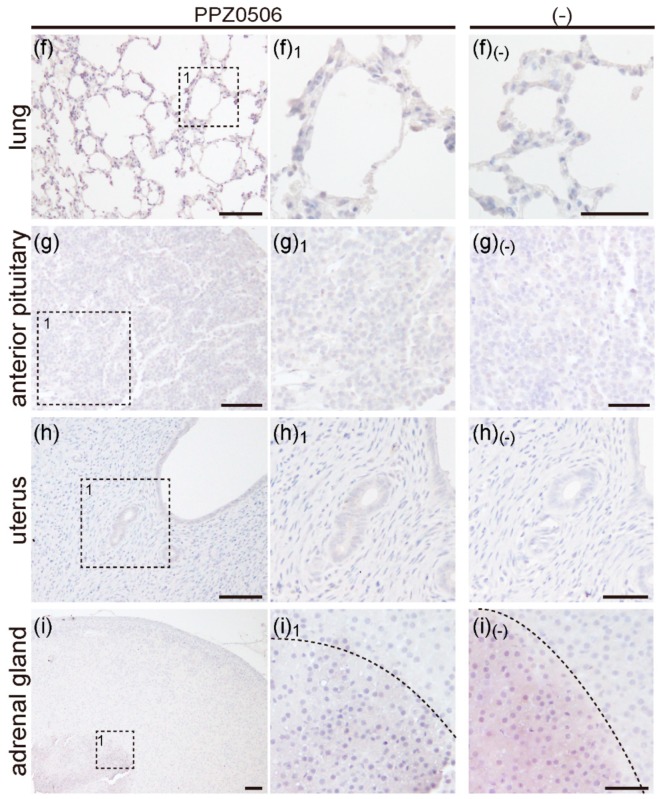
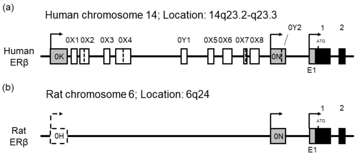
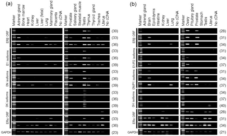
Several lines of controversial evidence concerning estrogen receptor β (ERβ) remain to be solved because of the unavailability of specific antibodies against ERβ. The recent validation analysis identified a monoclonal antibody (PPZ0506) with sufficient specificity against human ERβ. However, the specificity and cross-reactivity of PPZ0506 antibody against ERβ proteins from laboratory animals have not been confirmed. In the present study, we aimed to validate the applicability of PPZ0506 to rodent studies. The antibody exhibited specific cross-reactivity against mouse and rat ERβ proteins in immunoblot and immunocytochemical experiments using transfected cells. In immunohistochemistry for rat tissue sections, PPZ0506 showed immunoreactive signals in the ovary, prostate, and brain. These immunohistochemical profiles of rat ERβ proteins in rat tissues accord well with its mRNA expression patterns. Although the antibody was reported to show the moderate signals in human testis, no immunoreactive signals were observed in rat testis. Subsequent RT-PCR analysis revealed that this species difference in ERβ expression resulted from different expression profiles related to the alternative promoter usage between humans and rats. In conclusion, we confirmed applicability of PPZ0506 for rodent ERβ studies, and our results provide a fundamental basis for further examination of ERβ functions.
Keywords: ESR2; alternative promoter usage; alternative splicing; antibody validation.
Conflict of interest statement
The authors declare no conflict of interest.
Figures
Similar articles
-
Optimized Mouse-on-mouse Immunohistochemical Detection of Mouse ESR2 Proteins with PPZ0506 Monoclonal Antibody.Acta Histochem Cytochem. 2022 Oct 28;55(5):159-168. doi: 10.1267/ahc.22-00043. Epub 2022 Oct 25. Acta Histochem Cytochem. 2022. PMID: 36405553 Free PMC article.
-
Sex and interspecies differences in ESR2-expressing cell distributions in mouse and rat brains.Biol Sex Differ. 2023 Dec 18;14(1):89. doi: 10.1186/s13293-023-00574-z. Biol Sex Differ. 2023. PMID: 38111056 Free PMC article.
-
Immunohistochemical Detection of Estrogen Receptor-Beta (ERβ) with PPZ0506 Antibody in Murine Tissue: From Pitfalls to Optimization.Biomedicines. 2022 Dec 1;10(12):3100. doi: 10.3390/biomedicines10123100. Biomedicines. 2022. PMID: 36551855 Free PMC article.
-
Optimization of immunohistochemical detection of rat ESR2 proteins with well-validated monoclonal antibody PPZ0506.Mol Cell Endocrinol. 2021 Mar 1;523:111145. doi: 10.1016/j.mce.2020.111145. Epub 2021 Jan 2. Mol Cell Endocrinol. 2021. PMID: 33400952
-
The estrogen receptor beta subtype: a novel mediator of estrogen action in neuroendocrine systems.Front Neuroendocrinol. 1998 Oct;19(4):253-86. doi: 10.1006/frne.1998.0170. Front Neuroendocrinol. 1998. PMID: 9799586 Review.
Cited by
-
Optimized Mouse-on-mouse Immunohistochemical Detection of Mouse ESR2 Proteins with PPZ0506 Monoclonal Antibody.Acta Histochem Cytochem. 2022 Oct 28;55(5):159-168. doi: 10.1267/ahc.22-00043. Epub 2022 Oct 25. Acta Histochem Cytochem. 2022. PMID: 36405553 Free PMC article.
-
Sex and interspecies differences in ESR2-expressing cell distributions in mouse and rat brains.Biol Sex Differ. 2023 Dec 18;14(1):89. doi: 10.1186/s13293-023-00574-z. Biol Sex Differ. 2023. PMID: 38111056 Free PMC article.
-
A Phosphotyrosine Switch in Estrogen Receptor β Is Required for Mouse Ovarian Function.Front Cell Dev Biol. 2021 Apr 9;9:649087. doi: 10.3389/fcell.2021.649087. eCollection 2021. Front Cell Dev Biol. 2021. PMID: 33898441 Free PMC article.
-
Ovarian ERβ cistrome and transcriptome reveal chromatin interaction with LRH-1.BMC Biol. 2023 Nov 29;21(1):277. doi: 10.1186/s12915-023-01773-1. BMC Biol. 2023. PMID: 38031019 Free PMC article.
-
Immunohistochemical Detection of Estrogen Receptor-Beta (ERβ) with PPZ0506 Antibody in Murine Tissue: From Pitfalls to Optimization.Biomedicines. 2022 Dec 1;10(12):3100. doi: 10.3390/biomedicines10123100. Biomedicines. 2022. PMID: 36551855 Free PMC article.
References
-
- Nilsson S., Gustafsson J.A. Estrogen receptors: Their actions and functional roles in health and disease. In: Bunce C., Campbell M., editors. Nuclear Receptors, Proteins and Cell Regulation. Vol. 8. Springer; Berlin, Germany: 2010. pp. 91–141.
MeSH terms
Substances
Grants and funding
LinkOut - more resources
Full Text Sources
