. 2019 Dec 15;20(24):6330.
doi: 10.3390/ijms20246330.

Elucidating the Role of K+ Channels during In Vitro Capacitation of Boar Spermatozoa: Do SLO1 Channels Play a Crucial Role?

Affiliations

Elucidating the Role of K+ Channels during In Vitro Capacitation of Boar Spermatozoa: Do SLO1 Channels Play a Crucial Role?

Marc Yeste et al. Int J Mol Sci. .

Abstract

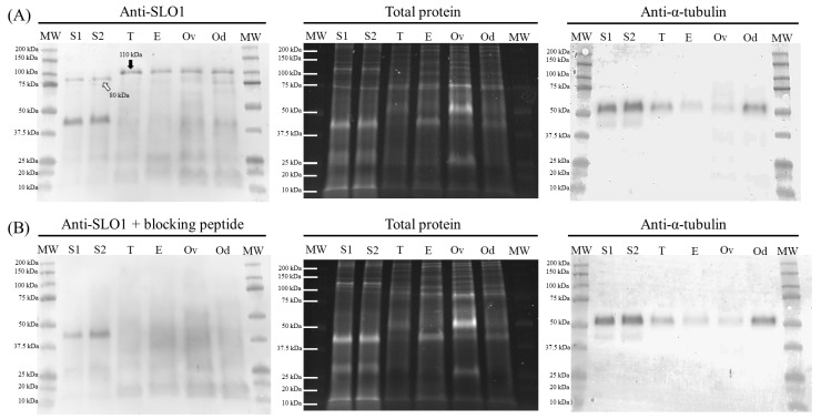
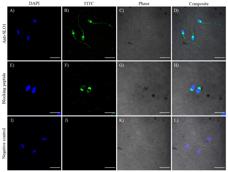
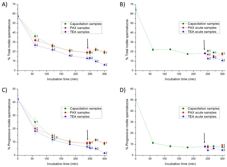
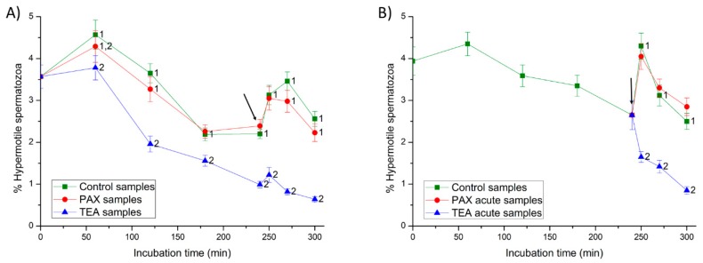
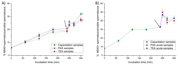
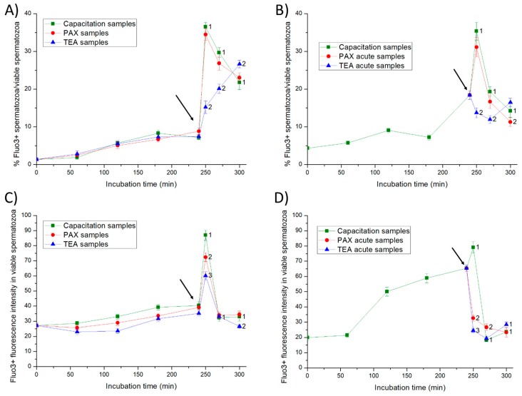
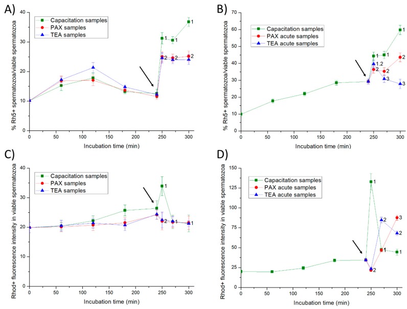
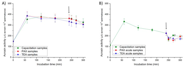
This study sought to identify and localize SLO1 channels in boar spermatozoa by immunoblotting and immunofluorescence, and to determine their physiological role during in vitro sperm capacitation. Sperm samples from 14 boars were incubated in a capacitation medium for 300 min in the presence of paxilline (PAX), a specific SLO1-channel blocker, added either at 0 min or after 240 min of incubation. Negative controls were incubated in capacitation medium, and positive controls in capacitation medium plus tetraethyl ammonium (TEA), a general K+-channel blocker, also added at 0 min or after 240 min of incubation. In all samples, acrosome exocytosis was triggered with progesterone after 240 min of incubation. Sperm motility and kinematics, integrity of plasma and acrosome membranes, membrane lipid disorder, intracellular calcium levels and acrosin activity were evaluated after 0, 60, 120, 180, 240, 250, 270 and 300 min of incubation. In boar spermatozoa, SLO1 channels were found to have 80 kDa and be localized in the anterior postacrosomal region and the mid and principal piece of the tail; their specific blockage through PAX resulted in altered calcium levels and acrosome exocytosis. As expected, TEA blocker impaired in vitro sperm capacitation, by altering sperm motility and kinematics and calcium levels. In conclusion, SLO1 channels are crucial for the acrosome exocytosis induced by progesterone in in vitro capacitated boar spermatozoa.

Keywords: PAX; SLO1 channels; TEA; boar spermatozoa; in vitro capacitation.


Conflict of interest statement

The authors declare no conflict of interest.

Figures

Figure 1
Figure 1
Representative immunoblot of sperm (S1 and S2) and tissue (positive controls; testicle [T], epididymis [E], ovary [Ov] and oviduct [Od]) samples for SLO1 channels (A), and its corresponding peptide competition assay (B). TGX™ (Tris-glycine extended) Stain Free (total protein) and α-tubulin (anti-α-tubulin) were performed as loading controls.
Figure 2
Figure 2
Immunolocalization of SLO1 (AD), with its corresponding peptide competition assay (EH) and negative control (IL). Nucleus is shown in blue (DAPI; 4′,6-diamidino-2-phenylindole) and SLO1 is shown in green (FITC; Fluorescein isothiocyanate). Phase images of sperm cells were collected using a transmitted light detector (Phase). Scale bars: (AD) and (IL): 20 μm; (EH): 13.5 μm.
Figure 3
Figure 3
Percentages of total and progressive motile spermatozoa throughout in vitro incubation of sperm in capacitation medium (negative control samples), capacitation medium plus TEA (tetraethyl ammonium) blocker (positive control samples), and capacitation medium plus PAX (paxilline) blocker. In experiment 1, TEA (TEA samples) or PAX (PAX samples) blockers were added to the capacitation medium at 0 min (A,C). In experiment 2, TEA (TEA acute samples) or PAX (PAX acute samples) blockers were added to the capacitation medium at 240 min of incubation (B,D). Different superscripts indicate significant differences (p < 0.05) between samples within the same time point. The arrow indicates the time at which 10 µg/mL of progesterone was added in all samples (i.e., 240 min of incubation).
Figure 4
Figure 4
Percentages of hypermotile spermatozoa (i.e., hyperactive spermatozoa with amplitude of lateral head displacement (ALH) > 2.5 µm and straightness (STR) > 85%) throughout in vitro incubation of sperm in capacitation medium (negative control samples), capacitation medium plus TEA blocker (positive control samples), and capacitation medium plus PAX blocker. In experiment 1, TEA (TEA samples) or PAX (PAX samples) blockers were added to the capacitation medium at 0 min (A). In experiment 2, TEA (TEA acute samples) or PAX (PAX acute samples) blockers were added to the capacitation medium at 240 min of incubation (B). Different superscripts indicate significant differences (p < 0.05) between samples within the same time point. The arrow indicates the time at which 10 µg/mL of progesterone was added in all samples (i.e., 240 min of incubation).
Figure 5
Figure 5
Percentages of viable spermatozoa with exocytosed acrosome (PNA-) in relation to total viable spermatozoa (EthD-1-) throughout in vitro incubation of sperm samples in capacitation medium (negative control samples), capacitation medium plus TEA blocker (positive control samples), and capacitation medium plus PAX blocker. In Experiment 1, TEA (TEA samples) or PAX (PAX samples) blockers were added to the capacitation medium at 0 min (A). In Experiment 2, TEA (TEA acute samples) or PAX (PAX acute samples) blockers were added to the capacitation medium at 240 min of incubation (B). Different superscripts indicate significant differences (p < 0.05) between samples within the same time point. The arrow indicates the time at which 10 µg/mL of progesterone was added in all samples (i.e., 240 min of incubation).
Figure 6
Figure 6
Percentages of viable spermatozoa with high lipid disorder (M540+) in relation to total viable spermatozoa (YO-PRO-1-) throughout in vitro incubation of sperm samples in capacitation medium (negative control samples), capacitation medium plus TEA blocker (positive control samples), and capacitation medium plus PAX blocker. In Experiment 1, TEA (TEA samples) or PAX (PAX samples) blockers were added to the capacitation medium at 0 min (A). In Experiment 2, TEA (TEA acute samples) or PAX (PAX acute samples) blockers were added to the capacitation medium at 240 min of incubation (B). Different superscripts indicate significant differences (p < 0.05) between samples within the same time point. The arrow indicates the time at which 10 µg/mL of progesterone was added in all samples (i.e., 240 min of incubation).
Figure 7
Figure 7
Percentages of viable spermatozoa with high intracellular calcium levels (Fluo3+) in relation to total viable spermatozoa (PI-) (A,B) and fluorescence intensity of Fluo3+ in viable spermatozoa with positive Fluo3-staining (C,D) throughout in vitro incubation of sperm samples in capacitation medium (negative control samples), capacitation medium plus TEA blocker (positive control samples), and capacitation medium plus PAX blocker. In experiment 1, TEA (TEA samples) or PAX (PAX samples) blockers were added to the capacitation medium at 0 min (A,C). In experiment 2, TEA (TEA acute samples) or PAX (PAX acute samples) blockers were added to the capacitation medium at 240 min of incubation (B,D). Different superscripts indicate significant differences (p < 0.05) between samples within the same time point. The arrow indicates the time at which 10 µg/mL of progesterone was added in all samples (i.e., 240 min of incubation).
Figure 8
Figure 8
Percentages of viable spermatozoa with high calcium levels evaluated through Rhod5 (Rh5+) in relation to total viable spermatozoa (YO-PRO-1-) and fluorescence intensity of Rhod5 in viable spermatozoa with positive Rhod5-staining (C,D) throughout in vitro incubation of sperm samples in capacitation medium (negative control samples), capacitation medium plus TEA blocker (positive control samples), and capacitation medium plus PAX blocker. In experiment 1, TEA (TEA samples) or PAX (PAX samples) blockers were added to the capacitation medium at 0 min (A,C). In experiment 2, TEA (TEA acute samples) or PAX (PAX acute samples) blockers were added to the capacitation medium at 240 min of incubation (B,D). Different superscripts indicate significant differences (p < 0.05) between samples within the same time point. The arrow indicates the time at which 10 µg/mL of progesterone was added in all samples (i.e., 240 min of incubation).
Figure 9
Figure 9
Acrosin activity (µIU/106 spermatozoa) throughout in vitro incubation of sperm samples in capacitation medium (negative control samples), capacitation medium plus TEA blocker (positive control samples), and capacitation medium plus PAX blocker. In experiment 1, TEA (TEA samples) or PAX (PAX samples) blockers were added to the capacitation medium at 0 min (A). In experiment 2, TEA (TEA acute samples) or PAX (PAX acute samples) blockers were added to the capacitation medium at 240 min of incubation (B). Different superscripts indicate significant differences (p < 0.05) between samples within the same time point. The arrow indicates the time at which 10 µg/mL of progesterone was added in all samples (i.e., 240 min of incubation).

References

    1. De la Vega-Beltran J.L., Sánchez-Cárdenas C., Krapf D., Hernandez-González E.O., Wertheimer E., Treviño C.L., Visconti P.E., Darszon A. Mouse sperm membrane potential hyperpolarization is necessary and sufficient to prepare sperm for the acrosome reaction. J. Biol. Chem. 2012;287:44384–44393. doi: 10.1074/jbc.M112.393488. - DOI - PMC - PubMed
    1. Mannowetz N., Niadoo N.M., Choo S.A.S., Smith J.F., Lishko P.V. Slo1 is the principal potassium channel of human spermatozoa. Elife. 2013;2:e01009. doi: 10.7554/eLife.01009. - DOI - PMC - PubMed
    1. Brenker C., Zhou Y., Müller A., Echeverry F.A., Trötschel C., Poetsch A., Xia X.M., Bönigk W., Lingle J.C., Kaupp U.B., et al. The Ca2+-activated K+ current of human sperm is mediated by Slo3. Elife. 2014;3:e01438. doi: 10.7554/eLife.01438. - DOI - PMC - PubMed
    1. Lishko P.V., Botchkina I.L., Fedorenko A., Kirichok Y. Acid extrusion from human spermatozoa is mediated by flagellar voltage-gated proton channels. Cell. 2010;140:327–337. doi: 10.1016/j.cell.2009.12.053. - DOI - PubMed
    1. Navarro B., Miki D.E., Clapham D.E. ATP-activated P2X2 current in mouse spermatozoa. Proc. Natl. Acad. Sci. USA. 2011;108:14342–14347. doi: 10.1073/pnas.1111695108. - DOI - PMC - PubMed

MeSH terms

LinkOut - more resources