. 2019 Nov 1;15(11):2229-2239.
doi: 10.1166/jbn.2019.2859.

A Nanobody Against Cytotoxic T-Lymphocyte Associated Antigen-4 Increases the Anti-Tumor Effects of Specific CD8+ T Cells

A Nanobody Against Cytotoxic T-Lymphocyte Associated Antigen-4 Increases the Anti-Tumor Effects of Specific CD8+ T Cells

Zhuoran Tang et al. J Biomed Nanotechnol. .

Abstract

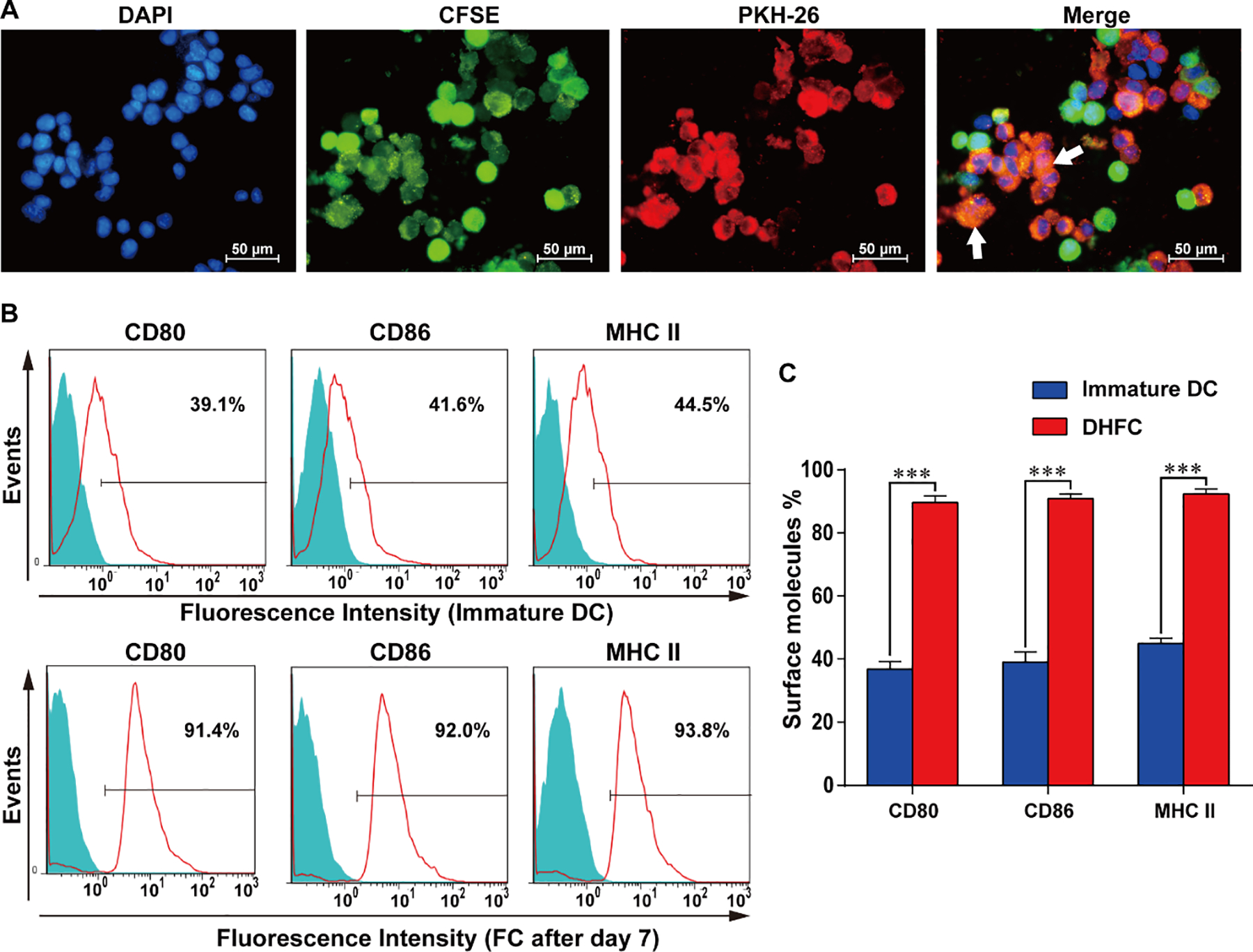
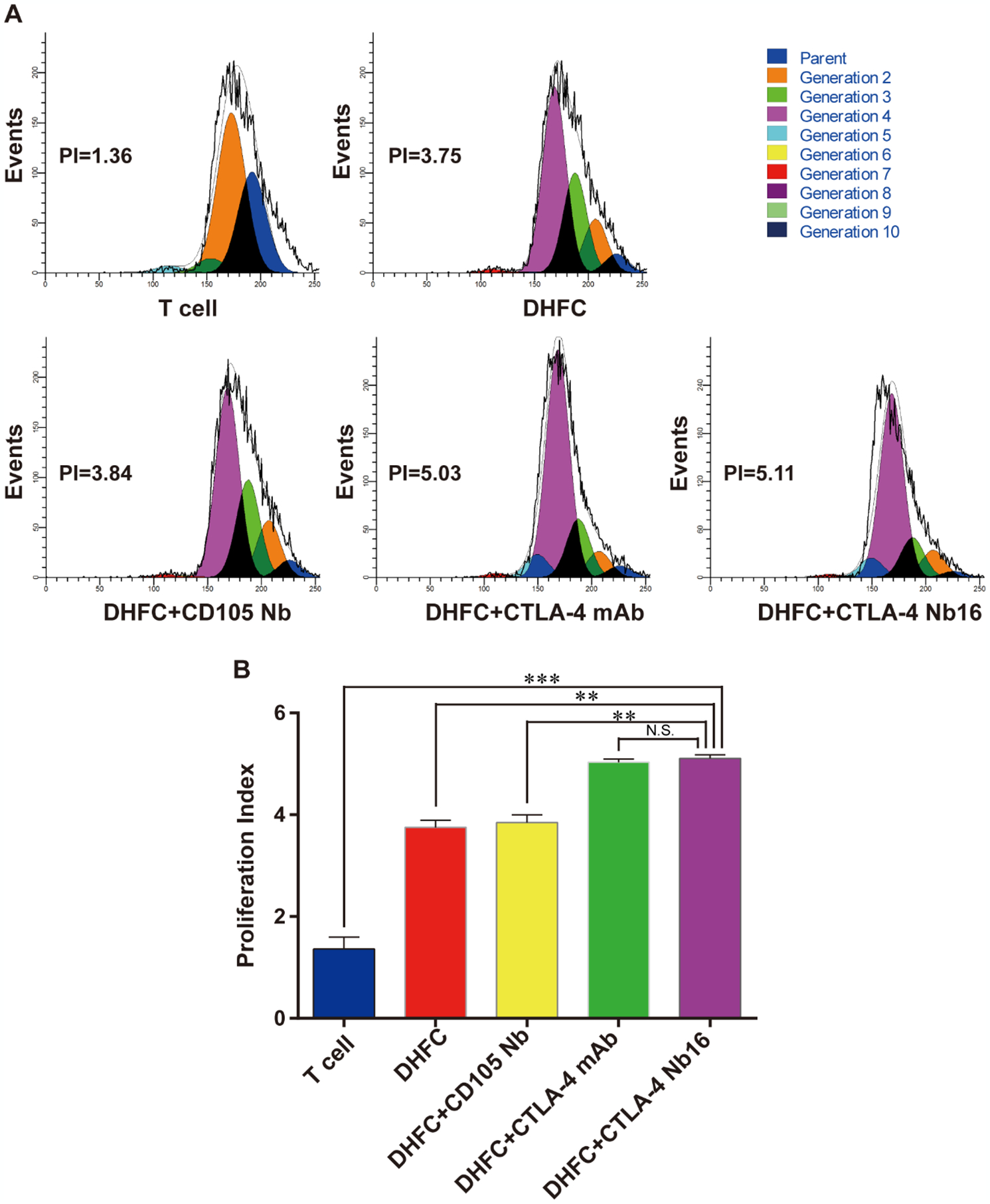
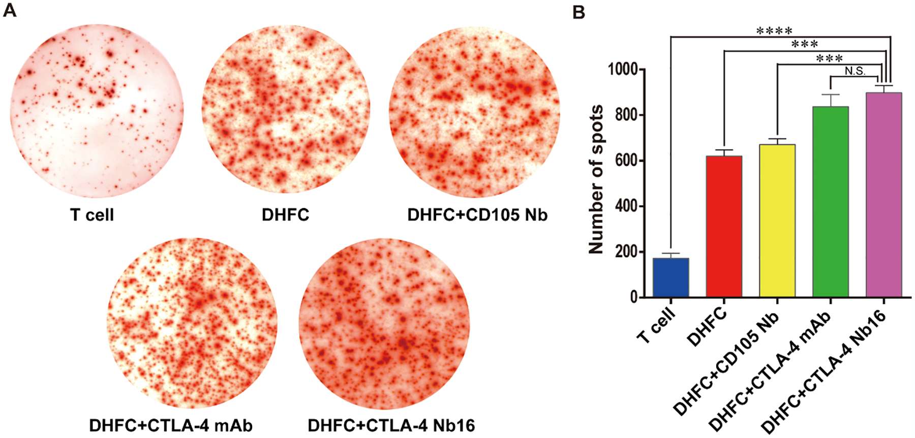
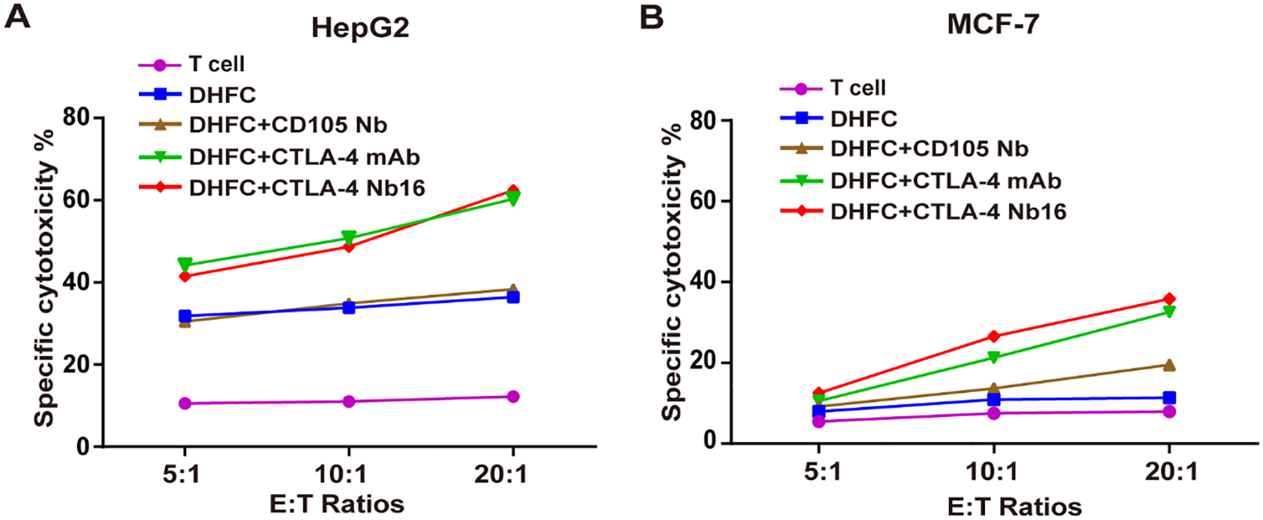
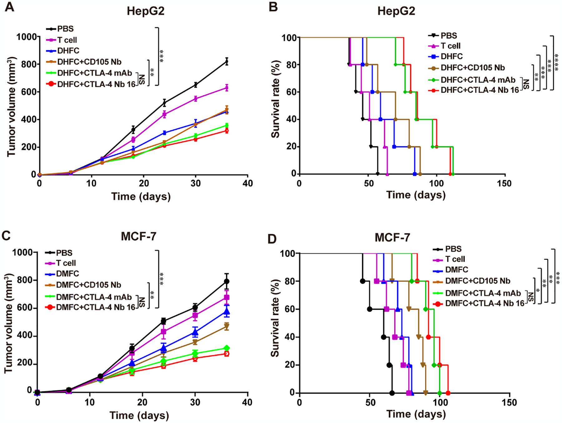
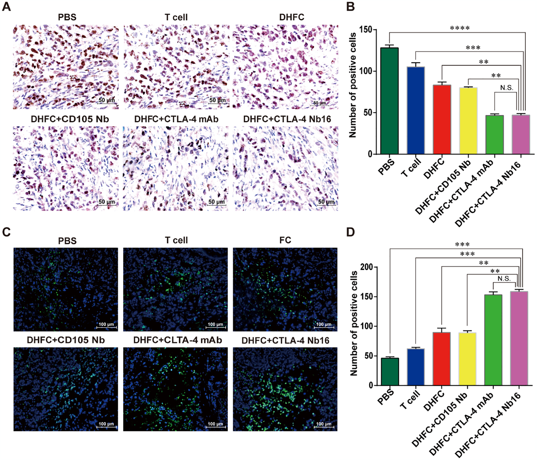
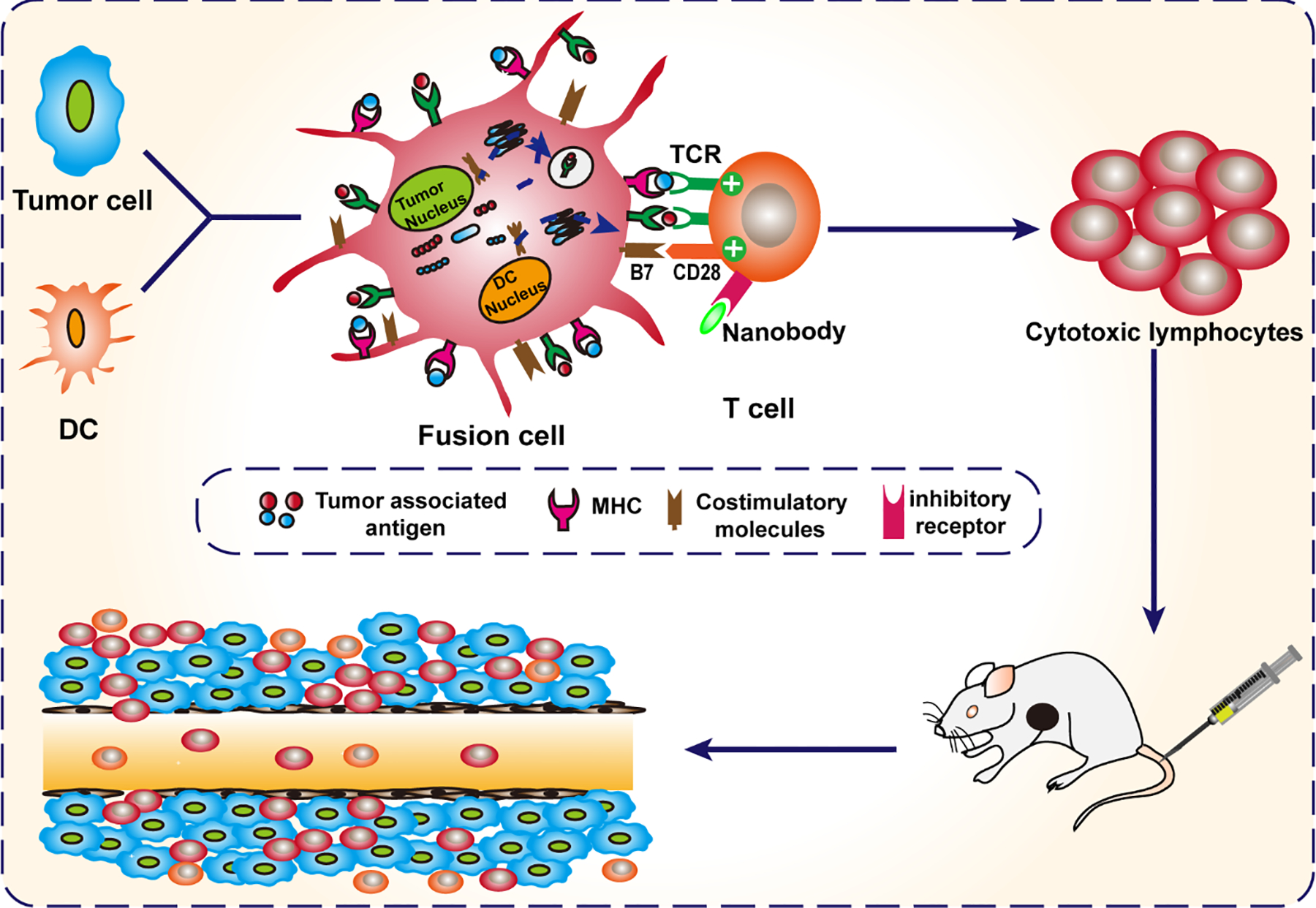
Adoptive cell-based immunotherapy typically utilizes cytotoxic T lymphocytes (CTLs), expanding these cells ex vivo. Such expansion is traditionally accomplished through the use of autologous APCs that are capable of interactions with T cells. However, incidental inhibitory program such as CTLA-4 pathway can impair T cell proliferation. We therefore designed a nanobody which is specific for CTLA-4 (CTLA-4 Nb 16), and we then used this molecule to assess its ability to disrupt CTLA-4 signaling and thereby overcome negative costimulation of T cells. With CTLA-4 Nb16 stimulation, dendritic cell/hepatocellular carcinoma fusion cells (DC/HepG2-FCs) enhanced autologous CD8+ T cell proliferation and production of IFN-γ in vitro, thereby leading to enhanced killing of tumor cells. Using this approach in the context of adoptive CD8+ immunotherapy led to a marked suppression of tumor growth in murine NOD/SCID hepatocarcinoma or breast cancer xenograft models. We also observed significantly increased tumor cell apoptosis, and corresponding increases in murine survival. These findings thus demonstrate that in response to nanobody stimulation, DC/tumor cells-FC-induced specific CTLs exhibit superior anti-tumor efficacy, making this a potentially valuable means of achieving better adoptive immunotherapy outcomes in cancer patients.


Conflict of interest statement

Disclosure of Potential Conflicts of Interest

No potential conflicts of interest were disclosed.

Figures

Figure 1.
Figure 1.. Identification of DC/HepG2 fusion cells and express increased CD80, CD86 and MHC II.
(A) The DC2.4/HepG2 fusion cell identification approach was as follows: CSFE (green) and PKH26 (red) were used for the respective labeling of DCs and HepG2 cells, after which PEG was used to mediate fusion. DAPI (blue) was then used for nuclear staining, followed by fluorescence microscopy with fused cells marked by white arrows (× 400). Images are representative of three independent experiments. (B) DCs exhibited MHC II and costimulatory molecule expression at day 7 in FCs. (C) The status of three membrane molecules, MHC II, CD80 and CD86 were high at day 7 in FCs. Data are means ± SD, n = 3. *** P < 0.001.
Figure 2.
Figure 2.. CTLA-4 Nb16 promoted CD8+ T cell proliferation.
(A) CFSE-labeled CD8+ cells were mixed with different antibodies following with DHFC, and then co-cultured for 5 days. Proliferation of CD8+ T cells was assessed via flow cytometry. (B) CD8+ T Cell proliferation Index in DHFC+CTLA-4 Nb16 group was higher than that in other’s groups, except DHFC+CTLA-4 mAb group. Data are means ± SD, n = 3. NS stand for P > 0.05, ** P < 0.01, *** P < 0.001.
Figure 3.
Figure 3.. CTLA-4 Nb16 increased the abundance of IFN-γ secreting CD8+ T lymphocytes.
(A) CD8+ cells were mixed with different antibodies following with DHFC, and then co-cultured for 7 days. ELISPOT assay was applied to examine the number of CD8+ T lymphocytes secreting IFN-γ. (B) The number of spots in DHFC+CTLA-4 Nb16 group increased significantly than other’s groups, except DHFC+CTLA-4 mAb groups. Data are means ± SD, n = 3. *** P < 0.001, **** P < 0.0001.
Figure 4.
Figure 4.. CTLA-4 Nb16 promoted the tumor cell killing effects of CD8+ lymphocytes.
(A) Comparison of cell killing effects of CD8+ T lymphocytes induced by indicated treatments on HepG2 in different E:T raitos of 5:1, 10:1 and 20:1. DHFC+CTLA-4 Nb16 enhanced cytotoxic T cell killing function on target HepG2 cells. (B) Comparison of cell killing effects of CD8+ T lymphocytes induced by indicated treatments on MCF-7. It had weaker killing effect on MCF-7 than HepG2. Data are means ± SD, n = 3.
Figure 5.
Figure 5.. CTLA-4 Nb16 increased the anti-tumor effects of CD8+ T lymphocyte by inhibiting HepG2 and MCF-7 tumor growth and enhancing survival.
(A) The Therapy with DHFC+CTLA-4 Nb16 significantly delayed the tumor growth in mice bearing liver cancer xenograft. (B) Kaplan-Meier survival curves of mice reported in DHFC+CTLA-4 Nb16 group therapy significantly increased survival. (C) The Therapy with DMFC+CTLA-4 Nb16 significantly delayed the tumor growth in mice bearing breast cancer xenograft. (D) Kaplan-Meier survival curves of mice reported in DMFC+CTLA-4 Nb16 group therapy significantly increased survival. Data are means ± SD or percentage survival (n = 5 mice/group). **P < 0.01, *** P < 0.001.
Figure 6.
Figure 6.. CTLA-4 Nb16 stimulation suppressed proliferation of tumor cells and promoted tumor cell apoptosis in mice.
(A) Immunohistochemical assay of Ki67 staining in different groups (x 40). Brown in nuclear indicates the positive staining. (B) The Ki67 expression of tumor cells in DHFC+CTLA4-Nb16 group was significant lower than in other groups, except in DHFC+CTLA4-mAb group. (C) TUNEL assay was used to monitor cell apoptosis in different groups. Blue indicates nuclear and green indicates the positive signal (×20). (D) The apoptosis of tumor cells in DHFC+CTLA4-Nb16 group was significant higher than that in other’s groups, except in DHFC+CTLA4-mAb group. Data are measn ± SD. **P < 0.01, *** P < 0.001, **** P < 0.0001.
Scheme 1.
Scheme 1.. Illustration of therapy with FC+ Nanobody.
DC fusion cells (DC/tumor fusion cells, FCs) are generated. Nanobody against CTLA-4 (CTLA-4 Nb16) eliminates immunosuppression via disrupting CTLA-4-mediated negative costimulation in T cells induced by dendritic cell/tumor fusion cells. Finally, the induced-CTLs were transferred to kill tumor cells.

References

    1. Liedtke C, and Kolberg HC, 2015. Current Medical Treatment of Patients with Non-Colorectal Liver Metastases: Primary Tumor Breast Cancer. Visceral Medicine, 31(6), pp.424–432. - PMC - PubMed
    1. Nam SJ, Yeo HY, Chang HJ, Kim BH, and Park JW, 2016. A new cell block method for multiple immunohistochemical analysis of circulating tumor cells in patients with liver cancer. Cancer research and treatment: official journal of Korean Cancer Association, 48(4), pp.1229–1242. - PMC - PubMed
    1. Hersey P, and Gallagher S, 2014. Intralesional immunotherapy for melanoma. Journal of surgical oncology, 109(4), pp.320–326. - PubMed
    1. Mona A, Sabry S, Eman K, Ahmed E, Mohamed S, & Motawa E, 2018. A novel potential effective strategy for enhancing the antitumor immune response in breast cancer patients using a viable cancer cell-dendritic cell-based vaccine. Oncology letters, 16(1), pp.529–535. - PMC - PubMed
    1. Zhang W, Zhu XD, Sun HC, Xiong YQ, Zhuang PY, Xu HX, and Tang ZY, 2010. Depletion of tumor-associated macrophages enhances the effect of sorafenib in metastatic liver cancer models by antimetastatic and antiangiogenic effects. Clinical Cancer Research, 16(13), pp.3420–3430. - PubMed