. 2020 Feb 14;295(7):2125-2134.
doi: 10.1074/jbc.RA119.010378. Epub 2019 Dec 17.

The conserved C-terminus of Sss1p is required to maintain the endoplasmic reticulum permeability barrier

Affiliations

The conserved C-terminus of Sss1p is required to maintain the endoplasmic reticulum permeability barrier

Christopher M Witham et al. J Biol Chem. .

Abstract

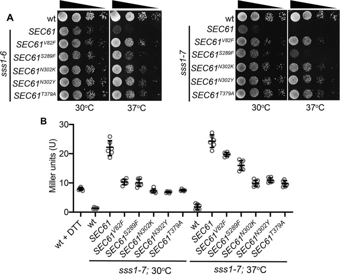
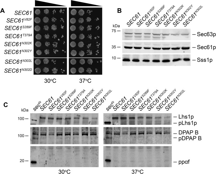
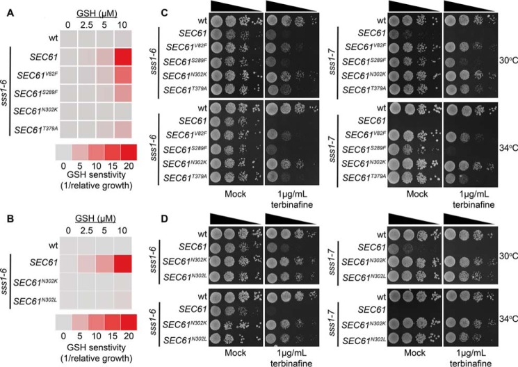
The endoplasmic reticulum (ER) is the entry point to the secretory pathway and major site of protein biogenesis. Translocation of secretory and integral membrane proteins across or into the ER membrane occurs via the evolutionarily conserved Sec61 complex, a heterotrimeric channel that comprises the Sec61p/Sec61α, Sss1p/Sec61γ, and Sbh1p/Sec61β subunits. In addition to forming a protein-conducting channel, the Sec61 complex also functions to maintain the ER permeability barrier, preventing the mass free flow of essential ER-enriched molecules and ions. Loss in Sec61 integrity is detrimental and implicated in the progression of disease. The Sss1p/Sec61γ C terminus is juxtaposed to the key gating module of Sec61p/Sec61α, and we hypothesize it is important for gating the ER translocon. The ER stress response was found to be constitutively induced in two temperature-sensitive sss1 mutants (sss1ts ) that are still proficient to conduct ER translocation. A screen to identify intergenic mutations that allow for sss1ts cells to grow at 37 °C suggests the ER permeability barrier to be compromised in these mutants. We propose the extreme C terminus of Sss1p/Sec61γ is an essential component of the gating module of the ER translocase and is required to maintain the ER permeability barrier.

Keywords: Sec61 complex; Sec61p/Sec61α; Sss1p/Sec61γ; endoplasmic reticulum (ER); endoplasmic reticulum stress (ER stress); endoplasmic reticulum–associated protein degradation (ERAD); gating; translocation; translocon gating.


Conflict of interest statement

The authors declare that they have no conflicts of interest with the contents of this article.

Figures

Figure 1.
Figure 1.
The Sss1p C terminus is highly conserved. A, ribbon diagram of the Sec61 homologue complex crystal structure (PDB ID: 2WWA) (32) was composed using Chimera software. Ssh1p, Sbh2p, and Sss1p are colored gray, blue, and red, respectively. The highly conserved KLIHIPI heptapeptide is highlighted in gold. B, the sequence of the extreme C terminus of Sec61γ and Sss1p are aligned using Clustal Omega sequence alignment software and the position of each double alanine scanning mutation indicated. C, BWY530 yeast transformed with either YCp HIS3, YCp SSS1, YCp SSS11I68A,K69A, YCp SSS1L70A,I71A, YCp SSS1H72A,I73A, or YCp SSS1P74A,I75A were streaked onto His selective medium and medium containing FOA and incubated at 30 °C for 2 days. D, cell extracts derived from WT cells or cells expressing either SSS1I68A,K69A, SSS1L70A,I71A, SSS1H72A,I73A, or SSS1P74A,I75A were immunoblotted with anti-Sss1p or anti-Sec61p antibodies.
Figure 2.
Figure 2.
sss1–6 and sss1–7 are inserted into the ER membrane. A, WT, sss1–6 or sss1–7 yeast were spotted on YPD agar in a 10-fold dilution series and incubated at 30 °C or 37 °C for 2 days. B, cell extracts derived from WT sss1–6 or sss1–7 yeast were immunoblotted with anti-Sss1p, anti-Sec61p, and anti-Sec63p antibodies. C, GFP-Sss1p, GFP-Sss1–6p, and GFP-Sss1–7p was visualized in cells grown at 30 °C and 37 °C and colocalized with Sec63p-RFP (5 μm bar).
Figure 3.
Figure 3.
ER translocation is not affected in sss1–6 and sss1–7. A, membranes derived from WT sss1–6 or sss1–7 yeast incubated with and without 1 mm DSS were immunoblotted with anti-Sss1p and anti-Sec61p antibodies. B, membranes prepared from WT sss1–6 or sss1–7 yeast were subject to ConA chromatography. An equal portion of each fraction was analyzed by immunoblotting with either anti-Sss1p, anti-Sec61p, or anti-Sec63p specific antibodies. The bound fraction is shown. C, cell extracts derived from WT sss1–6 or sss1–7 yeast were immunoblotted with anti-Sss1p, anti-Sec61p, anti-α factor, and anti-DPAP B antibodies. Secretory mutants, sec63–1 and sec65–1 (sects), were included as a negative control for α factor and DPAP B, respectively.
Figure 4.
Figure 4.
ER homeostasis is perturbed in sss1–6 and sss1–7. A, WT, sss1–6, and sss1–7 yeast transformed with pJT30 (UPRE-LACZ) were grown in Ura medium and β-galactosidase activity determined. WT cells treated with 5 mm DTT for 2 h were used as a positive control. B, WT, SEC61N302D, and sss1–6 yeast transformed with YEp HGT1 were grown in Ura selective medium with increasing concentrations of GSH. The relative growth of each strain determined and the GSH sensitivity (1/relative growth) presented. C, WT, sss1–6, and sss1–7 yeast were spotted on YPD agar or YPD agar containing 1 μg/ml terbinafine in a 10-fold dilution series and incubated at 30 °C or 34 °C for 2 days.
Figure 5.
Figure 5.
Mutations in residues of Sec61p located in important gating modules suppress sss1–6 and sss1–7 temperature sensitivity. A, WT, sss1–6, or sss1–7 yeast transformed with either YCp SEC61, YCp SEC61V82F, YCp SEC61S289F, YCp SEC61N302K, YCp SEC61N302Y, or YCp SEC61T379A were spotted on YPD agar in a 10-fold dilution series and incubated at 30 °C or 37 °C for 2 days. B, WT, sss1–6, or sss1–7 yeast transformed with either YCp SEC61, YCp SEC61V82F, YCp SEC61S289F, YCp SEC61N302K, YCp SEC61N302Y, or YCp SEC61T379A and with pJT30 (UPRE-LacZ) were grown in Ura selective medium and β-galactosidase activity determined. As a positive control WT cells were treated with 5 mm DTT for 2 h.
Figure 6.
Figure 6.
SEC61-dependent suppressors of sss1–6 and sss1–7 are functional. A, WT, SEC61V82F, SEC61S289F, SEC61N302K, SEC61N302Y, SEC61T379A, SEC61N302L, or SEC61N302D yeast were spotted on YPD agar in a 10-fold dilution series and incubated at 30 °C or 37 °C for 2 days. B, cell extracts derived from WT SEC61V82F, SEC61S289F, SEC61N302K, SEC61N302Y, SEC61T379A, or SEC61N302L yeast were immunoblotted with anti-Sss1p, anti-Sec61p, and anti-Sec63p antibodies. C, cell extracts derived from WT SEC61V82F, SEC61S289F, SEC61N302K, SEC61N302Y, SEC61T379A, or SEC61N302L yeast grown at 30 °C or 37 °C were immunoblotted with anti-Lhs1p, anti-DPAP B, and anti-α factor antibodies.
Figure 7.
Figure 7.
Mutations in the lumenal lateral gate genetically interact with sss1–6 and sss1–7. A, WT, and sss1–6 or sss1–7 yeast transformed with either YCp SEC61, YCp SEC61N302K, or YCp SEC61N302L were spotted on YPD agar in a 10-fold dilution series and incubated at 30 °C or 37 °C for 2 days. B, WT or sss1–7 yeast transformed with either YCp SEC61, YCp SEC61N302K, or YCp SEC61N302L and with pJT30 (UPRE-LacZ) were grown in Ura selective medium and β-galactosidase activity was determined. As a positive control WT cells were treated with 5 mm DTT for 2 h. C, CMY5 yeast were cotransformed with either YCp SSS1, YCp sss1–6, or YCp sss1–7 and either YCp SEC61 or YCp SEC61N302D. Transformants were streaked out onto either Leu, Trp selective medium or medium containing FOA and incubated at 30 °C for 3 days. D, CMY5 yeast cotransformed with YCp sss1–6 and either YCp SEC61 or YCp SEC61N302D recovered from FOA containing medium were spotted on YPD agar in a 10-fold dilution series and incubated at 30 °C or 37 °C for 2 days.
Figure 8.
Figure 8.
Mutations in residues of Sec61p located in important gating modules suppress ER permeability defects in sss1–6 and sss1–7. A, WT, or sss1–6 yeast transformed with either YCp SEC61, YCp SEC61V82F, YCp SEC61S289F, YCp SEC61N302K, or YCp SEC61T379A were transformed with YEp HGT1 and were grown in Ura selective medium with increasing concentrations of GSH. The relative growth of each strain determined and the GSH sensitivity (1/relative growth) presented. B, WT, sss1–6 or sss1–6 yeast transformed with either YCp SEC61N302K or YCp SEC61N302L and YEp HGT1 were grown in Ura medium with increasing concentrations of GSH, the relative growth of each strain determined and the GSH sensitivity (1/relative growth) presented. C, WT and sss1–6 or sss1–7 yeast transformed with either YCp SEC61, YCp SEC61V82F, YCp SEC61S289F, YCp SEC61N302K, or YCp SEC61T379A were spotted on YPD agar or YPD agar containing 1 μg/ml terbinafine in a 10-fold dilution series and incubated at 30 °C or 34 °C for 2 days. D, WT, sss1–6, sss1–7 or sss1–6 and sss1–7 yeast transformed with either YCp SEC61N302K or YCp SEC61N302L were spotted on YPD agar or YPD agar supplemented with 1 μg/ml terbinafine in a 10-fold dilution series and incubated at 30 °C or 34 °C for 2 days.

References

    1. Behnke J., Feige M. J., and Hendershot L. M. (2015) BiP and its nucleotide exchange factors Grp170 and Sil1: Mechanisms of action and biological functions. J. Mol. Biol. 427, 1589–1608 10.1016/j.jmb.2015.02.011 - DOI - PMC - PubMed
    1. Janda C. Y., Li J., Oubridge C., Hernández H., Robinson C. V., and Nagai K. (2010) Recognition of a signal peptide by the signal recognition particle. Nature 465, 507–510 10.1038/nature08870 - DOI - PMC - PubMed
    1. Rapoport T. A., Li L., and Park E. (2017) Structural and mechanistic insights into protein translocation. Annu. Rev. Cell Dev. Biol. 33, 369–390 10.1146/annurev-cellbio-100616-060439 - DOI - PubMed
    1. Voorhees R. M., and Hegde R. S. (2015) Structures of the scanning and engaged states of the mammalian SRP-ribosome complex. eLife 4, e07975 10.7554/eLife.07975 - DOI - PMC - PubMed
    1. Walter P., Ibrahimi I., and Blobel G. (1981) Translocation of proteins across the endoplasmic reticulum. I. Signal recognition protein (SRP) binds to in-vitro-assembled polysomes synthesizing secretory protein. J. Cell Biol. 91, 545–550 10.1083/jcb.91.2.545 - DOI - PMC - PubMed

Publication types

MeSH terms

Substances

Associated data

LinkOut - more resources