. 2020 Jan 21;117(3):1395-1403.
doi: 10.1073/pnas.1914380117. Epub 2019 Dec 17.

Retroconversion of estrogens into androgens by bacteria via a cobalamin-mediated methylation

Affiliations

Retroconversion of estrogens into androgens by bacteria via a cobalamin-mediated methylation

Po-Hsiang Wang et al. Proc Natl Acad Sci U S A. .

Abstract

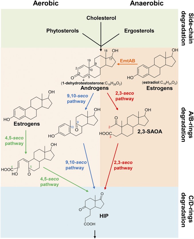
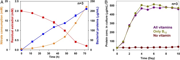
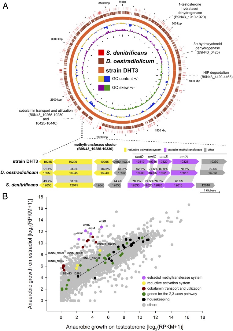
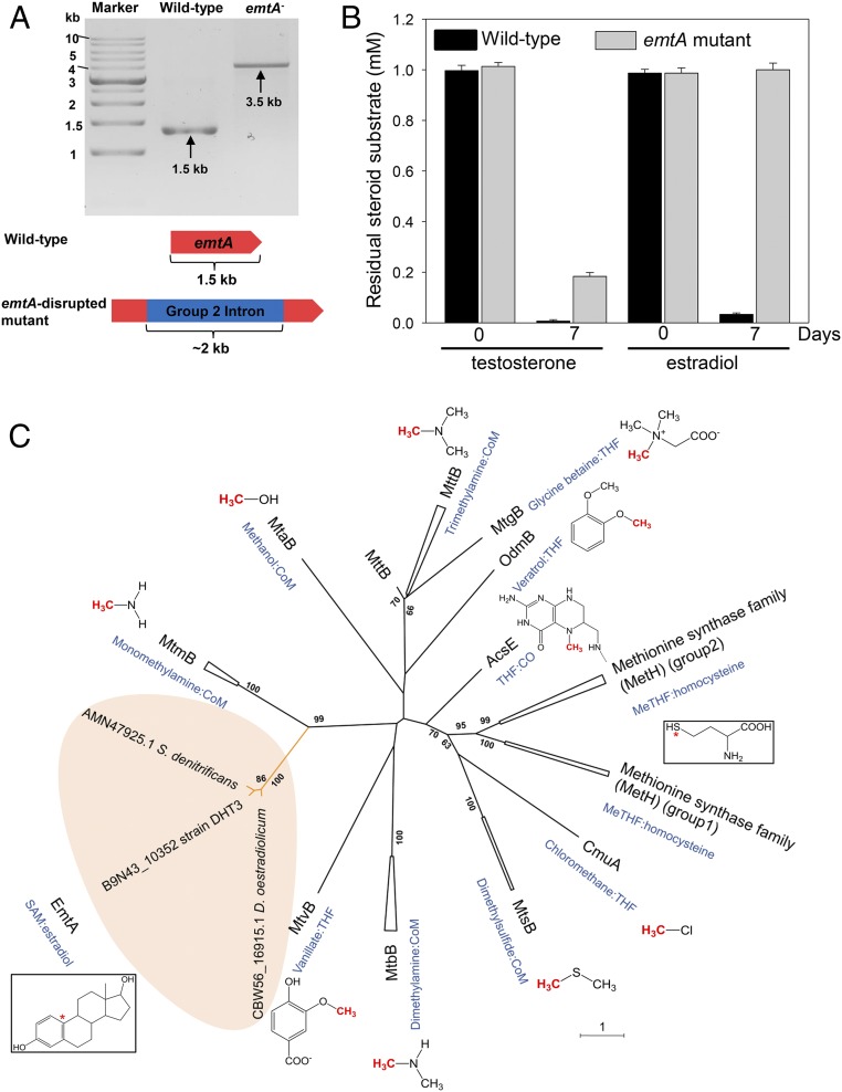
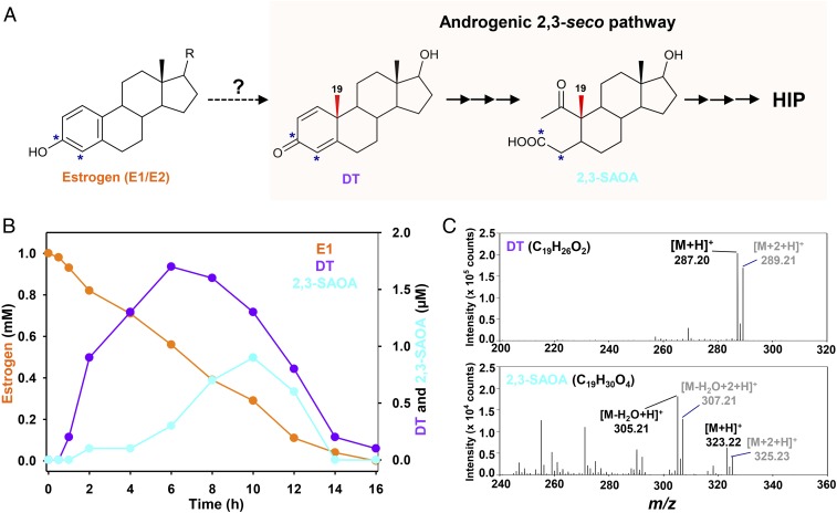
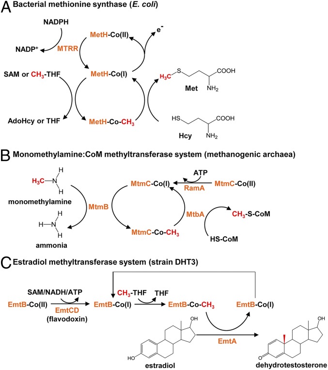
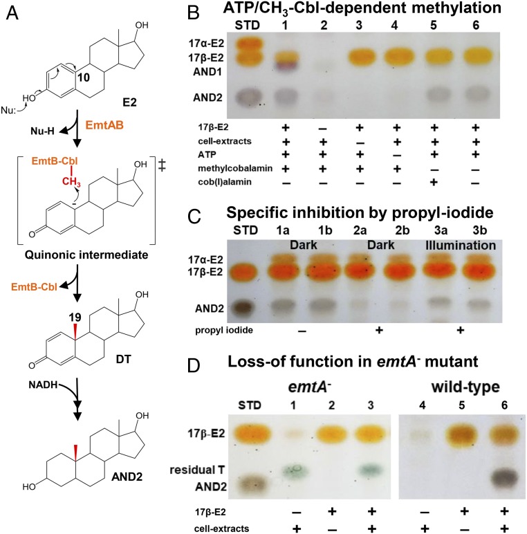
Steroid estrogens modulate physiology and development of vertebrates. Conversion of C19 androgens into C18 estrogens is thought to be an irreversible reaction. Here, we report a denitrifying Denitratisoma sp. strain DHT3 capable of catabolizing estrogens or androgens anaerobically. Strain DHT3 genome contains a polycistronic gene cluster, emtABCD, differentially transcribed under estrogen-fed conditions and predicted to encode a cobalamin-dependent methyltransferase system conserved among estrogen-utilizing anaerobes; an emtA-disrupted DHT3 derivative could catabolize androgens but not estrogens. These data, along with the observed androgen production in estrogen-fed strain DHT3 cultures, suggested the occurrence of a cobalamin-dependent estrogen methylation to form androgens. Consistently, the estrogen conversion into androgens in strain DHT3 cell extracts requires methylcobalamin and is inhibited by propyl iodide, a specific inhibitor of cobalamin-dependent enzymes. The identification of the cobalamin-dependent estrogen methylation thus represents an unprecedented metabolic link between cobalamin and steroid metabolism and suggests that retroconversion of estrogens into androgens occurs in the biosphere.

Keywords: biocatalysis; cobalamin-dependent methyltransferase; estrogens; microbial metabolism; steroids.


Conflict of interest statement

The authors declare no competing interest.

Figures

Fig. 1.
Fig. 1.
Central pathways for bacterial steroid catabolism. Bacteria adopt a convergent pathway (the 2,3-seco pathway) to catabolize different steroids under anaerobic conditions and adopt divergent pathways to catabolize estrogens (the 4,5-seco pathway) and other steroids (sterols and androgens; the 9,10-seco pathway) under aerobic conditions. All of the 3 steroid catabolic pathways converge at HIP. 2,3-SAOA, 17β-hydroxy-1-oxo-2,3-seco-androstan-3-oic acid (2,3-SAOA); HIP, 3aα-H-4α(3′-propanoate)-7aβ-methylhexahydro-1,5-indanedione.
Fig. 2.
Fig. 2.
Anaerobic growth of Denitratisoma sp. strain DHT3 with estradiol under denitrifying conditions and under different vitamin-supplementing conditions. (A) Anaerobic growth of strain DHT3 using estradiol as the sole substrate under denitrifying conditions. (B) Anaerobic growth of strain DHT3 on estradiol in the medium supplemented with different vitamins or without vitamins. Bacterial growth was measured based on the increasing total protein concentrations in the cultures. Results are representative of 3 individual experiments. Data shown are means ± SEM of 3 technical replicates.
Fig. 3.
Fig. 3.
Comparative genomic analysis and comparative transcriptomic analysis of Denitratisoma sp. strain DHT3. (A) Steroid catabolic genes and the putative estrogen catabolic genes on circular genomes of strain DHT3, D. oestradiolicum DSM 16959, and S. denitrificans DSM 18526. The gene cluster emtABCD encoding putative estradiol methyltransferase is polycistronically transcribed in strain DHT3 and is present in these 3 estrogen-degrading anaerobes. Homologous open reading frames (ORFs) (colored arrows) between different bacterial genomes are connected with gray-colored blocks. Percentage (%) indicates the shared identity of the deduced amino acid sequences. (B) Global gene expression profiles (RNA-Seq) of strain DHT3 anaerobically grown on estradiol or testosterone. Each spot represents a gene. The linear regression line is based on the data points of the selected housekeeping genes (SI Appendix, Table S3). Relative gene expression values were estimated by calculating reads per kilobase transcript per million mapped reads (RPKM).
Fig. 4.
Fig. 4.
EmtA is involved in the anaerobic estrogen catabolism in Denitratisoma sp. strain DHT3. (A) Confirmation of intragenic insertion of the group II intron (∼2 kb) into the emtA of the emtA-disrupted mutant (emtA) using PCR with the primers flanking this gene. (B) Testosterone and estradiol utilizations by the wild type or emtA-disrupted mutant of strain DHT3 (emtA). (C) Phylogenetic relationship of EmtA and other cobamide-dependent methyltransferases. The phylogenetic tree was constructed using the neighbor-joining method with Jukes–Cantor parameter and a bootstrap value of 1,000. The asterisk (*) represents the terminal methyl acceptors for MetH and EmtA.
Fig. 5.
Fig. 5.
Anaerobic estrogen catabolism by Denitratisoma sp. strain DHT3 via estrogen conversion into androgens. (A) Schematic of anaerobic estrogen catabolism in strain DHT3 through a step of androgen production and subsequent degradation via the established 2,3-seco pathway. *, 13C-labeled carbon. (B) Time-dependent estrone (E1) consumption and intermediate production in the strain DHT3 cultures incubated with estrone (1 mM). Data are averages (deviations <5%) of 3 experimental measurements. (C) UPLC–HRMS-based identification of the androgenic metabolites in the estrone-fed strain DHT3 culture. The estrogen substrate contained unlabeled estrone and [3,4C-13C]estrone mixed in a 1:1 molar ratio.
Fig. 6.
Fig. 6.
Proposed mechanisms involved in the catalytic cycles of EmtABCD based on the mechanisms of other characterized cobamide-dependent methyltransferases. (A) Methionine synthase MetH: In the catalytic cycle, the cobalamin prosthetic group is methylated by 5-methyl-tetrahydrofolate (CH3-THF), followed by the methyl transfer to homocysteine. For reductive activation of the cob(II)alamin prosthetic group, NAD(P)H and SAM serve as the electron donor and the methyl donor, respectively. AdoHcy, S-adenosylhomocysteine. (B) Monomethylamine:CoM methyltransferase MtmBC: the cobamide-binding subunit MtmC forms a heterotrimeric complex with MtbA (the CoM-binding subunit) and MtmB (the catalytic subunit). Reductive activation of the co(II)bamide prosthetic group proceeds through an ATP-dependent reduction catalyzed by RamA. (C) Proposed mechanism for the estradiol methylation to form 1-dehydrotestosterone by estradiol methyltransferase EmtABCD in strain DHT3. The cobalamin-binding subunit EmtB and the catalytic subunit EmtA are involved in the catalytic cycle of the estradiol methylation. Reductive activation of the co(II)balamin prosthetic group likely catalyzed by EmtCD (flavodoxin) at the cost of SAM, ATP, or NADH.
Fig. 7.
Fig. 7.
Proposed mechanism for the Emt-catalyzed, cobalamin-mediated estradiol methylation. (A) Proposed mechanism involved in estrogenic A-ring activation and subsequent cobalamin-mediated C-10 methylation to form androgens. (BD) TLC analysis of the cobalamin-mediated estradiol methylation in the strain DHT3 cell extracts. (B) ATP (lane 4) and methylcobalamin (lanes 5 and 6) are required for the estradiol (E2) methylation. (C) Specific inhibition of the E2 methylation in the strain DHT3 cell extracts by propyl iodide (lanes 2a/2b) in a reversible manner with daylight (lanes 3a/3b). Assays a and b are technical replicates in each treatment. All assays in C contain E2, cell extracts, ATP, NADH, and with or without propyl iodide. (D) Loss of E2 methylation activity in the cell extracts of the emtA-disrupted strain DHT3 mutant (lane 3). Assays 3 and 6 in D contain E2, cell extracts, ATP, NADH, and methylcobalamin. Abbreviations: AND1, 17β-hydroxyandrostan-3-one; AND2, 3β,17β-dihydroxyandrostane; DT, 1-dehydrotestosterone; Nu, nucleophile; STD, steroidal standards; and T, testosterone.

Comment in

References

    1. Albrecht E. D., Pepe G. J., Steroid hormone regulation of angiogenesis in the primate endometrium. Front. Biosci. 8, d416–d429 (2003). - PubMed
    1. Ryan K. J., Biochemistry of aromatase: Significance to female reproductive physiology. Cancer Res. 42 (suppl. 8), 3342s–3344s (1982). - PubMed
    1. Mechoulam R., Brueggemeier R., Denlinger D., Estrogens in insects. Cell. Mol. Life Sci. 40, 942–944 (1984).
    1. Tarrant A. M., Blomquist C. H., Lima P. H., Atkinson M. J., Atkinson S., Metabolism of estrogens and androgens by scleractinian corals. Comp. Biochem. Physiol. B Biochem. Mol. Biol. 136, 473–485 (2003). - PubMed
    1. Griffith D. R., Kido Soule M. C., Eglinton T. I., Kujawinski E. B., Gschwend P. M., Steroidal estrogen sources in a sewage-impacted coastal ocean. Environ. Sci. Process. Impacts 18, 981–991 (2016). - PubMed

Publication types

MeSH terms

LinkOut - more resources