. 2019 Dec 17;9(1):19261.
doi: 10.1038/s41598-019-55506-y.

Biosynthesis of heme O in intraerythrocytic stages of Plasmodium falciparum and potential inhibitors of this pathway

Affiliations

Biosynthesis of heme O in intraerythrocytic stages of Plasmodium falciparum and potential inhibitors of this pathway

Raquel M Simão-Gurge et al. Sci Rep. .

Abstract

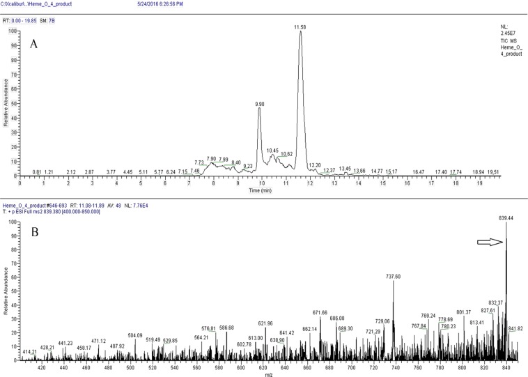
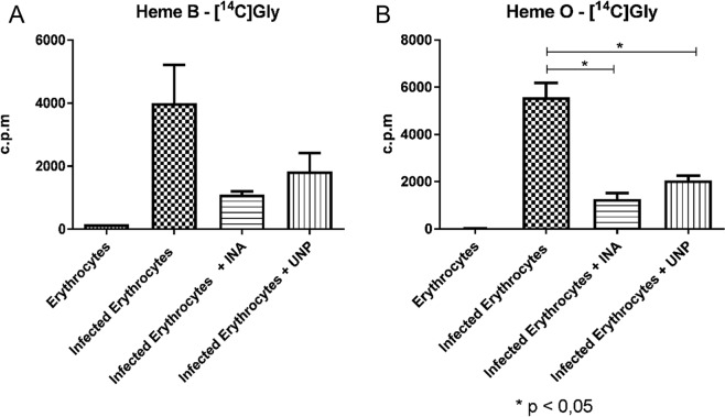
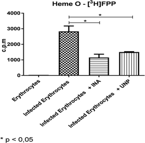
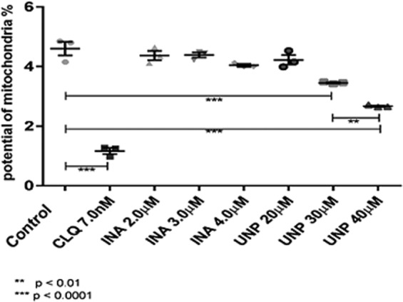
A number of antimalarial drugs interfere with the electron transport chain and heme-related reactions; however, the biosynthesis of heme derivatives in Plasmodium parasites has not been fully elucidated. Here, we characterized the steps that lead to the farnesylation of heme. After the identification of a gene encoding heme O synthase, we identified heme O synthesis in blood stage parasites through the incorporation of radioactive precursors. The presence of heme O synthesis in intraerythrocytic stages of Plasmodium falciparum was confirmed by mass spectrometry. Inabenfide and uniconazole-P appeared to interfere in heme synthesis, accordingly, parasite growth was also affected by the addition of these drugs. We conclude that heme O synthesis occurs in blood stage-P. falciparum and this pathway could be a potential target for antimalarial drugs.


Conflict of interest statement

The authors declare no competing interests.

Figures

Figure 1
Figure 1
P. falciparum synthesizes heme O. Parasitized erythrocytes and non-parasitized erythrocytes were labeled with [1-(n)-3H]-FPP or with [U-14C]-glycine, each extract was purified by affinity columns and the peaks were analyzed by a scintillator. The fraction eluted with 80% ACN, which elutes heme B, presents the radioactive incorporation of glycine and the fraction eluted with DMSO, contained radioactive heme O. Heme O-[3H]FPP is the extract of parasitized erythrocytes labeled with [1-(n)-3H]-FPP and eluted with DMSO; Heme B-[14C]Gly is the extract of parasitized erythrocytes labeled with [U-14C]-glycine eluted with 80% ACN; Heme O-[14C]Gly is the extract of parasitized erythrocytes labeled with [U-14C]-glycine eluted with DMSO; Erythrocytes Heme O-[3H]FPP is the extract erythrocytes labeled with [1-(n)-3H]-FPP and eluted with DMSO; Erythrocytes Heme B-[14C]Gl is the extract of erythrocytes labeled with [U-14C]-glycine and eluted with 80% ACN; Erythrocytes Heme O-[14C]Gl is the extract of erythrocytes labeled with [U-14C]-glycine and eluted with DMSO.
Figure 2
Figure 2
Identification of heme O by LC-MS/MS in schizonts extracts. Heme O was separated using C18 Vac columns and eluted with DMSO and analyzed by LC-MS / MS. (A) Heme O separation was performed using a Hypersil Gold C18 Column (see material and methods: Mass Spectrometry; LC-MS/MS) and the peak with retention time of 11.58 was analyzed by MS/MS. (B) MS/MS of peak of 11.58 showing the m/z of heme O (arrow, 839.4, compatible with its calculated mass).
Figure 3
Figure 3
Identification of heme O by MALDI-TOF/TOF in schizonts extracts. Heme O was separated using C18 Vac columns eluted with DMSO, and analyzed in MALDI-TOF/TOF. Heme O represented by the parent mass m/z 839.405 (compatible with its calculated mass), followed by its fragments with masses m/z 721.060 and m/z 693.951.
Figure 4
Figure 4
(A) The cyclic voltammograms of glass carbon electrodes modified with (a) uninfected erythrocytes and (b) P. falciparum-infected erythrocytes in contact with 0.1 M PBS at pH 7.4. The potential scan rate was 50 mV/s. Semi-derivative convolution was performed to increase signal resolution. (B) The square wave voltammograms of glassy carbon electrodes modified with (a) uninfected erythrocytes, (b) P. falciparum-infected erythrocytes. The potential scan was initiated at +0.2 V in the negative direction; the potential step increment was 4 mV. The square wave amplitude was 25 mV at a frequency of 5 Hz.
Figure 5
Figure 5
The square wave voltammograms of glassy carbon electrodes modified with (a) uninfected erythrocytes treated with INA at 2.0 µM, (b) uninfected erythrocytes treated with UNP at 20.0 µM, (c) Plasmodium-infected erythrocytes treated with INA at 2.0 µM, and (d) Plasmodium-infected erythrocytes treated with UNP at 20.0 µM, in contact with 0.1 M PBS at pH 7.4. The potential scan was initiated at +0.2 V in the negative direction; there was a potential step increment of 4 mV. The square wave amplitude was 25 mV with a frequency of 5 Hz.
Figure 6
Figure 6
INA or UNP lead to the inhibition of heme B and heme O biosynthesis in parasites treated for 48 h. Cultures of P. falciparum were treated with 2.0 μM INA or 20 μM UNP and labeled with [U-14C]-glycine. The same amounts of treated and untreated parasites were applied to the column. The extract of the labeled schizonts was analyzed using C18 Vac columns and heme B was eluted with 80% of ACN while heme O was eluted with DMSO. (A) Heme B was detected in untreated parasites (Infected Erythrocytes) and in parasites treated with INA (Infected Erythrocytes + INA). The inhibition was approximately 75%. In parasites treated with UNP (Infected Erythrocytes + UNP), the inhibition was approximately 54%. No significant differences were observed in the inhibition of heme B. (B) heme O was detected in untreated parasites (Infected Erythrocytes). In parasites treated with INA (Infected Erythrocytes + INA), the inhibition was approximately 76% (p < 0.05), and in parasites treated with UNP (Infected Erythrocytes + UNP), the inhibition was approximately 64% (p < 0.05). The results presented correspond to three experiments. Erythrocytes: uninfected erythrocytes.
Figure 7
Figure 7
INA or UNP inhibit heme O biosynthesis in parasites treated for 48 h. Cultures of P. falciparum were treated with INA at 2.0 μM or 20 μM UNP and labeled with [3H]-FPP. The same amounts of treated and untreated parasites were applied to the column. The extract of labeled schizonts was analyzed with C18 Vac columns and heme O was eluted with DMSO. Heme O was detected in untreated parasites (Infected Erythrocytes). In parasites treated with INA (Infected Erythrocytes + INA), the inhibition was approximately 58% (p < 0.05), and in parasites treated with UNP (Infected Erythrocytes + UNP), the inhibition was approximately 52.5% (p < 0.05). The results correspond to three independent experiments. Erythrocytes: uninfected erythrocytes.
Figure 8
Figure 8
UNP interferes with the mitochondrial potential. Parasites treated with the indicated drug concentrations were incubated with the chromophore JC-1 and fluorescence was analyzed using a FACScalibur cytometer and the CellQuest Pro program, version 5.2. After a 24-hour incubation interval, we observed a significant difference in relation to the control. CLQ (chloroquine) was used at 7.0 nM and INA at 2, 3 and 4 μM. A significant difference between treatment with UNP at 30 and 40 μM, indicating dose dependence, was observed. Significance was assessed using Student’s t test. The results correspond to three independent experiments.

References

    1. WHO. World malaria report 2014. World Health WHO/HTM/GM (2014).
    1. Eastman RT, Fidock DA. Artemisinin-based combination therapies: a vital tool in efforts to eliminate malaria. Nat. Rev. Microbiol. 2009;7:864–874. doi: 10.1038/nrmicro2239. - DOI - PMC - PubMed
    1. Borrmann S, et al. Fosmidomycin-clindamycin for the treatment of Plasmodium falciparum malaria. J. Infect. Dis. 2004;190:1534–1540. doi: 10.1086/424603. - DOI - PubMed
    1. Cassera MB, et al. The methylerythritol phosphate pathway is functionally active in all intraerythrocytic stages of Plasmodium falciparum. J. Biol. Chem. 2004;279:51749–51759. doi: 10.1074/jbc.M408360200. - DOI - PubMed
    1. Chakrabarti D, et al. Protein farnesyltransferase and protein prenylation in Plasmodium falciparum. J. Biol. Chem. 2002;277:42066–42073. doi: 10.1074/jbc.M202860200. - DOI - PubMed

Publication types

MeSH terms