Comparative Study
. 2019 Dec 17;9(1):19240.
doi: 10.1038/s41598-019-55871-8.

Predictors of neovascular activity during neovascular age-related macular degeneration treatment based on optical coherence tomography angiography

Affiliations
Comparative Study

Predictors of neovascular activity during neovascular age-related macular degeneration treatment based on optical coherence tomography angiography

Kunho Bae et al. Sci Rep. .

Abstract

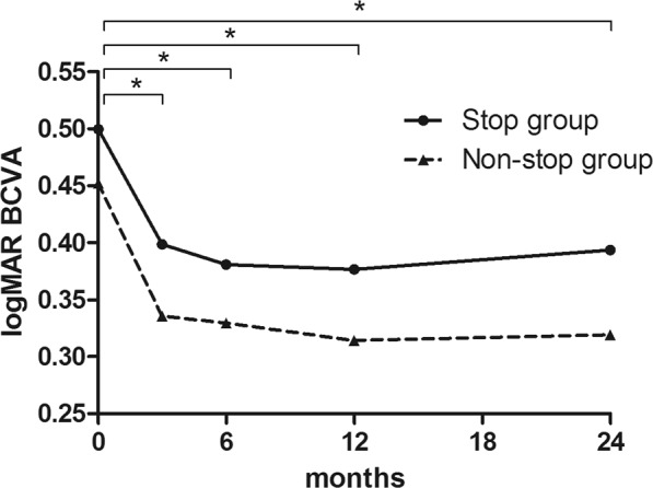
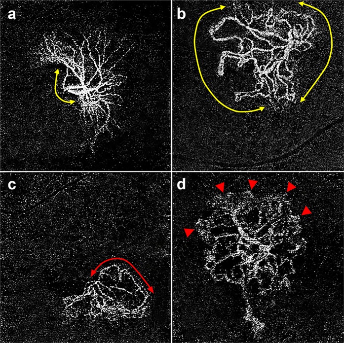
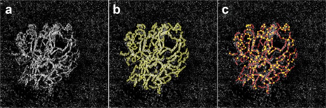
The advent of anti-vascular endothelial growth factor (VEGF) therapies has remarkably improved the functional outcomes of neovascular age-related macular degeneration (nAMD) patients. However, there are guidelines on how to start treatment, the guidelines for discontinuing treatment are not yet clear. In this respect, the treat-extend-stop (TES) protocol have showed us the possibility of discontinuing treatment. In this study, we tried to investigate optical coherence tomography angiography (OCTA) biomarkers related to recurrence of neovascular activity in eyes with nAMD undergoing treatment using TES protocol. A total of 134 eyes with nAMD were divided into two groups (stop, non-stop) depending on whether they met criteria for stopping anti-VEGF treatment. Quantitative and qualitative OCTA parameters including the morphologic pattern of choroidal neovascularization (CNV) were compared between groups. Of these, 44 eyes (32.8%) were in the stop group and 90 eyes (67.2%) were in the non-stop group. In multivariate regression analysis, closed-circuit pattern of CNV and the presence of peripheral loop were associated with the non-stop group (all p < 0.001). Our results imply that the morphologic appearance of CNV on OCTA after anti-VEGF treatment may be a useful biomarker to predict weaning from treatment.


Conflict of interest statement

The authors declare no competing interests.

Figures

Figure 1
Figure 1
Changes in best-corrected visual acuity (BCVA) of eyes with an age-related macular degeneration managed with a Treat-Extend-Stop protocol at 3, 6, 12, and 24 months after initiating anti-vascular endothelial growth factor treatment. BCVA improved in both groups and showed no significant differences between the two groups according to follow-up period (all p < 0.05, asterisk).
Figure 2
Figure 2
Optical coherence tomography angiography (OCTA) images (3 × 3 mm) with corresponding OCT B-scans of choroidal neovascular lesions in neovascular age-related macular degeneration at baseline (a), 4 weeks (b), 8 weeks (c), 14 weeks (d), and 26 weeks (e) after undergoing intravitreal aflibercept treatment using a treat-extend-stop protocol. Note the disappearance of immature capillary fringes (dotted circle) after initial anti-vascular endothelial growth factor (VEGF) therapy and formation of peripheral loop (arrow head) in the same location. There were no significant changes in overall morphology after the initial loading phase of anti-VEGF (d,e), however, dilation of central vessels and formation of anastomosing connections (arrow) intensified during periodic anti-VEGF treatment.
Figure 3
Figure 3
Optical coherence tomography angiography (OCTA) images (3 × 3 mm) with corresponding OCT B-scans of choroidal neovascular (CNV) lesions in age-related macular degeneration. (a) En face OCTA image without detectable CNV at 47 months after last treatment. This case was classified in the stop group and received a total of 8 injections of ranibizumab. (b) En face OCTA image of an open-circuit pattern patient at 7 months after the last treatment. This case was classified in the stop group and a total of 21 injections of aflibercept were performed. (c) En face OCTA image of a closed-circuit pattern patient at 2 months after the last treatment. This case was classified in the non-stop group and a total of 17 injections of ranibizumab were performed, after which the patient was switched to bevacizumab. Treatment was gradually discontinued according to the Treat-Extend-Stop protocol, but the inner retinal cyst recurred every time the treatment interval was extended over 3 months. Note the relatively large portion of anastomotic margin of CNV compared to patients with open-circuit pattern. Inner retinal cyst was observed on OCT B-scans, despite the fact that the patient was currently undergoing maintenance therapy every 2 months. (d) Another case with a closed-circuit pattern in the non-stop group. This is an en face OCTA image taken 2 months after the last treatment, after a total of 26 injections of bevacizumab after being switched from ranibizumab. Note the peripheral loop (arrows) as well as the anastomotic margin of CNV. Since the inner retinal cyst recurred when the treatment interval was prolonged for more than 3 months, treatment was administered every 2 months.
Figure 4
Figure 4
Optical coherence tomography angiography (OCTA) images (3 × 3 mm) with corresponding OCT B-scans of choroidal neovascular lesions in neovascular age-related macular degeneration at baseline (a), 4 weeks (b), 8 weeks (c), and 14 weeks (d) undergoing intravitreal aflibercept treatment using a Treat-Extend-Stop protocol. Note the size of the choroidal neovascularization (CNV) decreased during the treatment every 4 weeks and is not detected eventually (c). When the treatment interval is extended to 6 weeks, the flow of CNV was detected in OCTA with inner retinal cyst in structural OCT.
Figure 5
Figure 5
Morphologic patterns of choroidal neovascularization (CNV) on optical coherence tomography angiography were classified into two categories: CNV with an open circuit, and closed circuit. Closed circuit was defined as anastomotic vessels bounding the outer border of the vascular lesion for more than 50% of the entire CNV margin. Note the relatively small portion (less than 50%) of the anastomotic margin (yellow line with arrow heads) included in the open-circuit pattern (a) compared to the closed-circuit pattern (b). Images were additionally analyzed for presence of peripheral loop (c, red line with arrow heads) and capillary fringe (d, arrow heads). Peripheral loop was defined as looping vessels in the periphery branching into vascular arcades between the vessel termini, and capillary fringe was defined as a fine peripheral network or flush of vessels on the margin of the CNV.
Figure 6
Figure 6
Optical coherence tomographic angiography (OCTA) image of exudative age-related macular degeneration in the “outer retina” (a). Outline of a neovascular lesion (b) and skeletonization (c) to calculate area, total length, and number of junctions of a neovascular lesion using semi-automated software.

References

    1. Wong WL, et al. Global prevalence of age-related macular degeneration and disease burden projection for 2020 and 2040: a systematic review and meta-analysis. Lancet Glob Health. 2014;2:e106–116. doi: 10.1016/S2214-109X(13)70145-1. - DOI - PubMed
    1. Smith W, et al. Risk factors for age-related macular degeneration: Pooled findings from three continents. Ophthalmology. 2001;108:697–704. doi: 10.1016/S0161-6420(00)00580-7. - DOI - PubMed
    1. Kawasaki R, et al. The prevalence of age-related macular degeneration in Asians: a systematic review and meta-analysis. Ophthalmology. 2010;117:921–927. doi: 10.1016/j.ophtha.2009.10.007. - DOI - PubMed
    1. Wong TY, et al. The natural history and prognosis of neovascular age-related macular degeneration: a systematic review of the literature and meta-analysis. Ophthalmology. 2008;115:116–126. doi: 10.1016/j.ophtha.2007.03.008. - DOI - PubMed
    1. Age-Related Eye Disease Study Research G. Risk factors associated with age-related macular degeneration. A case-control study in the age-related eye disease study: Age-Related Eye Disease Study Report Number 3. Ophthalmology. 2000;107:2224–2232. doi: 10.1016/S0161-6420(00)00409-7. - DOI - PMC - PubMed

Publication types

MeSH terms

Substances