Review
. 2020 Apr 1;143(4):1073-1087.
doi: 10.1093/brain/awz382.

Lipid metabolic pathways converge in motor neuron degenerative diseases

Affiliations
Review

Lipid metabolic pathways converge in motor neuron degenerative diseases

Olivia J Rickman et al. Brain. .

Abstract

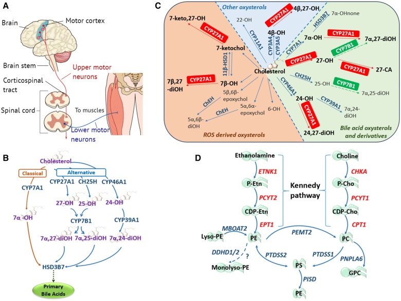
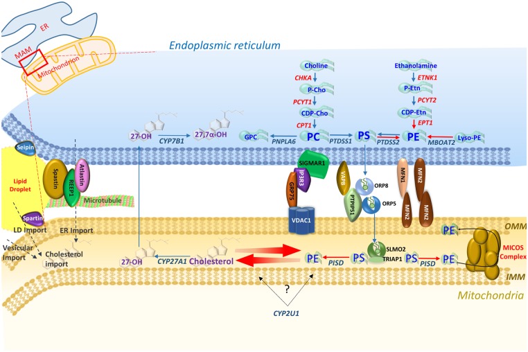
Motor neuron diseases (MNDs) encompass an extensive and heterogeneous group of upper and/or lower motor neuron degenerative disorders, in which the particular clinical outcomes stem from the specific neuronal component involved in each condition. While mutations in a large number of molecules associated with lipid metabolism are known to be implicated in MNDs, there remains a lack of clarity regarding the key functional pathways involved, and their inter-relationships. This review highlights evidence that defines defects within two specific lipid (cholesterol/oxysterol and phosphatidylethanolamine) biosynthetic cascades as being centrally involved in MND, particularly hereditary spastic paraplegia. We also identify how other MND-associated molecules may impact these cascades, in particular through impaired organellar interfacing, to propose 'subcellular lipidome imbalance' as a likely common pathomolecular theme in MND. Further exploration of this mechanism has the potential to identify new therapeutic targets and management strategies for modulation of disease progression in hereditary spastic paraplegias and other MNDs.

Keywords: HSP; MND; cholesterol; lipidome imbalance; mitochondria.


Figures

Figure 1
Figure 1
The primary motor neuron components and lipidome pathways in MND/HSP. (A) Simplified schematic of the core components of the primary motor pathway, including upper and lower motor neurons, for the transmission of voluntary commands from the primary motor cortex to skeletal muscles. (B) Classic and alternative pathways through which cholesterol can be converted into primary bile acids. The classical pathway is initiated by cholesterol 7α-hydroxylase (CYP7A1) located in the endoplasmic reticulum (ER) of the liver, catalysing the conversion of cholesterol to 7α-OH. The alternative pathways are initiated by three enzymes: (i) the mitochondrial sterol 27-hydroxylase (CYP27A1) requiring mitochondrial cholesterol import, primarily via the ER and lipid droplets, mediated by steroidogenic acute regulatory protein (StAR) and StAR-related lipid transfer domain (StarD) proteins (Norlin et al., 2000; Scharwey et al., 2013); (ii) the liver microsomal sterol 25-hydroxylase (CH25H); and (iii) the brain sterol 24-hydroxylase (CYP46A1), forming 27-OH, 25-OH and 24-OH, respectively. Oxysterol 7α-hydroxylase (CYP7B1) catalyses the 7α-hydroxylation of both 25- and 27-OH to 7α,25- and 7α,27-diOH, whereas liver microsomal oxysterol 7α-hydroxylase II (CYP39A1) catalyses the 7α-hydroxylation of 24-OH to 7α,24-diOH (Li-Hawkins et al., 2000). In the liver the 7α-hydroxylated oxysterols converge in the common bile acid pathway, initiated by 3β-hydroxysteroid dehydrogenase (HSD3B7), where they are further modified to form the primary bile acids. (C) Enzymatically (blue and green) and ROS (orange) derived oxysterols, highlighting the importance and multiple enzymatic roles of the mitochondrial sterol 27-hydroxylase (CYP27A1) in oxysterol metabolism. CA = cholestanoic acid; ChEH = cholesterol epoxide hydrolase; HSD = hydroxysteroid dehydrogenase; none = cholestenone; ROS = reactive oxygen species. Figure adapted from Mutemberezi et al. (2016). (D) Biosynthetic pathways for phosphatidylethanolamine (PE) synthesis. The primary routes of PE synthesis include the Kennedy pathway (enzymes shown in red) and the phosphatidylserine (PS) decarboxylase pathway in mitochondria. PE is formed from ethanolamine via the CDP-ethanolamine branch of the Kennedy pathway comprising three enzymatic steps. Two additional pathways of PE production: the acylation of lyso-PE catalysed by lyso-PE-acyltransferase (MBOAT2), and the synthesis from PS via PS-synthase-2 (PTDSS2). The CDP-choline branch of the Kennedy pathway forms phosphatidylcholine (PC), also via three enzymatic steps. PE and PC synthesized via the Kennedy pathway is catalysed by PS-synthase-1 and 2 (PTDSS1/2) to PS, which can be transported to the inner mitochondrial membrane where PSD (encoded by PISD) decarboxylates it to PE. Phosphatidylethanolamine N-methyltransferase 2 (PEMT2) methylates PE to form PC. DDHD1/2 may also play a role in maintaining PE homeostasis.
Figure 2
Figure 2
PE and oxysterol pathways and the mitochondria-associated ER membrane (MAM). Overviewing enzymes in each pathway, three of the four main tethering complexes of the MAM-mitochondrial region, and other HSP/MND-associated molecules with putative roles in these areas. PS synthesized in the ER membrane is then transported across the MAM via ORP5 and ORP8 that interact with protein tyrosine phosphatase interacting protein 51 (PTPIP51) on the outer mitochondrial membrane (OMM) (Galmes et al., 2016). PS is then transported to the inner mitochondrial membrane (IMM) via the SLMO2-TRIAP1 lipid transfer complex for delivery to PSD where it is decarboxylated to PE. The mitochondrial contact site and cristae organizing system (MICOS) complex spans the mitochondrial membrane and is crucial for the distribution of PE to the OMM and ER (Tatsuta and Langer, 2017).

Comment in

  • Defective phosphatidylethanolamine biosynthesis leads to a broad ataxia-spasticity spectrum.
    Kaiyrzhanov R, Wortmann S, Reid T, Dehghani M, Vahidi Mehrjardi MY, Alhaddad B, Wagner M, Deschauer M, Cordts I, Fernandez-Murray JP, Treffer V, Metanat Z, Pitman A, Houlden H, Meitinger T, Carroll C, McMaster CR, Maroofian R. Kaiyrzhanov R, et al. Brain. 2021 Apr 12;144(3):e30. doi: 10.1093/brain/awaa442. Brain. 2021. PMID: 33454747 Free PMC article. No abstract available.

References

    1. Aaltonen MJ, Friedman JR, Osman C, Salin B, di Rago J-P, Nunnari J, et al.MICOS and phospholipid transfer by Ups2-Mdm35 organize membrane lipid synthesis in mitochondria. J Cell Biol 2016; 213: 525–34. - PMC - PubMed
    1. Abdel-Khalik J, Crick PJ, Yutuc E, DeBarber AE, Duell PB, Steiner RD, et al.Identification of 7α,24-dihydroxy-3-oxocholest-4-en-26-oic and 7α,25-dihydroxy-3-oxocholest-4-en-26-oic acids in human cerebrospinal fluid and plasma. Biochimie 2018; 153: 86–98. - PMC - PubMed
    1. Ahmed MY, Al-Khayat A, Al-Murshedi F, Al-Futaisi A, Chioza BA, Pedro Fernandez-Murray J, et al.A mutation of EPT1 (SELENOI) underlies a new disorder of Kennedy pathway phospholipid biosynthesis. Brain 2017; 140: 547–54. - PMC - PubMed
    1. Ajroud-Driss S, Siddique T. Sporadic and hereditary amyotrophic lateral sclerosis (ALS). Biochim Biophys Acta 2015; 1852: 679–84. - PubMed
    1. Al-Saif A, Al-Mohanna F, Bohlega S. A mutation in sigma-1 receptor causes juvenile amyotrophic lateral sclerosis. Ann Neurol 2011; 70: 913–9. - PubMed

Publication types

MeSH terms