. 2019 Dec 18;18(1):426.
doi: 10.1186/s12936-019-3055-9.

Optimization of an in vivo model to study immunity to Plasmodium falciparum pre-erythrocytic stages

Affiliations

Optimization of an in vivo model to study immunity to Plasmodium falciparum pre-erythrocytic stages

Yevel Flores-Garcia et al. Malar J. .

Abstract

Background: The circumsporozoite protein (CSP) of Plasmodium is a key surface antigen that induces antibodies and T-cells, conferring immune protection in animal models and humans. However, much of the work on CSP and immunity has been developed based on studies using rodent or non-human primate CSP antigens, which may not be entirely translatable to CSP expressed by human malaria parasites, especially considering the host specificity of the different species.

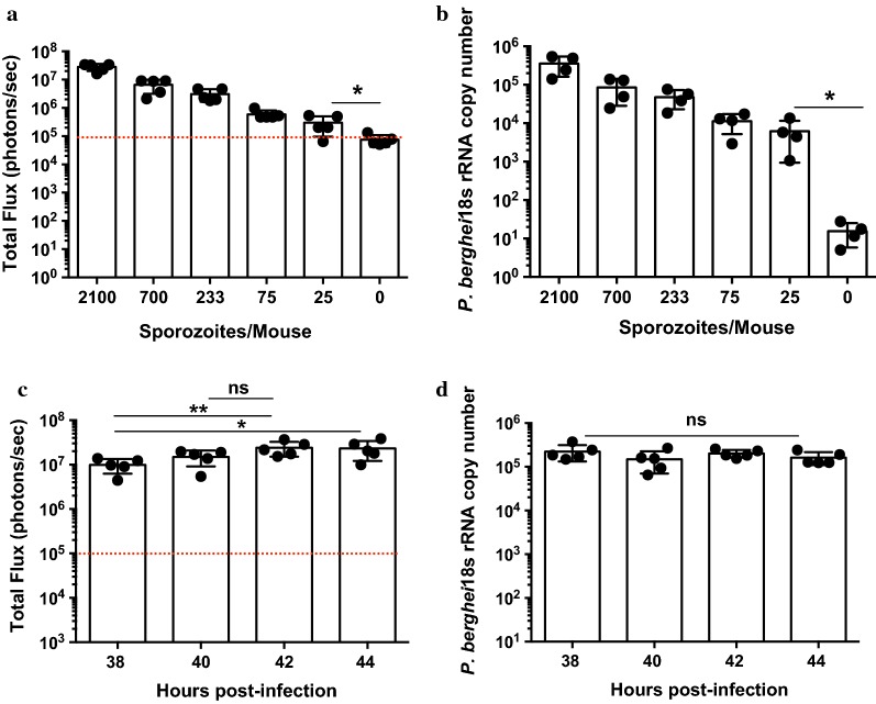
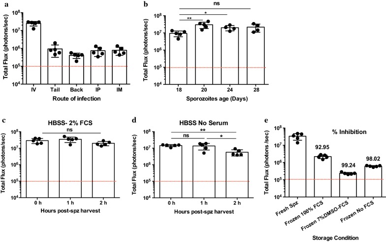
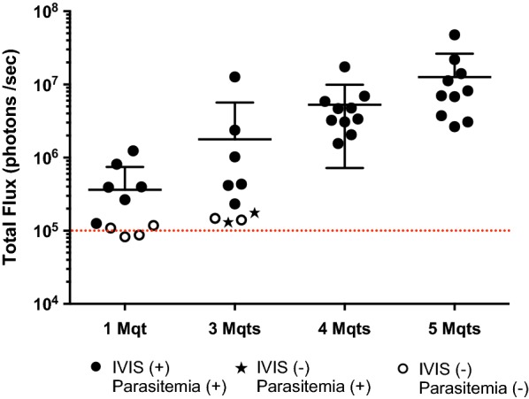
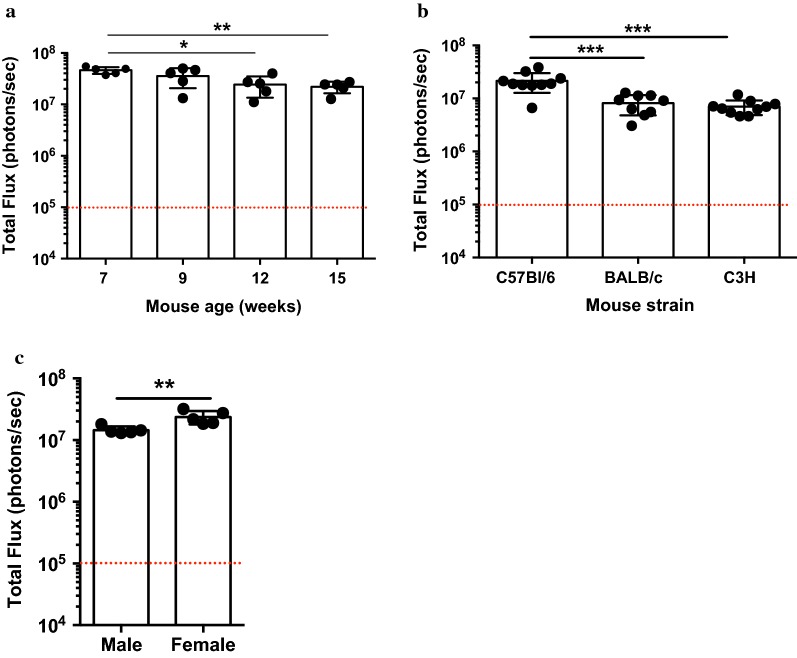
Methods: Using a genetically engineered strain of Plasmodium berghei that expresses luciferase, GFP and the Plasmodium falciparum orthologue of CSP, the effect of laboratory preparation, mosquito treatment and mouse factors on sporozoite infectivity was assessed using an in vivo bioluminescence assay on mice. This assay was compared with a PCR-based protection assay using an already described monoclonal antibody that can provide sterile protection against sporozoite challenge.

Results: Bioluminescence assay demonstrated similar detection levels of the quantity and kinetics of liver-stage infection, compared to PCR-based detection. This assay was used to evaluate treatment of sporozoite and delivery method on mouse infectivity, as well as the effects of age, sex and strain of mice. Finally, this assay was used to test the protective capacity of monoclonal antibody AB317; results strongly recapitulate the findings of previous work on this antibody.

Conclusions: The PbGFP-Luc line and in vivo bioluminescence imaging provide highly sensitive read-outs of liver-stage infection in mice, and this method can be useful to reliably evaluate potency of pre-erythrocytic interventions.

Keywords: Bioluminescence; Malaria; Plasmodium falciparum; Transgenic parasite; Vaccine.


Conflict of interest statement

The authors declare that they have no competing interests.

Figures

Fig. 1
Fig. 1
Sensitivity of the bioluminescence assay for liver-stage infection. a Luciferin-based luminescence in mice as measured by total flux (photons/second) compared to a titration of the number of sporozoites injected. Naïve non-infected mice treated with d-luciferin served as a control for baseline signal. b Measurement of liver burden in the same mice used for luminescence using RT-qPCR P. berghei 18s RNA with the same sporozoite titration as in a. Both experiments demonstrate dose dependency with a dynamic range of approximately 2 logs. c Luminescence measurements of mouse livers at various time points post infection. Data indicate that the peak of infection is approximately 42 h post infection. d RT-qPCR measurements of liver infection as in d, however when liver burden is measured by RT-qPCR, no statistical significance is observed at different time points tested. *p < 0.05; **p < 0.01, Mann–Whitney test. Dashed lines represent background bioluminescence levels obtained from uninfected mice injected with luciferin
Fig. 2
Fig. 2
The effect of sporozoite treatment prior to injection. a Sporozoite infectivity is highest when delivered intravenously as compared to subcutaneous (tail, back), intraperitoneal and intramuscular injection. b Luminescence of mice infected with sporozoites isolated on different days of mosquito infection. Data indicate that sporozoites isolated on day 20 of mosquito infection are most infective. Luminescence readings were taken 42 h after mouse infection. The red dotted line indicates the background signal as determined in Fig. 1a. b, c Mouse luminescence of sporozoites dissected in HBSS supplemented with 2% FCS (b) or without FCS (c) and injected at various times after dissection. Dissection in serum provides greater sporozoite infectivity than dissection without serum. Dissecting without serum also results in a time-dependent loss of infectivity. d Comparison of infectivity of fresh sporozoites to sporozoites frozen under various different media compositions. All frozen sporozoites are > 90% less infective than freshly isolated sporozoites. *p < 0.05; **p < 0.01, Mann–Whitney test. Dashed lines represent background bioluminescence levels obtained from uninfected mice injected with luciferin
Fig. 3
Fig. 3
Mosquito bite challenge. Challenge by bites from 1 or 3 infectious mosquitoes does not guarantee successful infection, while bites from 4 or greater mosquitoes do guarantee successful infection. Filled circle indicates mice that were IVIS positive and positive for blood stage infection; open circle indicates mice that were negative for both IVIS imaging and blood stage infection; filled star indicates mice that were negative for IVIS imaging, but positive for blood stage infection. Dashed lines represent background bioluminescence levels obtained from uninfected mice injected with luciferin
Fig. 4
Fig. 4
The effect of mouse variables on sporozoite infectivity. a Luminescence of C57Bl/6 mice of different ages challenged with the same number of sporozoites. Mice challenged at 7 weeks of age were most susceptible to infection, and this susceptibility decreases with age, with statistical significance at days 12 and 15. b Luminescence data of C57Bl/6, BALB/c, and C3H mice infected with sporozoites. C57Bl/6 mice are the most susceptible of the three strains. c Comparison of infectivity of male and female C57Bl/6 mice. Female mice demonstrate higher susceptibility to infection than males. *p < 0.05; **p < 0.01; ***p < 0.0005, Mann–Whitney test. Dashed lines represent background bioluminescence levels obtained from uninfected mice injected with luciferin
Fig. 5
Fig. 5
Using IVIS imaging for protection models. a Monoclonal antibody protection prior to infection. Treatment with 300 μg of AB317 before IV injection of parasites, confers > 99% inhibition of liver stage development as measured by IVIS. There is dose dependent reduction of liver stage development as AB317 treatment decreases. The numbers on top of each histogram represents the percent inhibition compared to the parasite load measured in naïve mice. b Treatment with AB-317 before and after sporozoite challenge. Treatment before challenge inhibits liver stage development, while treatment after challenge has no effect. c Sterile immunity, as assessed by lack of blood parasitaemia, after mAb treatment followed by exposure to mosquito bites. Kaplan–Meier curves demonstrate dose-dependent protection by AB317, with complete protection with 300 μg. 1245, a mAb directed against an irrelevant antigen, is used as a negative control for mAb treatment. Dashed lines represent background bioluminescence levels obtained from uninfected mice injected with luciferin

References

    1. Nussenzweig RS, Vanderberg J, Most H, Orton C. Protective immunity produced by the injection of x-irradiated sporozoites of Plasmodium berghei. Nature. 1967;216:160–162. doi: 10.1038/216160a0. - DOI - PubMed
    1. Cockburn IA, Seder RA. Malaria prevention: from immunological concepts to effective vaccines and protective antibodies. Nat Immunol. 2018;19:1199–1211. doi: 10.1038/s41590-018-0228-6. - DOI - PubMed
    1. Longley RJ, Salman AM, Cottingham MG, Ewer K, Janse CJ, Khan SM, et al. Comparative assessment of vaccine vectors encoding ten malaria antigens identifies two protective liver-stage candidates. Sci Rep. 2015;5:11820. doi: 10.1038/srep11820. - DOI - PMC - PubMed
    1. Espinosa DA, Christensen D, Munoz C, Singh S, Locke E, Andersen P, et al. Robust antibody and CD8(+) T-cell responses induced by P. falciparum CSP adsorbed to cationic liposomal adjuvant CAF09 confer sterilizing immunity against experimental rodent malaria infection. NPJ Vaccines. 2017;2:e10. doi: 10.1038/s41541-017-0011-y. - DOI - PMC - PubMed
    1. Gimenez AM, Lima LC, Francoso KS, Denapoli PMA, Panatieri R, Bargieri DY, et al. Vaccine containing the three allelic variants of the Plasmodium vivax circumsporozoite antigen induces protection in mice after challenge with a transgenic rodent malaria parasite. Front Immunol. 2017;8:1275. doi: 10.3389/fimmu.2017.01275. - DOI - PMC - PubMed