

Baseline Predictive Factors of Visual Outcome and Persistence of Subretinal Fluid Based on Morphologic Changes in Spectral Domain Optical Coherence Tomography in Patients with Idiopathic Central Serous Chorioretinopathy
- PMID: 31849441
- PMCID: PMC6910099
- DOI: 10.2147/OPTH.S233273
Baseline Predictive Factors of Visual Outcome and Persistence of Subretinal Fluid Based on Morphologic Changes in Spectral Domain Optical Coherence Tomography in Patients with Idiopathic Central Serous Chorioretinopathy
Abstract
Background: To determine the influence of spectral domain optical coherence tomography (OCT) changes on visual outcome and persistence of subretinal fluid (SRF) in patients with idiopathic central serous chorioretinopathy (CSCR).
Materials and methods: In a retrospective study done in 48 eyes of 45 patients diagnosed as CSCR, all eyes were subjected to fundus photography, spectral domain OCT, and fluorescein angiography (FA) in selected cases.
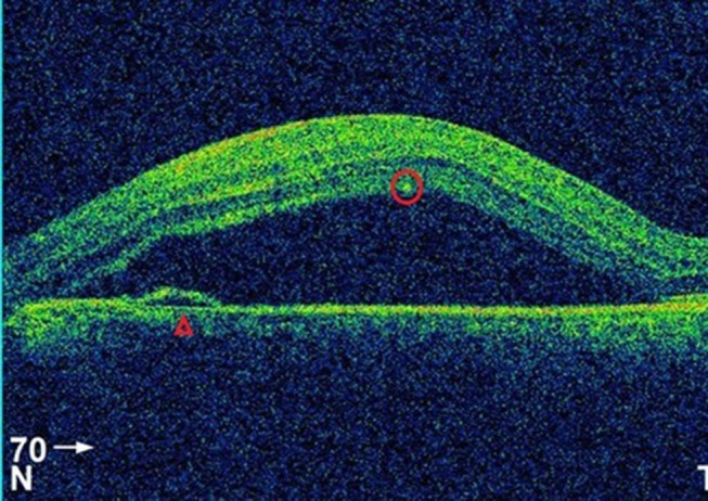
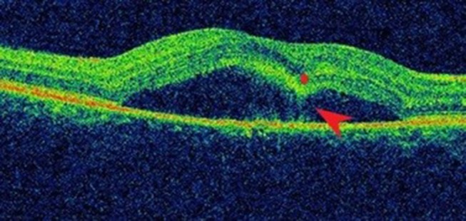
Results: Retinal pigment epithelium detachment was present in 22.91% of the cases at presentation. The logMar best corrected visual acuity improved from 0.46±0.29 at presentation to 0.18±0.22 at 3 months (P-value<0.01). The mean foveal thickness was 486.81±146.06 µm at presentation and 259±94.5 µm at 3 months (P-value<0.01) (paired T-test). OCT factors that were associated with poor visual outcome (BCVA>0.3 logMar) were disruption in the inner segment (IS)/outer segment (OS) junction or external limiting membrane (ELM) line and hyper-reflective dots in the intra/subretinal layer (P-value<0.05) (Fischer's Exact test). Out of the total 48 eyes, 26 had persistent SRF at 3 months. The presence of discontinuation in IS/OS junction and hyper-reflective dots in the intra/subretinal layer were the only two OCT factors that were associated with the persistence of SRF (P-value<0.01) (Pearson's Chi-square test).
Conclusion: Visual outcome and persistence of subretinal fluid at 3 months can be predicted on the basis of early morphologic changes in OCT. This will aid in counseling patients regarding its course and may guide us in its management.
Keywords: central serous chorioretinopathy; optical coherence tomography; persistent subretinal fluid; visual outcome.
© 2019 Suwal et al.
Conflict of interest statement
The authors declare no competing interests.
Figures

References
-
- Coscas G. Optical coherence tomography in age-related macular degeneration (OCT in AMD) In: Chapter 7: OCT Interpretation. G. Coscas, F. Coscas, S.Vismara, A. Zourdani, C. I. Li Calzi Heidelberg, Germany: Springer Medizin Verlag; 2009:159–166.
LinkOut - more resources
Full Text Sources
Miscellaneous
