

Choroidal Neovascularization: Mechanisms of Endothelial Dysfunction
- PMID: 31849644
- PMCID: PMC6895252
- DOI: 10.3389/fphar.2019.01363
Choroidal Neovascularization: Mechanisms of Endothelial Dysfunction
Abstract
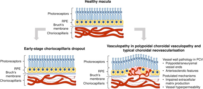
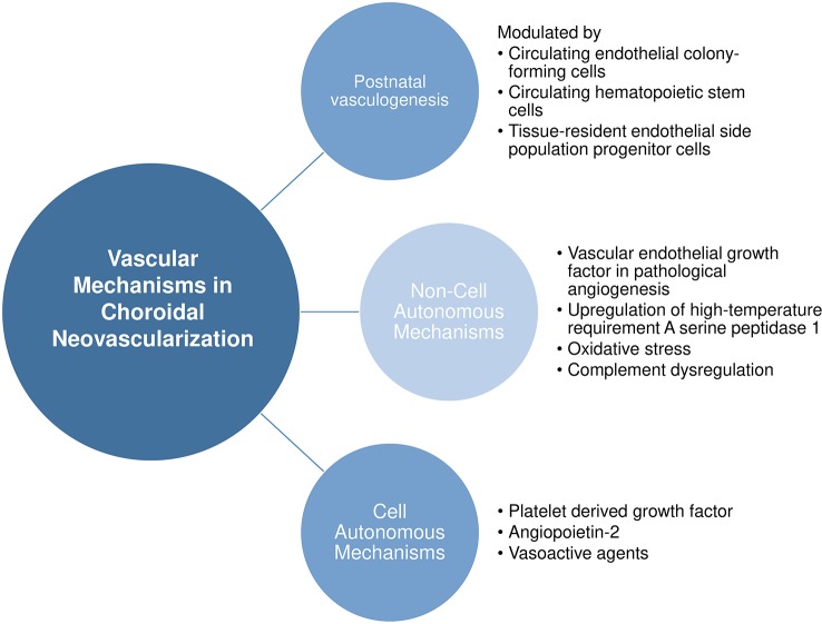
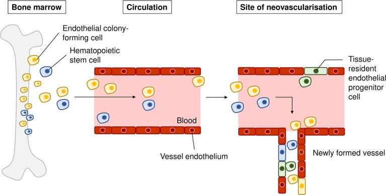
Many conditions affecting the heart, brain, and even the eyes have their origins in blood vessel pathology, underscoring the role of vascular regulation. In age-related macular degeneration (AMD), there is excessive growth of abnormal blood vessels in the eye (choroidal neovascularization), eventually leading to vision loss due to detachment of retinal pigmented epithelium. As the advanced stage of this disease involves loss of retinal pigmented epithelium, much less attention has been given to early vascular events such as endothelial dysfunction. Although current gold standard therapy using inhibitors of vascular endothelial growth factor (VEGF) have achieved initial successes, some drawbacks include the lack of long-term restoration of visual acuity, as well as a subset of the patients being refractory to existing treatment, alluding us and others to hypothesize upon VEGF-independent mechanisms. Against this backdrop, we present here a nonexhaustive review on the vascular underpinnings of AMD, implications with genetic and systemic factors, experimental models for studying choroidal neovascularization, and interestingly, on both endothelial-centric pathways and noncell autonomous mechanisms. We hope to shed light on future research directions in improving vascular function in ocular disorders.
Keywords: age-related macular degeneration; choroidal neovascularization; disease models; endothelial; vascular mechanisms.
Copyright © 2019 Yeo, Chan and Cheung.
Figures
References
-
- Akpek E. K., Smith R. A. (2013). Overview of age-related ocular conditions. Am. J. Manag Care 19 (5 Suppl), S67–S75. - PubMed
-
- Anand-Apte B., Hollyfield J. G. (2010). Developmental anatomy of the retinal and choroidal vasculature. In: Besharse Joseph C., Bok Dean, eds. The Retina and Its Disorders. (San Diego, CA: Academic Press; ) 179–185. 10.1016/B978-0-12-374203-2.00169-X - DOI
Publication types
LinkOut - more resources
Full Text Sources
Research Materials