. 2019 Nov 28:10:2758.
doi: 10.3389/fmicb.2019.02758. eCollection 2019.

Expression Level of HIV-1 Vif Can Be Fluctuated by Natural Nucleotide Variations in the vif-Coding and Regulatory SA1D2prox Sequences of the Proviral Genome

Affiliations

Expression Level of HIV-1 Vif Can Be Fluctuated by Natural Nucleotide Variations in the vif-Coding and Regulatory SA1D2prox Sequences of the Proviral Genome

Naoya Doi et al. Front Microbiol. .

Abstract

Vif is required for HIV-1 replication in natural target cells by counteracting host restriction factors, APOBEC3 (A3) proteins. We recently demonstrated that Vif expression level can be changed by naturally occurring single-nucleotide variations within SA1D2prox of the HIV-1 genome. We also found that levels for vif/vpr mRNAs are inversely correlated. While amino acid sequence per se is critical for functionality, Vif expression level modulated by signal sequences in its coding region is likely to be important as well. There are two splicing sites in the region involved in vpr expression. To reveal possible fluctuations of Vif-expression level, we examined SA1D2prox and vif gene by chimeric approaches using HIV-1 subtypes B and C with distinct anti-A3 activity. In this report, recombinant clones in subtype B backbone carrying chimeric sequences with respect to SA1D2prox/vif and those within the vif-coding region were generated. Of these, clones containing vif-coding sequence of subtype C, especially its 3' region, expressed vif/Vif at a decreased level but did at an increased level for vpr/Vpr. Clones with reduced vif/Vif level grew similarly or slightly better than a parental clone in weakly A3G-positive cells but more poorly in highly A3G-expressing cells. Three clones with this property were also tested for their A3-degrading activity. One of the clones appeared to have some defect in addition to the poor ability to express vif/Vif. Taken all together, our results show that natural variations in the SA1D2prox and vif-coding region can change the Vif-expression level and affect the HIV-1 replication potential.

Keywords: HIV-1; SA1D2prox; expression level; natural variation; subtype; vif.


Figures

Figure 1
Figure 1
Sequence alignment and genome organization around the vif gene of HIV-1 proviral clones used in this study. Proviral clones of HIV-1 subtype B (NL4-3, HIV-1NL4-3; GenBank: AF324493) (Adachi et al., 1986) and subtype C (Indie, HIV-1IndieC1; GenBank: AB023804) (Mochizuki et al., 1999) were used in this study. Dots in the nucleotide and amino acid sequences of Indie show the nucleotides and residues identical to those of NL4-3. Sequence identities between regions of NL4-3 and Indie are as follows. (1) Nucleotide identity. SA1D2prox, 89%. Vif: nos. 1 to 324 (amino acid no. 108), 87%; no. 325 to 3′end, 87%; nos. 1 to 93 (amino acid no. 31), 91%; no. 94 to 3′end, 86%. (2) Amino acid identity for Vif. Nos.1 to 108, 78%; no. 109 to C-terminus, 78%; nos. 1 to 31, 80%; no. 32 to C-terminus, 78%. (A) Nucleotide sequence alignment of SA1D2prox region. SA1 and SD2 sites are indicated. Based on our previous results (Nomaguchi et al., 2014, 2016), single-nucleotide variations within the SA1D2prox in NL4-3 that decrease and increase vif production levels are indicated by green and red letters, respectively, in the Indie sequence. Single-nucleotide variations for which their effects on the vif production are not much or have not been determined yet are represented as blue and black letters, respectively, in the Indie sequence. Reported SREs, i.e., ESEVif (Exline et al., 2008), ESEM1/M2 (Kammler et al., 2006), G4 motif (Exline et al., 2008), ESS2b (Brillen et al., 2017), ESE2b (Brillen et al., 2017), and GI2-1 (Widera et al., 2013) are indicated. See also a review (Sertznig et al., 2018). (B) Alignment of the vif-coding sequence (positions from 301 to 579). SA2, SD3, and amino acid residue H at position 108 of the Vif protein are indicated. Known splicing silencers, ESSV (Madsen and Stoltzfus, 2005), HIVE3D3 (Tsuruno et al., 2011), and GI3-2 (Widera et al., 2014), and a splicing enhancer, ESEvpr (Erkelenz et al., 2013) are shown as reference. (C) Alignment of Vif amino acid sequence. Red letters indicate residues (17 K and 31 V) reported to be responsible for high anti-A3G activity of Vif derived from HIV-1 subtype C (Iwabu et al., 2010). Numbers 31 and 108 marked above sequences show the amino acid positions that were utilized to generate chimeric Vif. Domains that are important for proteasomal degradation of A3s are indicated for reference (Feng et al., 2014; Nakashima et al., 2015). (D) Genome organization around the vif gene of the HIV-1 proviral clones constructed in this study. SA1D2prox regions derived from NL4-3 and Indie are shown in green and orange, respectively. SA1, SD2, SA2, and SD3 are indicated in all clones as shown. Above the chimeric vif gene, the corresponding amino acid residues at positions 31/32 and 108/109 of Vif are indicated. Recombinant viral clones between NL4-3 and Indie were generated by amplifying chimeric regions with overlapping PCR as indicated at amino acid positions and then by introducing resultant PCR fragments into NL4-3 using unique sites (SbfI in pol and EcoRI in vpr).
Figure 2
Figure 2
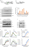
Virological characteristics of NL4-3 and chimeric viral clones newly constructed. Most experiments here were performed similarly as described previously (Nomaguchi et al., 2014, 2016, 2017). (A) Semiquantitative PCR analysis. Indicated proviral clones (2.5 μg) were transfected into 293T cells by Lipofectamine 2000 (Thermo Fischer Scientific), and cell lysates were made at 18 to 20 h post-transfection. Total RNAs were prepared and subjected to cDNA synthesis with oligo(dT) primer. Vif/vpr mRNAs were amplified simultaneously in one reaction using the cDNA as template and a specific primer pair indicated at the top. The reverse primer 5,622-3 was designed for the 100% matched sequence between NL4-3 and Indie. Exon7 (amplified by a primer pair indicated at the top) and gapdh were used as a transfection control (total level of HIV-1 mRNAs) and an internal control, respectively. Representative data from three independent experiments are presented in the lower left portion of this panel. Vif and vpr mRNA levels relative to those of NL4-3 are presented in the lower right portion of this panel. Expression levels of vif and vpr mRNAs in each sample were normalized by those of all HIV-1 mRNAs (exon7) and gapdh. Mean values ± standard errors from three independent experiments are shown. (B) Western blotting analysis. 293T cells were transfected with 3.5 μg of proviral clones indicated by Lipofectamine 2000, and on day 1 post-transfection, cell lysates were prepared. To detect Vif and Vpr proteins, the polyclonal anti-Vif peptide antibody (Akari et al., 1999) and anti-Vpr peptide antibody (#3951, NIH Research and References Reagent Program) were used, respectively. These rabbit polyclonal antibodies were raised against a synthetic NL4-3 Vif peptide (amino acids 170-184; amino acid identity between NL4-3 and Indie is 9/15 = 60%) and against a synthetic NL4-3 Vpr peptide (amino acids 1-46; amino acid identity between NL4-3 and our chimeric clones NLpInV, InpInV, NL/InV1, and NL/InV3 is 45/46 = 98%). β-actin was used as an internal control. Representative data from at least two independent experiments are shown. (C) Comparative analysis of the A3G-degrading activity. Ability of virus clones to degrade A3G in cells was assessed by the co-transfection experiment as previously described (Yamashita et al., 2008, 2010). A flag-tagged A3G expression vector (0.1 μg) and a proviral clone (2.5 μg) were co-transfected into 293T cells, and the A3G expression level in the cells at 48 h post-transfection was monitored by Western blotting analysis as described in (B). For detection of the flag-tagged A3G and control Gag (precursor p55) proteins, anti-Flag (Sigma) and anti-Gag (#3537, NIH Research and References Reagent Program) antibodies were used, respectively. As a negative control, NL-Nd (∆Vif) which lacks the Vif expression (Adachi et al., 1991) was used. Representative data from two independent experiments are shown. (D) Growth kinetics in CEM-SS and H9 cells. Viruses were prepared from 293T cells transfected with indicated proviral clones (2.5–5.0 μg) by Lipofectamine 2000 or calcium-phosphate co-precipitation method, and virus amounts were determined by the virion-associated reverse transcriptase (RT) assays (Willey et al., 1988; Nomaguchi et al., 2013b). Equal amounts of viruses (104 RT units) were inoculated into a weakly A3G-positive cell line CEM-SS and a highly A3G-expressing cell line H9 (105 cells). Culture supernatants were collected every 3 days, and virus replication was monitored by RT assays. All viruses were examined for their growth properties in the same single experiment. Results obtained for chimeric clones were separately presented in the upper and lower portions of this panel for clarity, and the same NL4-3 data were shown in both graphs for easy comparison. Representative data from at least three independent infection experiments performed using virus samples prepared by separate transfections are shown. (E) Growth kinetics in CEM-SS cells. A series of infection experiments were performed as described in the legend to (D), and results from two independent experiments for InpInV, NL/InV1, and NL/InV3, other than those shown in (D), are presented at upper and lower panels.

Similar articles

Cited by

References

    1. Adachi A., Gendelman H. E., Koenig S., Folks T., Willey R., Rabson A., et al. . (1986). Production of acquired immunodeficiency syndrome-associated retrovirus in human and nonhuman cells transfected with an infectious molecular clone. J. Virol. 59, 284–291. PMID: - PMC - PubMed
    1. Adachi A., Ono N., Sakai H., Ogawa K., Shibata R., Kiyomasu T., et al. . (1991). Generation and characterization of the human immunodeficiency virus type 1 mutants. Arch. Virol. 117, 45–58. 10.1007/BF01310491, PMID: - DOI - PubMed
    1. Akari H., Uchiyama T., Fukumori T., Iida S., Koyama A. H., Adachi A. (1999). Pseudotyping human immunodeficiency virus type 1 by vesicular stomatitis virus G protein does not reduce the cell-dependent requirement of vif for optimal infectivity: functional difference between Vif and Nef. J. Gen. Virol. 80, 2945–2949. 10.1099/0022-1317-80-11-2945, PMID: - DOI - PubMed
    1. Amendt B. A., Si Z. H., Stoltzfus C. M. (1995). Presence of exon splicing silencers within human immunodeficiency virus type 1 tat exon 2 and tat-rev exon 3: evidence for inhibition mediated by cellular factors. Mol. Cell. Biol. 15, 4606–4615. 10.1128/MCB.15.8.4606, PMID: - DOI - PMC - PubMed
    1. Aydin H., Taylor M. W., Lee J. E. (2014). Structure-guided analysis of the human APOBEC3-HIV restrictome. Structure 22, 668–684. 10.1016/j.str.2014.02.011, PMID: - DOI - PubMed

LinkOut - more resources