. 2019 Nov 26:10:2722.
doi: 10.3389/fimmu.2019.02722. eCollection 2019.

Elucidating the Immune Evasion Mechanisms of Borrelia mayonii, the Causative Agent of Lyme Disease

Affiliations

Elucidating the Immune Evasion Mechanisms of Borrelia mayonii, the Causative Agent of Lyme Disease

Lea Walter et al. Front Immunol. .

Abstract

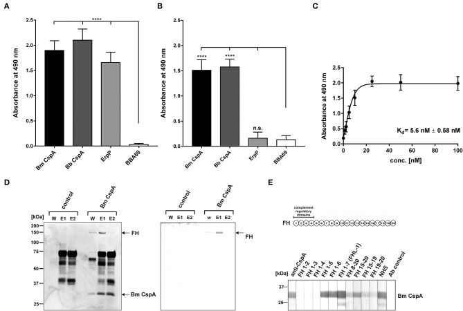
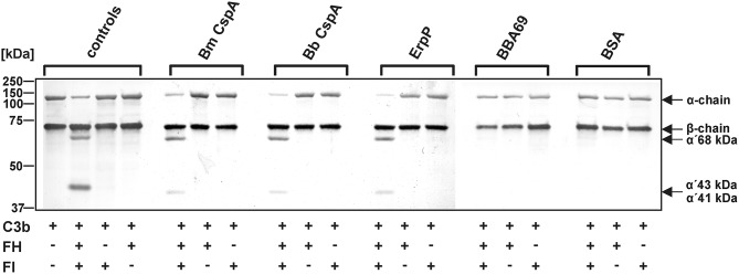
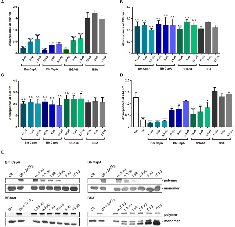
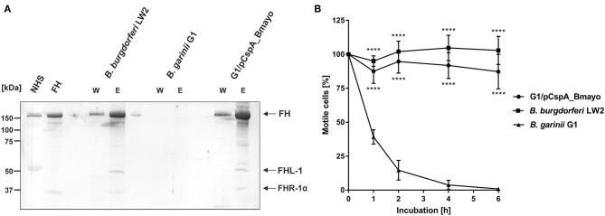
Borrelia (B.) mayonii sp. nov. has recently been reported as a novel human pathogenic spirochete causing Lyme disease (LD) in North America. Previous data reveal a higher spirochaetemia in the blood compared to patients infected by LD spirochetes belonging to the B. burgdorferi sensu lato complex, suggesting that this novel genospecies must exploit strategies to overcome innate immunity, in particular complement. To elucidate the molecular mechanisms of immune evasion, we utilized various methodologies to phenotypically characterize B. mayonii and to identify determinants involved in the interaction with complement. Employing serum bactericidal assays, we demonstrated that B. mayonii resists complement-mediated killing. To further elucidate the role of the key regulators of the alternative pathway (AP), factor H (FH), and FH-like protein 1 (FHL-1) in immune evasion of B. mayonii, serum adsorption experiments were conducted. The data revealed that viable spirochetes recruit both regulators from human serum and FH retained its factor I-mediated C3b-inactivating activity when bound to the bacterial cells. In addition, two prominent FH-binding proteins of approximately 30 and 18 kDa were detected in B. mayonii strain MN14-1420. Bioinformatics identified a gene, exhibiting 60% identity at the DNA level to the cspA encoding gene of B. burgdorferi. Following PCR amplification, the gene product was produced as a His-tagged protein. The CspA-orthologous protein of B. mayonii interacted with FH and FHL-1, and both bound regulators promoted inactivation of C3b in the presence of factor I. Additionally, the CspA ortholog counteracted complement activation by inhibiting the alternative and terminal but not the classical and Lectin pathways, respectively. Increasing concentrations of CspA of B. mayonii also strongly affected C9 polymerization, terminating the formation of the membrane attack complex. To assess the role of CspA of B. mayonii in facilitating serum resistance, a gain-of-function strain was generated, harboring a shuttle vector allowing expression of the CspA encoding gene under its native promotor. Spirochetes producing the native protein on the cell surface overcame complement-mediated killing, indicating that CspA facilitates serum resistance of B. mayonii. In conclusion, here we describe the molecular mechanism utilized by B. mayonii to resists complement-mediated killing by capturing human immune regulators.

Keywords: borrelia; borrelia mayonii; complement; host cell interaction; immune evasion; innate immunity; lyme disease; spirochetes.


Figures

Figure 1
Figure 1
B. mayonii resists complement-mediated killing in human serum. Survival of B. mayonii MN14-1420, B. burgdorferi LW2, and B. garinii G1 in 50% NHS was monitored by dark-field microscopy. Viability and motility of borrelial cells were determined at 1, 2, 4, and 6 h. At least four independent experiments were conducted, each with very similar results. For clarity, only data from a representative experiment is shown. All experiments were performed at least four times. ****p ≤ 0.0001, **p ≤ 0.0021, *p ≤ 0.0002; one-way ANOVA with Bonferroni post test (confidence interval = 95%). ns, no statistical significance.
Figure 2
Figure 2
Interaction of B. mayonii with complement regulator FH. (A) Binding of purified FH to immobilized spirochetes was assessed by ELISA. Bound FH was detected using a polyclonal anti-FH antiserum (1:1,000 dilution). All experiments were performed at least four times, with each individual test carried out in triplicate. ****p ≤ 0.0001, one-way ANOVA with Bonferroni post test (confidence interval = 95%). (B) Determination of FH binding to viable spirochetes. B. mayonii MN14-1420, B. burgdorferi LW2, and B. garinii G1 were incubated in NHS-EDTA and cell-bound proteins were eluted. The last wash (w) and the eluate (e) fractions were separated and transferred to nitrocellulose. For detection of molecules of the FH protein family, a polyclonal anti-FH antibody (dilution 1:1,000) was applied. The mobilities of molecular mass standards are shown to the left of the panel. For better visualization of faint signals, the contrast has been increased as indicated in Supplementary Figure 4. (C) Determination of the regulatory activity of cell-bound FH. Factor I-mediated inactivation of C3b was assessed by the detection of C3b cleavage products. Spirochetes were incubated with purified FH and afterwards with C3b and factor I. For control purposes, all reactions were also performed in the absence of FH. The C3b fragments were visualized by Western blotting using an anti-human C3 antiserum (dilution 1:1,000). As additional controls, samples containing C3b and factor I were incubated with (+) or without (-) purified FH, respectively. The mobility of the α'- and the β-chain of C3 and the cleavage products of the α'-chain the α'68,' α'46 and α'43 fragments are indicated as well as the mobilities of molecular mass standards. A full scan of the original membrane is presented in Supplementary Figure 4.
Figure 3
Figure 3
Identification and surface exposure of FH/FHL-1-binding proteins in B. mayonii MN14-1420. (A,B) Detection of FH/FHL-1-binding proteins by Far Western blot analysis using NHS as source of FH and purified FHL-1 (750 ng/ml). Cell lysates obtained from B. mayonii MN14-1420, B. burgdorferi LW2, B. burgdorferi B31, B. burgdorferi PKa-1, B. garinii G1, and transformant G1/pCspA_Bmayo were separated by 10% Tris/tricine-SDS-PAGE and transferred onto a nitrocellulose membrane. Flagellin (FlaB) was detected with the monoclonal antibody L41 1C11. The FH-binding proteins (A) were visualized by applying an anti-FH antiserum and FHL-1-binding proteins (B) were detected by using an anti-CCP1-4 antiserum. The corresponding to CspA protein of B. mayonii (Bm) MN14-1420, CspA of B. burgdorferi (Bb) LW2, CspZ, ErpP, and ErpA of B. burgdorferi s.s. are indicated at the right. (C) in situ protease accessibility assay. Native spirochetes were incubated with or without proteinase K or trypsin, then lysed by sonication and total proteins were separated by 10% Tris/tricine-SDS-PAGE. The band corresponding to CspA of B. mayonii is indicated on the right. The mobilities of molecular mass standards are indicated on the left. A full scan of the original membranes is presented in Supplementary Figure 5.
Figure 4
Figure 4
Interaction of CspA of B. mayonii MN14-1420 with FH and FHL-1 and mapping of the FH/FHL-1 binding site. (A) Binding of recombinant borrelial proteins to FH. Microtiter plates were coated with His6-tagged proteins, incubated with purified FH, and antigen-antibody complexes were detected using an anti-FH antiserum. All experiments were performed at least three times, with each individual test carried out in triplicate. ****p ≤ 0.0001, one-way ANOVA with Bonferroni post test (confidence interval = 95%). (B) Binding of recombinant proteins to FHL-1. Binding of purified FHL-1 was assessed by ELISA as described in (A). (C) Dose-dependent binding of FH to CspA of B. mayonii MN14-1420. Recombinant CspA of B. mayonii MN14-1420 (5 μg/ml) was immobilized and incubated with increasing concentrations of FH. Binding curve and dissociation constant were approximated via non-linear regression, using a one-site, specific binding model. Data represent means and standard deviation of at least three different experiments, each conducted in triplicate. (D) FH adsorption to immobilized CspA of B. mayonii MN14-1420. Recombinant CspA of B. mayonii MN14-1420 immobilized onto magnetic particles was incubated with NHS. Empty beads were also incubated under the same conditions and used as a control to identify non-specific binding of serum proteins. After extensive washing, bound proteins were eluted with 100 mM glycine and eluate fraction e1 and e2 were separated by 10% Tris/tricine SDS-PAGE, following silver staining and Western blotting using a polyclonal anti-FH antibody. Mobilities of molecular mass standards are indicated to the left. For better visualization of faint signals, the background of the original silver stained gels was reduced as indicated in Supplementary Figure 8. (E) Mapping of the binding domain in FH. Schematic representation of the FH molecule (upper panel). The complement regulatory domains (CCP domains 1-4) are indicated. Localization of the CspA interacting domain in FH by Far Western blotting (lower panel). Purified CspA of B. mayonii (Bm) MN14-1420 was separated by 10% Tris/tricine SDS-PAGE, and transferred to nitrocellulose. The membrane strips were then incubated with constructs of FH containing different CCP domains FH1-2, FH1-3, FH1-4, FH1-5, FH1-6, FH1-7/FHL-1, FH8-20, FH15-20, FH15-19, FH19-20, NHS, and with the secondary Ab (Ab control). Bound proteins were visualized using polyclonal anti-FH antibody. A full scan of the original gel and membrane is presented in Supplementary Figure 6.
Figure 5
Figure 5
Determination of the C3b inactivation capacity of FH bound to CspA of B. mayonii MN14-1420. Purified proteins immobilized to microtiter plates were incubated with purified FH. After washing, C3b and factor I were added. As controls, all reactions were performed in the absence of FH or factor I (FI). All samples were subjected to Tris/tricine SDS-PAGE and transferred onto a nitrocellulose membrane. As additional controls, reaction mixtures containing different combinations of purified complement proteins were assessed. The various C3b fragments were visualized by Western blotting using an anti-human C3 antiserum (dilution 1:1,000). The mobility of the α'- and the β-chain of C3 and the cleavage products of the α'-chain the α'68,' α'46, and α'43 fragments are indicated as well as the mobilities of molecular mass standards. A full scan of the original membranes is presented in Supplementary Figure 7. For better visualization of faint signals, the contrast has been increased as indicated. Bm, B. mayonii; Bb, B. burgdorferi.
Figure 6
Figure 6
CspA of B. mayonii MN14-1420 terminates activation of the AP and TP and inhibits C9 polymerization. (A–C) Assessment of the inhibitory capacity of CspA of B. mayonii MN14-1420 on complement activation by an ELISA-based assay. Microtiter plates immobilized with LPS for the AP (A), IgM for the CP (B), and mannan for the LP (C) were incubated with NHS pre-incubated with increasing concentrations of recombinant proteins or BSA. After washing, formation of the MAC was detected by a monoclonal anti-C5b-9 antibody (dilution 1:500). All experiments were performed at least three times, with each individual test carried out in triplicate. ****p ≤ 0.0001, one-way ANOVA with Bonferroni post test (confidence interval = 95%). n.s., no statistical significance. (D) CspA-mediated inhibition of the TP. Sensitized sheep erythrocytes covered with the preforming C5b-6 complex were incubated with a reaction mixture containing C7, C8, and C9 that was pre-incubated with increasing concentrations of recombinant proteins or BSA. Following incubation, hemolysis of erythrocytes was detected at 414 nm. Means of three independent experiments are shown and error bars correspond to SD. Raw data were analyzed using one-way ANOVA with Bonferroni post test (confidence interval = 95%). ****p ≤ 0.0001; ***p < 0.001; **p < 0.01; *p < 0.05. (E) CspA MN14-1420 inhibits C9 polymerization. C9 was incubated with increasing concentrations of recombinant proteins or BSA and ZnCl2 were then added to the samples to induced polymerization. C9 incubated with and without ZnCl2 was used as controls. Following incubation, reactions mixtures were separated by 7.5% SDS-PAGE and C9 monomers and high molecular weight polymers were visualized by silver staining. A full scan of the original gels is presented in Supplementary Figure 8. For better visualization of faint signals in (E), the contrast has been increased as indicated in Supplementary Figure 8. Bm, B. mayonii; Bb, B. burgdorferi.
Figure 7
Figure 7
CspA of B. mayonii MN14-1420 facilitates resistance of Borrelia to complement-mediated killing. (A) Determination of FH binding to vital spirochetes. B. burgdorferi LW2, B. garinii G1, and B. garinii strain G1 producing CspA of B. mayonii MN14-1420 (G1/pCspA_Bmayo) were incubated in NHS-EDTA and cell-bound proteins were eluted using 0.1 M glycine. The last wash (w) and the eluate (e) fractions were separated by glycine-SDS-PAGE and transferred to nitrocellulose. For detection of molecules of the FH protein family, a polyclonal anti-FH antibody (dilution 1:1,000) was applied. The mobilities of molecular mass standards are shown to the left of the panel. A full scan of the original membranes is presented in Supplementary Figure 9. (B) Survival of G1/pCspA_Bmayo, B. burgdorferi LW2, and B. garinii G1 in 50% NHS was monitored by dark-field microscopy. Viability and motility of borrelial cells were determined at 1, 2, 4, and 6 h. At least four independent experiments were conducted, each with very similar results. ****p ≤ 0.0001; one-way ANOVA with Bonferroni post test (confidence interval = 95%).
Figure 8
Figure 8
Structural model of the CspA of B. mayonii MN14-1420 dimer and superimposed structures of CspA of B. burgdorferi. (A) The two subunits are represented as orange or green ribbons, regions involved in binding of FHL-1 or FH are colored magenta (151–159) and violet (240–246) in the orange subunit and light blue and blue in the green subunit, respectively. Helices are marked by a capital letter, mutated residues are shown in sticks, and an apostrophe denotes the second subunit. Molecular graphics images are produced using the UCSF Chimera (92). (B) Superimposed structures of the CspA homodimer of both orthologs (1w33, 4bl4, 5a2u) show the different orientations of the C-terminal helix and the second subunit. The subunits in the structures of 1w33, 4bl4, and 5a2u are colored light-and dark-gray, light- and dark-magenta and light- and dark-blue, respectively. We modeled the CspA of B. mayonii MN14-1420 structure according to the crystal structure of CspA of B. burgdorferi B31 (1w33) using the Swiss-Model automated comparative protein modeling server (93).

References

    1. Pritt BS, Mead PS, Johnson DKH, Neitzel DF, Respicio-Kingry LB, Davis JP, et al. . Identification of a novel pathogenic Borrelia species causing Lyme borreliosis with unusually high spirochaetaemia: a descriptive study. Lancet Infect Dise. (2016) 16:556–64. 10.1016/S1473-3099(15)00464-8 - DOI - PMC - PubMed
    1. Pritt BS, Respicio-Kingry LB, Sloan LM, Schriefer ME, Replogle AJ, Bjork J, et al. . Borrelia mayonii sp. nov., a member of the Borrelia burgdorferi sensu lato complex, detected in patients and ticks in the upper midwestern United States. Int J Syst Evol Microbiol. (2016) 66:4878–80. 10.1099/ijsem.0.001445 - DOI - PMC - PubMed
    1. Eisen L, Breuner NE, Hojgaard A, Hoxmeier JC, Pilgard MA, Replogle AJ, et al. . Comparison of vector efficiency of Ixodes scapularis (Acari: Ixodidae) from the northeast and upper midwest of the United States for the Lyme disease spirochete Borrelia mayonii. J Med Entomol. (2017) 54:239–42. 10.1093/jme/tjw160 - DOI - PMC - PubMed
    1. Boyer PH, De Martino SJ, Hansmann Y, Zilliox L, Boulanger N, Jaulhac B. No evidence of Borrelia mayonii in an endemic area for Lyme borreliosis in France. Parasit Vectors. (2017) 10:282. 10.1186/s13071-017-2212-7 - DOI - PMC - PubMed
    1. Breuner NE, Hojgaard A, Eisen L. Lack of evidence for transovarial transmission of the Lyme disease spirochete Borrelia mayonii by infected female Ixodes scapularis (Acari: Ixodidae) ticks. J Med Entomol. (2018) 55:739–741. 10.1093/jme/tjx248 - DOI - PMC - PubMed

Publication types

MeSH terms

Substances

Supplementary concepts