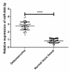
Expression of miR-664-3p in Osteosarcoma and Its Effects on the Proliferation and Apoptosis of Osteosarcoma Cells
- PMID: 31850259
- PMCID: PMC6908901
Expression of miR-664-3p in Osteosarcoma and Its Effects on the Proliferation and Apoptosis of Osteosarcoma Cells
Abstract
Background: To explore the expression level of miR-664-3p in osteosarcoma and its effects on the proliferation and apoptosis of osteosarcoma cells.
Methods: Specimens of osteosarcoma tissues were collected from 41 cases undergoing surgical treatment in the Orthopedics Department of Wuhan Puai Hospital, Wuhan, China from January 2015 to February 2018. Another 40 cases of normal bone tissue were collected. The expression of miR-664-3p were detected using quantificational real-time polymerase chain reaction. miR-664-3p mimics, miR-664-3p inhibitor and miR-664-3p negative control (NC) were used to transfect U2-OS, which were named as mimics group, inhibitor group and NC group, respectively. MTT assay was adopted to detect the effects of microRNA-664-3p on the proliferation of U2-OS after 24, 48, 72, 96 and 120 hours of transfection. Flow cytometry was applied to measure the apoptosis rate of U2-OS after miR-664-3p transfection. Finally, Western Blot was employed to detect the expression of proteolipid protein 2 (PLP2).
Results: The total apoptosis rate of cells in the inhibitor group was obviously higher than those in the mimics group and the NC group (P<0.001). The relative expression level of PLP2 in the inhibitor group was significantly lower than those in the mimics group and the NC group (P<0.001).
Conclusion: MiR-664-3p may be involved in the occurrence and development of osteosarcoma, and can regulate the proliferation and apoptosis of U2-OS cells, and the expression of PLP2. Besides, miR-664-3p may become a novel molecular biological indicator for the diagnosis, targeted treatment and prognosis assessment of osteosarcoma.
Keywords: Apoptosis; Osteosarcoma; Proliferation; U2-OS; miR-664-3p.
Copyright© Iranian Public Health Association & Tehran University of Medical Sciences.
Conflict of interest statement
Conflict of interests The authors declare that there is no conflict of interest.
Figures


References
-
- Mishra CB, Mongre RK, Kumari S, Jeong DK, Tiwari M. (2017). Novel Triazole-Piperazine Hybrid Molecules Induce Apoptosis via Activation of the Mitochondrial Pathway and Exhibit Antitumor Efficacy in Osteosarcoma Xenograft Nude Mice Model. ACS Chem Biol, 12: 753–768. - PubMed
-
- Angelini A, Ceci F. (2017). The role of (18)FFDG PET/CT in the detection of osteosarcoma recurrence. Eur J Nucl Med Mol Imaging, 44: 1712–1720. - PubMed
-
- Guenther LM, Rowe RG, Acharya PT, et al. (2018). Response Evaluation Criteria in Solid Tumors (RECIST) following neoadjuvant chemotherapy in osteosarcoma. Pediatr Blood Cancer, 65(4). - PubMed
LinkOut - more resources
Full Text Sources
