Review
. 2019 Nov 28:7:824.
doi: 10.3389/fchem.2019.00824. eCollection 2019.

Bacterial Biofilm Eradication Agents: A Current Review

Affiliations
Review

Bacterial Biofilm Eradication Agents: A Current Review

Anthony D Verderosa et al. Front Chem. .

Abstract

Most free-living bacteria can attach to surfaces and aggregate to grow into multicellular communities encased in extracellular polymeric substances called biofilms. Biofilms are recalcitrant to antibiotic therapy and a major cause of persistent and recurrent infections by clinically important pathogens worldwide (e.g., Pseudomonas aeruginosa, Escherichia coli, and Staphylococcus aureus). Currently, most biofilm remediation strategies involve the development of biofilm-inhibition agents, aimed at preventing the early stages of biofilm formation, or biofilm-dispersal agents, aimed at disrupting the biofilm cell community. While both strategies offer some clinical promise, neither represents a direct treatment and eradication strategy for established biofilms. Consequently, the discovery and development of biofilm eradication agents as comprehensive, stand-alone biofilm treatment options has become a fundamental area of research. Here we review our current understanding of biofilm antibiotic tolerance mechanisms and provide an overview of biofilm remediation strategies, focusing primarily on the most promising biofilm eradication agents and approaches. Many of these offer exciting prospects for the future of biofilm therapeutics for a large number of infections that are currently refractory to conventional antibiotics.

Keywords: antibiotics; bacteria; biofilm; biofilm antibiotic tolerance; biofilm eradication agent; infection; resistance.


Figures

Figure 1
Figure 1
A model showing the typical stage-wise development of a bacterial biofilm accompanied by transmitted light microscopy images showing these different stages for a P. aeruginosa biofilm. Republished with permission of Annual Reviews, Inc. (Stoodley et al., 2002); permission conveyed through Copyright Clearance Center, Inc.
Figure 2
Figure 2
Chemical structure of two predominant types of small molecule autoinducers involved in quorum sensing.
Figure 3
Figure 3
Quorum sensing illustration. During planktonic cell growth (blue ovals), the relative amount of autoinducers (red triangles) is proportionally low. As cells enter a densely populated mode of growth (green ovals) the relative proportion of autoinducers increases.
Figure 4
Figure 4
Proposed mechanisms contributing to biofilm antimicrobial tolerance (BAT). The biofilm shown is comprised of bacteria (circles and ovals), which are encapsulated by the extracellular polymeric substance (EPS) (dark-brown line surrounding biofilm and multi-colored background within biofilm). Red stars represent antibiotics which are in contact with the biofilm. Restricted penetration of antibiotics through the biofilm EPS is depicted by the black arrows (indicating antibiotics failing to penetrate the EPS of the biofilm) and the red stars at the surface of the biofilm (indicating antibiotics that have failed to diffuse past surface-residing cells). Orange circles and ovals surrounded with yellow/tan background represent surface-residing cells which are in contact with the antibiotics (red stars). Green circles and ovals surrounded by a blue/gray background are indicative of microenvironments within the biofilm (areas of reduced oxygen concentration and reduced cell replication). Purple circles indicate persister cells present within the biofilm (small subpopulation of cells within the biofilm that enter a protected metabolically quiescent state recalcitrant to the action of antimicrobials). Image adapted from Penesyan et al. (2015).
Figure 5
Figure 5
Helical wheel illustration of residues 11–28 of the mature LL-37. Republished with permission of American Society for Microbiology (Turner et al., 1998); permission conveyed through Copyright Clearance Center, Inc.
Figure 6
Figure 6
Chemical structure of Oritavancin.
Figure 7
Figure 7
Chemical structure of tris-QAC-10.
Figure 8
Figure 8
Chemical structures of XF-70 and XF-73.
Figure 9
Figure 9
Chemical structures of glycerol monolaurate, docosahexaenoic acid, and eicosapentaenoic acid.
Figure 10
Figure 10
Chemical structures of mitomycin C and cisplatin.
Figure 11
Figure 11
Chemical structures of bromopheazine-8 and halogenated phenazine-14.
Figure 12
Figure 12
Chemical structures of halogenated quinoline-3 and halogenated quinoline-4.
Figure 13
Figure 13
Chemical structure of cephalosporin-3′-diazeniumdiolate.
Figure 14
Figure 14
Chemical structures of ciprofloxacin-nitroxide-10 and ciprofloxacin-nitroxide-16.
Figure 15
Figure 15
Chemical structures of ciprofloxacin-nitroxide-23, ciprofloxacin-nitroxide-25, and ciprofloxacin-nitroxide-27.

References

    1. Abouelhassan Y., Garrison A. T., Burch G. M., Wong W., Norwood V. M., Huigens R. W., III. (2014). Discovery of quinoline small molecules with potent dispersal activity against methicillin-resistant Staphylococcus aureus and Staphylococcus epidermidis biofilms using a scaffold hopping strategy. Bioorg. Med. Chem. Lett. 24, 5076–5080. 10.1016/j.bmcl.2014.09.009 - DOI - PubMed
    1. Ali H. M., El-Shikh M. S., Salem M. Z., M M. (2016). Isolation of bioactive phenazine-1-carboxamide from the soil bacterium Pantoea agglomerans and study of its anticancer potency on different cancer cell lines. J. AOAC Int. 99, 1233–1239. 10.5740/jaoacint.16-0090 - DOI - PubMed
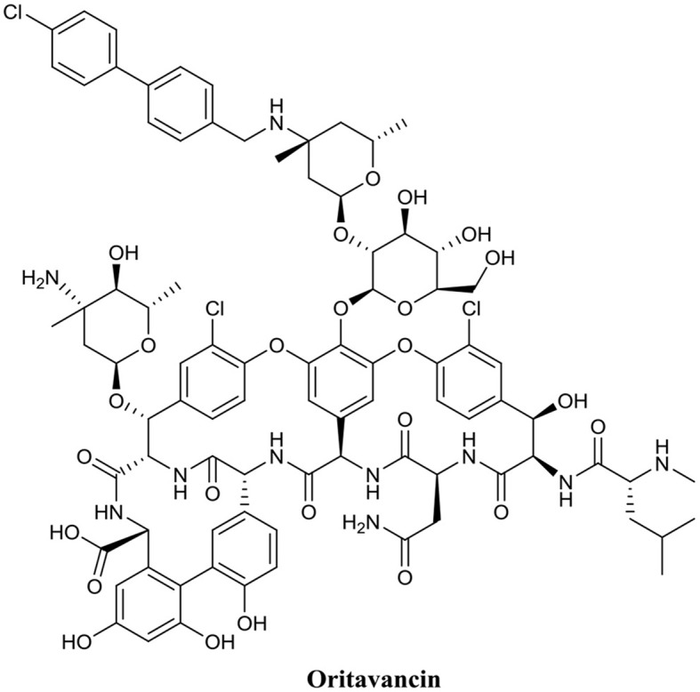
    1. Allen N. E. (2010). From vancomycin to oritavancin: the discovery and development of a novel lipoglycopeptide antibiotic. Antiinfect. Agents Med. Chem. 9, 23–47. 10.2174/187152110790886745 - DOI
    1. Anderl J. N., Franklin M. J., Stewart P. S. (2000). Role of antibiotic penetration limitation in Klebsiella pneumoniae biofilm resistance to ampicillin and ciprofloxacin. Antimicrob. Agents Chemother. 44, 1818–1824. 10.1128/AAC.44.7.1818-1824.2000 - DOI - PMC - PubMed
    1. Anderl J. N., Zahller J., Roe F., Stewart P. S. (2003). Role of nutrient limitation and stationary-phase existence in Klebsiella pneumoniae biofilm resistance to ampicillin and ciprofloxacin. Antimicrob. Agents Chemother. 47, 1251–1256. 10.1128/AAC.47.4.1251-1256.2003 - DOI - PMC - PubMed

LinkOut - more resources