Editorial
. 2020 Jul;52(1):117-128.
doi: 10.1002/jmri.27004. Epub 2019 Dec 18.

Dual-Venc acquisition for 4D flow MRI in aortic stenosis with spiral readouts

Affiliations
Editorial

Dual-Venc acquisition for 4D flow MRI in aortic stenosis with spiral readouts

Sean Callahan et al. J Magn Reson Imaging. 2020 Jul.

Abstract

Background: Single Venc 4D flow MRI with Cartesian readout is hampered by poor velocity resolution and noise when imaging during diastole. Dual Venc acquisitions typically require the acquisition of two distinct datasets, which leads to longer scan times.

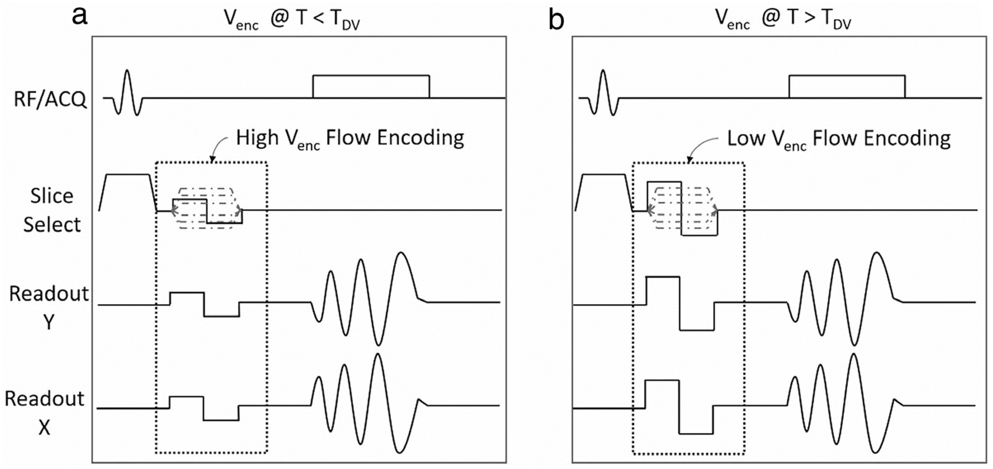
Purpose/hypothesis: To design and develop a 4D Spiral Dual Venc sequence. The sequence allows for separate systolic and diastolic Venc s as part of a single acquisition with a prescribed switch time. The implemented sequence was hypothesized to be comparable to Cartesian 4D flow, but with increased velocity resolution in the diastolic phase and with better scan efficiency and reduced noise.

Study type: Prospective.

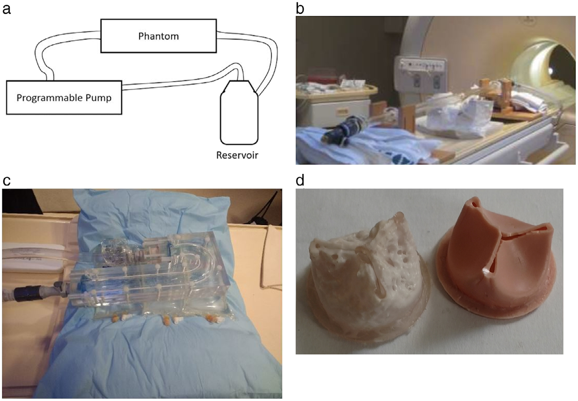
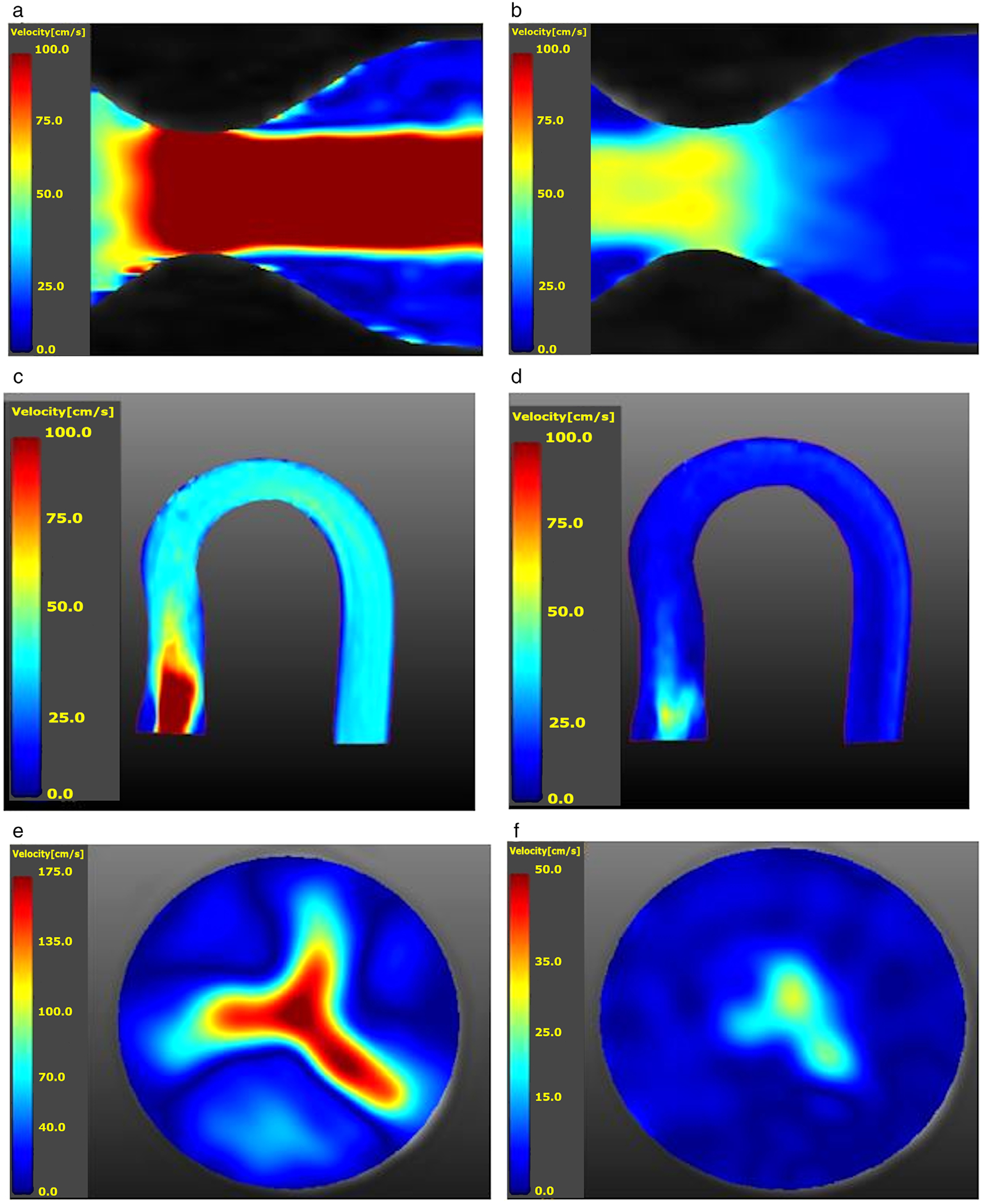
Population: The studied populations were two phantoms-a straight pipe with a stenotic narrowing and a phantom of the aortic arch which included a calcific polymeric valve-under both steady and pulsatile flows, six healthy volunteers, and eight patients with severe aortic stenosis (AS).

Field strength/sequence: 1.5T, Dual Venc 4D flow with spiral readouts.

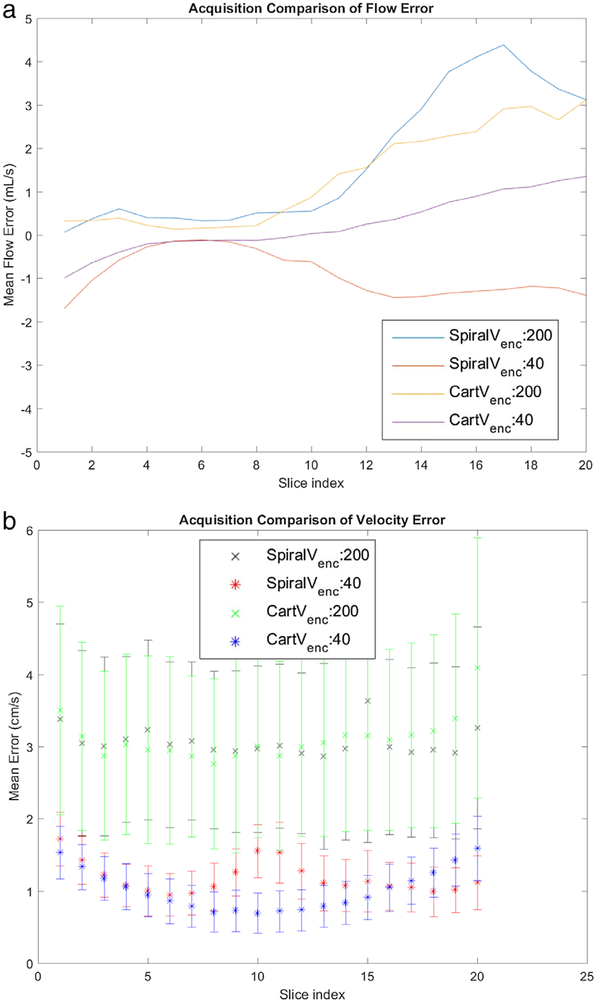
Assessment: Data from the proposed sequence were compared with data from 4D Cartesian Dual Venc and Single Venc acquisitions. Noise was assessed from the acquired velocity data with the pump turned off and by varying Venc . Steady acquisitions were compared to the proximal slice of the lowest Single Venc acquisition.

Statistical tests: Steady flows were compared using relative-root-mean-squared-error (RRMSE). For in vivo flows and pulsatile in vitro flows, net flow for corresponding timepoints were compared with the Pearson correlation test (P < 0.01).

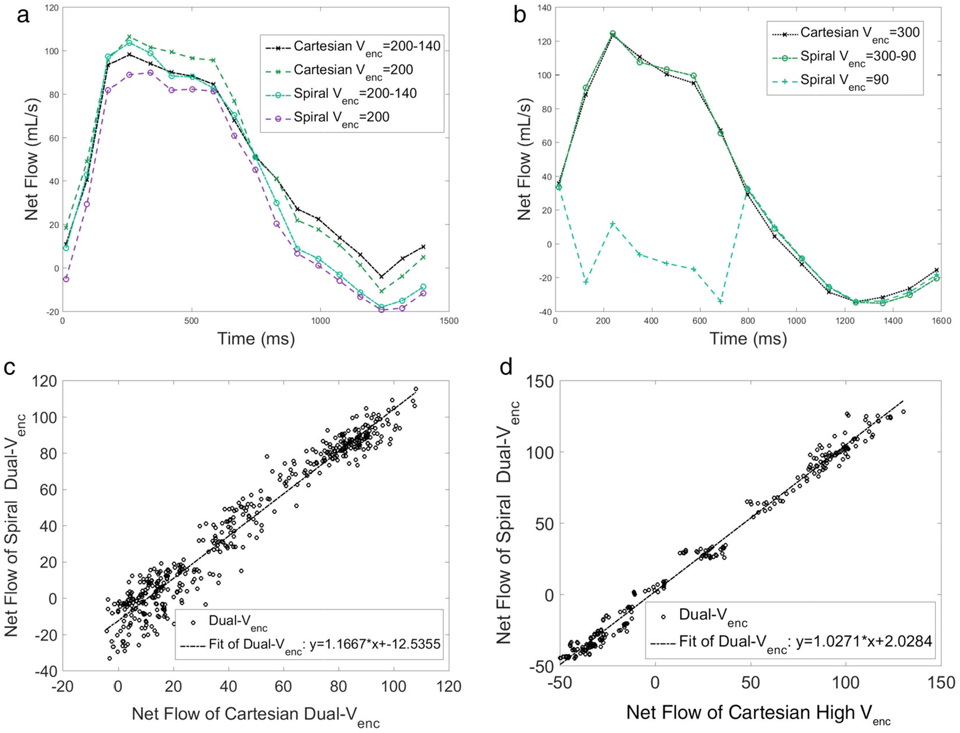
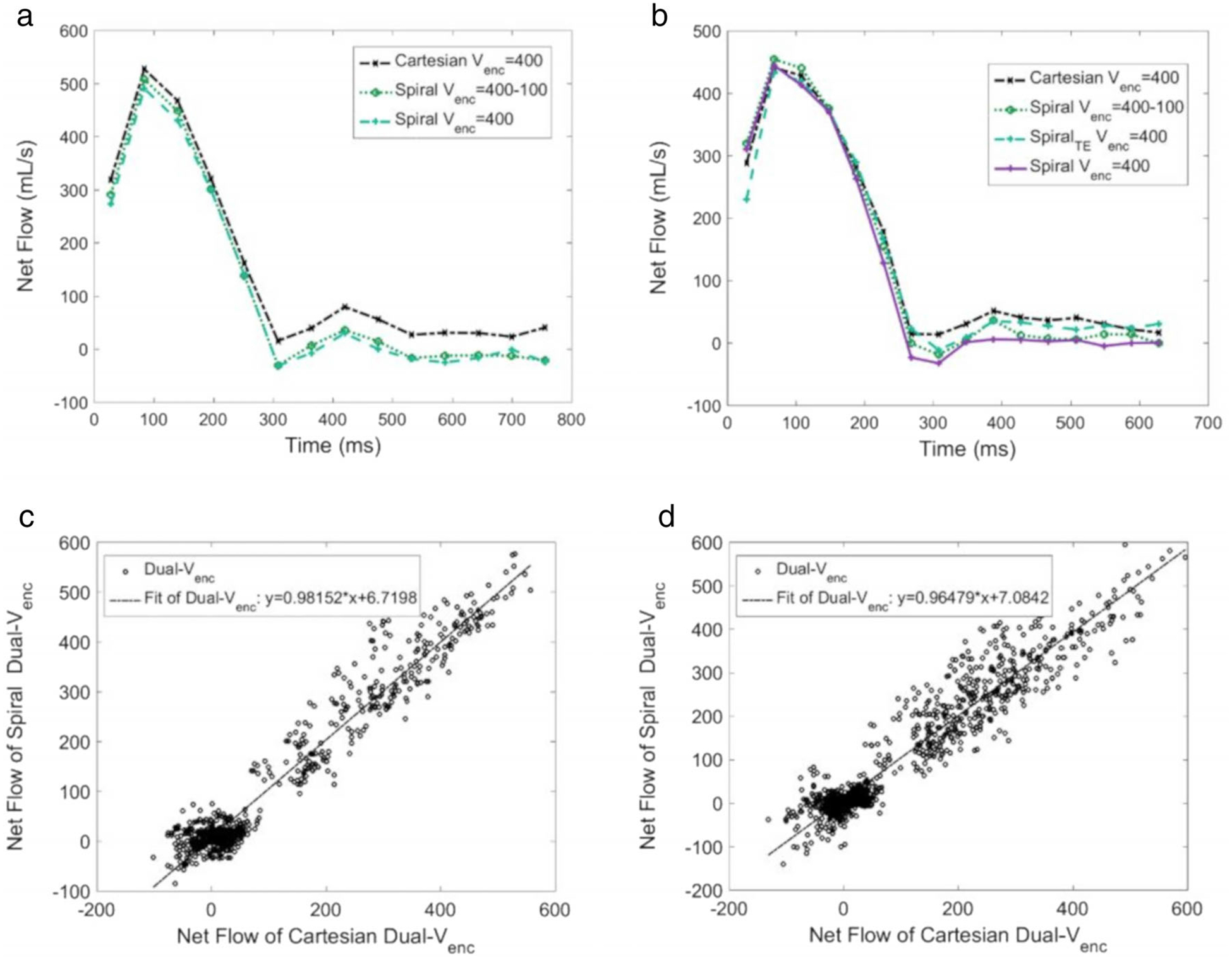
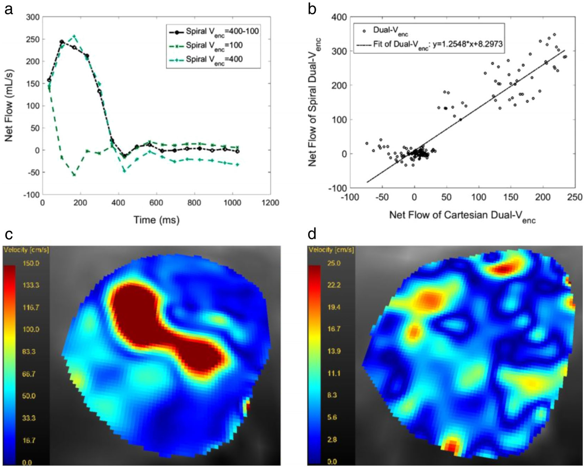
Results: For steady flows, RRMSEs for Single Venc s ranged from 17.6% to 19.4%, and 9.6% to 16.5% for Dual Venc s. The net flow correlation coefficient for the aortic arch phantom was 0.975, and 0.995 for the stenotic phantom. Normal volunteer and patient comparisons yielded a correlation of 0.970 and 0.952, respectively. in vitro and in vivo pulsatile flow waveforms closely matched.

Data conclusion: The Dual Venc offers improved noise properties and velocity resolution, while the spiral trajectory offers a scan efficient acquisition with short echo time yielding reduced flow artifacts.

Level of evidence: 2 Technical Efficacy Stage: 1 J. Magn. Reson. Imaging 2020;52:117-128.

Keywords: 4D flow MRI; Dual Venc; non-Cartesian trajectory; phase contrast MRI; spiral acquisition; stenotic flow.


Figures

FIGURE 1:
FIGURE 1:
The Dual Venc pulse sequence. The bipolar gradients change the maximum based on the set Venc. The Venc of the sequence on the left is higher than the one on the right. The switch to the low Venc happens at TDV, which is a user defined time in the R-R interval.
FIGURE 2:
FIGURE 2:
(a) The flow circuit. The open-air reservoir holds the blood mimicking fluid, which circulates through the system. The pump pulls the fluid from the reservoir, through a flexible plastic tubing. The pump then pushes the fluid in the flow circuit as prescribed by the programmed flow waveform. The fluid then exits the phantom and returns to the reservoir. (b) A picture of the stenotic phantom on the MRI table; the acrylic phantom has a 1” inner diameter, which narrows to 0.35” at the throat for an 87% area occlusion. (c) The aortic arch phantom precision machined with 1” diameter from clear acrylic. The setup permits changing polymeric valves in a separate block close to the inlet. (d) Calcific polymeric valves, at 50% (left) and at 0% (right) calcification. The valves, which are 1” in diameter, are made of polyurethane with various levels of calcium phosphate.
FIGURE 3:
FIGURE 3:
(a) Velocity magnitude map of a sagittal cut of the stenotic phantom distal to the throat or region of interest during systole. Qmax = 150 ml/s (range: 100.0 cm/s to 0.0 cm/s. (b) Velocity magnitude map in diastole for the same study (range: 100.0 cm/s to 0.0 cm/s). (c) Velocity magnitude map of the aortic arch phantom with a 0% calcification synthetic valve at peak systole (Qmax = 150 ml/s). The valve is located on the lower left portion of the picture; the high-speed jet may be seen immediately distal to the valve (range: 100.0 cm/s to 0.0 cm/s). (d) The same sagittal slice as in (c), but during diastole (range: 100.0 cm/s to 0.0 cm/s). (e) peak systolic axial slice of velocity magnitudes corresponding to (c) and immediately distal to the valve. The valve opening clearly visible with its trileaflets (range: 175.0 cm/s to 0.0 cm/s). (f) Corresponding diastolic axial slice of velocity magnitudes (range: 50.0 cm/s to 0.0 cm/s).
FIGURE 4:
FIGURE 4:
No-flow Single Venc results. Four sequences are compared. These are: Spiral High Venc (200 cm/s), Spiral Low Venc (40 cm/s), Cartesian High Venc (200 cm/s), and Cartesian Low Venc (40 cm/s). (a) flow error by slice. (b) Mean and standard deviation of velocity error as a function of Slice 5 is approximately at the isocenter and is at the stenosis throat.
FIGURE 5:
FIGURE 5:
(a) Flow waveform of the aortic arch phantom (Fig. 2c) with a synthetic valve, with 0% occlusion. The slice chosen was 5 mm distal to the valve. (b) Flow waveform in the stenotic phantom (Fig. 2b) calculated from a slice location 4 mm distal to the stenosis throat. Note the Spiral Single Venc waveform is artifactual because of velocity aliasing. In this figure the Single Venc acquisitions are plotted with dotted line, while the Dual Venc results are plotted with a solid line. The Cartesian has a data marker of an x, while Spiral has a circular marker. (c) Comparison of Dual Venc Spiral and Dual Venc Cartesian flow data points for the slice immediately distal to the 50% polymeric valve in the aortic arch phantom. The Pearson correlation coefficient for the scatterplot is 0.975. (d) Comparison of the same Dual Venc Spiral, and High Venc Cartesian in the stenotic phantom. Pearson correlation coefficient for this scatterplot is 0.995. See text for additional description.
FIGURE 6:
FIGURE 6:
(a) Flow waveform in a normal subject distal to the aortic valve from Spiral and Cartesian Single Venc and Spiral Dual Venc acquisitions. (b) Waveforms measured with the same sequences in a different normal subject. (c) Scatterplot of flow data for all healthy volunteers (n = 6, Pearson correlation coefficient of 0.970). (d) Scatterplot of flow data for all patients with severe aortic stenosis (n = 7, Pearson correlation coefficient of 0.952). For both (c,d), flow rates for a specific slice, timepoint, and volunteer are single points.
FIGURE 7:
FIGURE 7:
(a) Flow waveform in a patient with severe AS as measured with a Dual Venc, a high Venc and a low Venc spiral acquisition for the entire R-R interval. The TDV was set to 400 msec. Note that the low Venc acquisition leads to velocity aliasing in systole and an artifactual flow waveform. (b) The scatterplot of the Dual Venc vs. High Venc acquisition in the same patient. All slice positions and timepoints were included. The Pearson correlation coefficient was 0.930, (c) (range: 150 cm/s to 0.0 cm/s) and (d) (range: 25.0 cm/s to 0.0 cm/s) show magnitude of velocities in an axial slice at the aortic valve level in the same patient at peak systole and in mid-diastole.

References

    1. Pellikka PA, Sarano ME, Nishimura RA, et al. Outcome of 622 adults with asymptomatic, hemodynamically significant aortic stenosis during prolonged follow-up. Circulation 2005;111:3290–3295. - PubMed
    1. Nishimura RA, Otto CM, Bonow RO, et al. 2014 AHA/ACC guideline for the management of patients with valvular heart disease: A report of the American College of Cardiology/American Heart Association Task Force on Practice Guidelines. J Am Coll Cardiol 2014;63:e57–e185. - PubMed
    1. Bonow RO, Carabello BA, Chatterjee K, et al. 2008 focused update incorporated into the ACC/AHA 2006 guidelines for the management of patients with valvular heart disease: A report of the American College of Cardiology/American Heart Association Task Force on Practice Guidelines (Writing Committee to revise the 1998 guidelines for the management of patients with valvular heart disease) Endorsed by the Society of Cardiovascular Anesthesiologists, Society for Cardiovascular Angiography and Interventions, and Society of Thoracic Surgeons. J Am Coll Cardiol 2008;52:e1–e142. - PubMed
    1. Otto CM, Lind BK, Kitzman DW, Gersh BJ, Siscovick DS. Association of aortic-valve sclerosis with cardiovascular mortality and morbidity in the elderly. N Engl J Med 1999;341:142–147. - PubMed
    1. Stewart BF, Siscovick D, Lind BK, et al. Clinical factors associated with calcific aortic valve disease fn1. J Am Coll Cardiol 1997;29:630–634. - PubMed

Publication types