. 2020 Feb 1;79(2):144-162.
doi: 10.1093/jnen/nlz122.

Early-Onset Dementia in War Veterans: Brain Polypathology and Clinicopathologic Complexity

Affiliations

Early-Onset Dementia in War Veterans: Brain Polypathology and Clinicopathologic Complexity

Diego Iacono et al. J Neuropathol Exp Neurol. .

Abstract

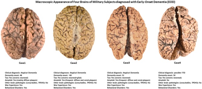
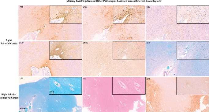
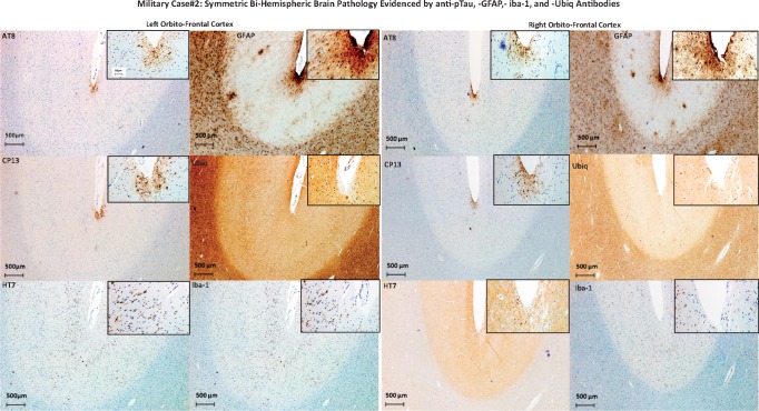
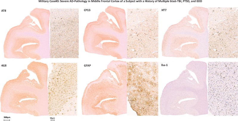
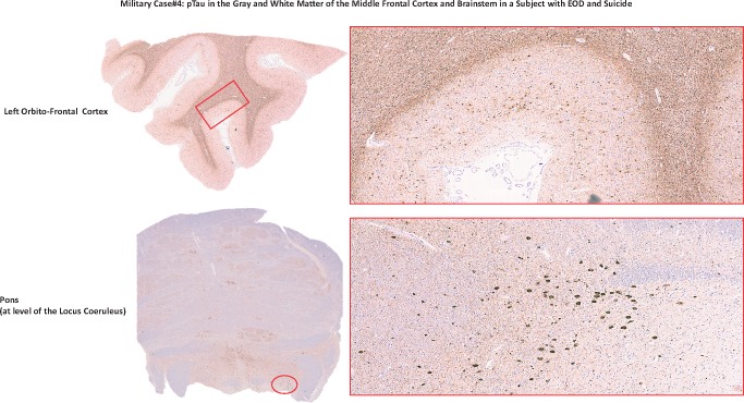
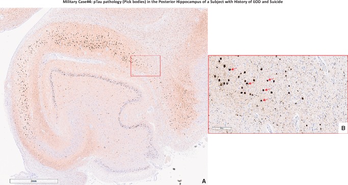
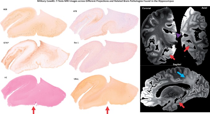
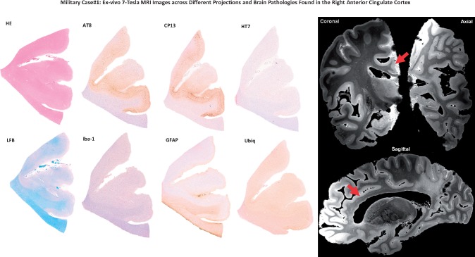
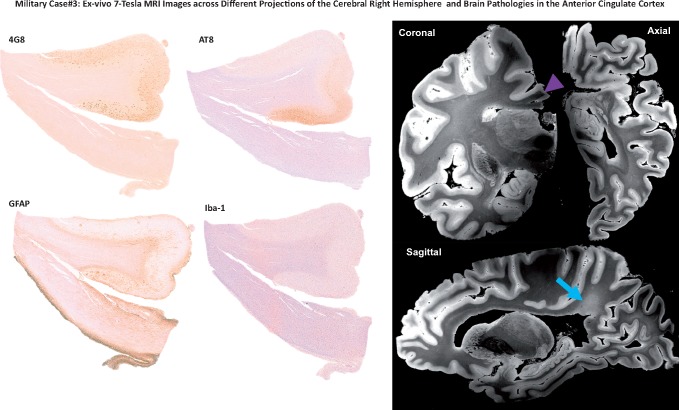
The neuropathology associated with cognitive decline in military personnel exposed to traumatic brain injury (TBI) and chronic stress is incompletely understood. Few studies have examined clinicopathologic correlations between phosphorylated-tau neurofibrillary tangles, β-amyloid neuritic plaques, neuroinflammation, or white matter (WM) lesions, and neuropsychiatric disorders in veterans. We describe clinicopathologic findings in 4 military veterans with early-onset dementia (EOD) who had varying histories of blunt- and blast-TBI, cognitive decline, behavioral abnormalities, post-traumatic stress disorder, suicidal ideation, and suicide. We found that pathologic lesions in these military-EOD cases could not be categorized as classic Alzheimer's disease (AD), chronic traumatic encephalopathy, traumatic axonal injury, or other well-characterized clinicopathologic entities. Rather, we observed a mixture of polypathology with unusual patterns compared with pathologies found in AD or other dementias. Also, ultrahigh resolution ex vivo MRI in 2 of these 4 brains revealed unusual patterns of periventricular WM injury. These findings suggest that military-EOD cases are associated with atypical combinations of brain lesions and distribution rarely seen in nonmilitary populations. Future prospective studies that acquire neuropsychiatric data before and after deployments, as well as genetic and environmental exposure data, are needed to further elucidate clinicopathologic correlations in military-EOD.

Keywords: Brain co-occurring pathologies; Chronic stress; Combat-TBI; Histologic distribution; Short- and long-terms neuropsychiatric manifestations in veterans; War settings.


Figures

FIGURE 1.
FIGURE 1.
Macroscopic appearance of 4 brains of military subjects diagnosed with early onset dementia. All brains looked relatively normal except for Case 4 (see right cerebral hemisphere at level of the frontal pole) and Case 1 in which a moderate level of diffuse cortical atrophy is evident (see intersulcal enlargements).
FIGURE 2.
FIGURE 2.
Military Case 1: pTau and other pathologies assessed across different brain regions. The figure shows images of AT8 and CP13 (pTau), HT7 (all Tau), GFAP, Ubiq, LFB staining of the right parietal cortex (2 upper rows of images), and LFB, H&E and AT8 staining of the right inferior temporal cortex of Case 1. Each image contains a larger image at lower magnification and an inset (upper right corner of each image) of the same cortical area at higher magnification.
FIGURE 3.
FIGURE 3.
Military Case 2: Symmetric bihemispheric brain pathology evidenced by anti-pTau, -GFAP, -Iba-1, and -Ubiq antibodies. The figure shows left and right orbito-frontal cortical areas of Case 2 brain stained for pTau (AT8, CP13), all Tau (HT7), astroglial cells (GFAP), ubiquitin (Ubiq), and microglia (Iba-1). Each image contains a larger image at lower magnification and an inset (upper right corner of each image) of the same cortical area at higher magnification.
FIGURE 4.
FIGURE 4.
Military Case 3: Severe AD pathology in middle frontal cortex of a subject with a history of multiple blast-TBI, PTSD, and EOD. The figure shows positivity for pTau (AT8, CP13), all tau (HT7), β-amyloid lesions, and immunoreactivity for GFAP (astroglial cells) and Iba-1 (microglial cells) in Case 3.
FIGURE 5.
FIGURE 5.
pTau in the gray and white matter of the middle frontal cortex and brainstem in a subject with EOD and suicide. The figure shows left orbito-frontal cortex and pons area of the Case 4 brain stained for pTau (AT8). Photographs were taken at lower (left) and higher (right) magnification. The red rectangle and circle on the lower magnification photographs correspond to the area on the right photographed at higher magnification.
FIGURE 6.
FIGURE 6.
Military Case 4: pTau pathology (Pick bodies) in the posterior hippocampus of a subject with EOD and suicide. (A) The frequency and distribution of rounded pTau lesions (Pick bodies) across the entire posterior hippocampus region (CA subregions and dentate gyrus; left hemisphere) as observed at a lower magnification (×2; scale bar in lower left corner equal to 2 mm distance); (B) Rounded intracytoplasmic pTau-positive lesions (Pick bodies; red arrows) as observed at a higher magnification (×20; scale bar in lower left corner equal to 200 μm distance) localized in the CA1 region of the posterior hippocampus of Case 4 (red square in A).
FIGURE 7.
FIGURE 7.
A 7-Tesla MRI data detect hippocampal and white matter pathologies. The figure shows immunohistochemistry images of the hippocampal area (right hemisphere) of Case 1 stained for amyloid (4G8), pTau (AT8), astrocytes (GFAP), microglia (Iba-1), intraneuronal inclsuions (Ubiq), and possible vascular lesions (H&E). Right panel shows coronal, axial, and sagittal images from the ex vivo 7-Tesla MRI dataset at the level of the hippocampal area analyzed in the neuropathologic analyses. The ex vivo MRI data reveal hippocampal atrophy (red arrows), as well as thinning of the corpus callosum (purple arrow head) and rarefaction of the parietal periventricular white matter (blue arrow).
FIGURE 8.
FIGURE 8.
A 7-Tesla MRI investigation of pathology in the right anterior cingulate cortex. Immunohistochemistry images of the anterior cingulate cortex (right hemisphere) of Case 1 stained with H&E, AT8 and CP13 (pTau), HT7 (all Tau), LFB (myelin), Iba-1 (microglia), astrocytes (GFAP), and possible intracytoplasmic inclusions (Ubiq). Right panel shows ex vivo MRI results from the 7 Tesla scan in coronal, axial, and sagittal projections of the right anterior cingulate cortex (red arrows) that included the anterior cingulate cortical area used for the neuropathologic analyses. In this region, ex vivo MRI does not reveal the burden of pathology seen with immunohistochemistry.
FIGURE 9.
FIGURE 9.
A 7-Tesla MRI investigation of pathology in the right cerebral hemisphere and the anterior cingulate cortex. Left panel shows immunohistochemistry images of the anterior cingulate cortex (right hemisphere) of Case 3 stained with 4G8 (β-amyloid), AT8 (pTau), GFAP (astroglial cells), and Iba-1 (microglial cells). Right panel shows ex vivo MRI results obtained from the 7 Tesla scan in coronal, axial, and sagittal projections of the right anterior cingulate cortex. The region of the anterior cingulate cortex analyzed in the immunohistochemistry images is indicated by the purple arrow. The ex vivo MRI data demonstrate rarefaction of the parietal periventricular white matter (blue arrow).

References

    1. Ursano RJ, Kessler RC, Stein MB, et al. Suicide attempts in the US Army during the wars in Afghanistan and Iraq, 2004 to 2009. JAMA Psychiatry 2015;72:917–26 - PMC - PubMed
    1. Ursano RJ, Kessler RC, Naifeh JA, et al. Frequency of improvised explosive devices and suicide attempts in the U.S. Army. Mil Med 2017;182:e1697–703 - PMC - PubMed
    1. Ursano RJ, Kessler RC, Naifeh JA, et al. Risk factors associated with attempted suicide among US Army soldiers without a history of mental health diagnosis. JAMA Psychiatry 2018;75:1022–32 - PMC - PubMed
    1. Kenney K, Iacono D, Edlow BL, et al. Dementia after moderate-severe traumatic brain injury: Coexistence of multiple proteinopathies. J Neuropathol Exp Neurol 2018;77:50–63 - PMC - PubMed
    1. McKhann GM, Knopman DS, Chertkow H, et al. The diagnosis of dementia due to Alzheimer's disease: Recommendations from the National Institute on Aging-Alzheimer's Association workgroups on diagnostic guidelines for Alzheimer's disease. Alzheimers Dement 2011;7:263–9 - PMC - PubMed