. 2020 Feb 1;123(2):529-547.
doi: 10.1152/jn.00939.2017. Epub 2019 Dec 18.

Role of muscle coactivation in adaptation of standing posture during arm reaching

Affiliations

Role of muscle coactivation in adaptation of standing posture during arm reaching

Alison Pienciak-Siewert et al. J Neurophysiol. .

Abstract

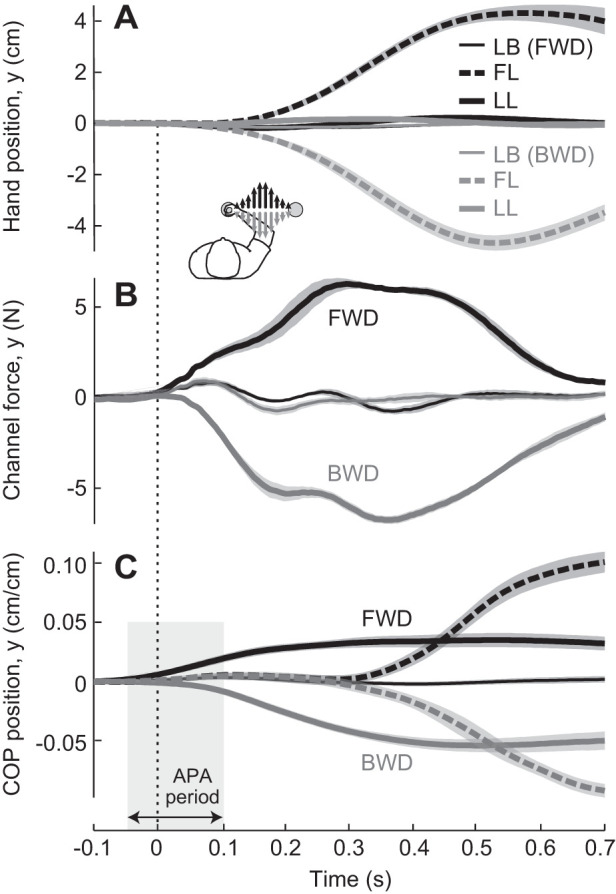
The ability to maintain stable, upright standing in the face of perturbations is a critical component of daily life. A common strategy for resisting perturbations and maintaining stability is muscle coactivation. Although arm muscle coactivation is often used during adaptation of seated reaching movements, little is known about postural muscle activation during concurrent adaptation of arm and standing posture to novel perturbations. In this study we investigate whether coactivation strategies are employed during adaptation of standing postural control, and how these strategies are prioritized for adaptation of standing posture and arm reaching, in two different postural stability conditions. Healthy adults practiced planar reaching movements while grasping the handle of a robotic arm and standing on a force plate; the robotic arm generated a velocity-dependent force field that created novel perturbations in the forward (more stable) or backward (less stable) direction. Surprisingly, the degree of arm and postural adaptation was not influenced by stability, with similar adaptation observed between conditions in the control of both arm movement and standing posture. We found that an early coactivation strategy can be used in postural adaptation, similar to what is observed in adaptation of arm reaching movements. However, the emergence of a coactivation strategy was dependent on perturbation direction. Despite similar adaptation in both directions, postural coactivation was largely specific to forward perturbations. Backward perturbations led to less coactivation and less modulation of postural muscle activity. These findings provide insight into how postural stability can affect prioritization of postural control objectives and movement adaptation strategies.NEW & NOTEWORTHY Muscle coactivation is a key strategy for modulating movement stability; this is centrally important in the control of standing posture. Our study investigates the little-known role of coactivation in adaptation of whole body standing postural control. We demonstrate that an early coactivation strategy can be used in postural adaptation, but muscle activation strategies may differ depending on postural stability conditions.

Keywords: anticipatory postural adjustment; coactivation; motor adaptation; neuromechanics; postural base of support.


Conflict of interest statement

No conflicts of interest, financial or otherwise, are declared by the authors.

Figures

Fig. 1.
Fig. 1.
Experimental setup and protocol. A: Experimental apparatus and setup; visual feedback is provided on computer screen. B: subjects experienced either a forward (FWD) or backward (BWD) perturbation during the learning block. C: experimental protocol; a rightward reach (+x) and perpendicular forces (±y) are illustrated. COP, center of pressure.
Fig. 2.
Fig. 2.
Group mean movement trajectories. Late baseline (LB), first learning (FL), and late learning (LL) phases are shown for subjects experiencing either a forward (FWD group; black) or backward perturbation (BWD group; gray). A: perpendicular hand position (force trials only). Inset shows the direction of perturbing forces for each group. B: channel force (channel trials only). C: perpendicular center-of-pressure (COP) position (force trials only). Note: trajectories were averaged across trials in each phase for each subject and then averaged across subjects in each group. Gray box indicates the anticipatory postural adjustment (APA) period. Line shading indicates SE across subjects. Time 0 represents movement onset of the arm.
Fig. 3.
Fig. 3.
Hand error and anticipatory force metrics. A–C and D–F show hand error and anticipatory force, respectively. A and D show each metric vs. batch (5 trials). In each plot, 2 traces show group means (solid lines) ± SE (shading) for the forward (FWD; black) and backward perturbation (BWD; gray) groups. Bar plots in B and E show the absolute magnitude of the change from late baseline (LB) to first learning (FL), early learning (EL), and late learning (LL) phases, for FWD group (black) vs. BWD group (gray), with error bars showing SE. Bar plots in C and F show the absolute magnitude of the change from LB for each subject. +P < 0.050, statistically significant change within group for hand error, from LB to FL or from FL to LL, and for anticipatory force, from LB to LL. G and H show exponential fits of hand error and anticipatory force data from the learning block, respectively. In each plot, 2 traces show bootstrapped mean data for FWD (black) and BWD (gray) across 1,000 bootstrap-resampled groups (solid lines are means, shading indicates ±SE).
Fig. 4.
Fig. 4.
Arm muscle activity. A: average time traces of muscle activity (mean normalized electromyographic activity) for the pectoralis major (Pec), posterior deltoid (PDelt), biceps brachii (Biceps), and long head of the triceps (Triceps), in the late baseline (LB) and late learning (LL) phases, for the forward (FWD; black) and backward perturbation (BWD; gray) groups. Line shading indicates SE across subjects. Time 0 represents movement onset of the arm. B: group mean values (root-mean-square value of normalized EMG activity from movement onset to movement end) at LB and LL for FWD (black) vs. BWD group (gray), with error bars showing SE across subjects. +P < 0.050, statistically significant change within a group from LB to LL.
Fig. 5.
Fig. 5.
Coactivation in arm muscle pairs. A: coactivation vs. batch (5 trials) for the indicated muscle pairs. In each plot, 2 traces show group means (solid lines) ± SE (shading) for the forward (FWD; black) and backward perturbation (BWD; gray) groups. B: group mean values at late baseline (LB), early learning (EL), and late learning (LL) for FWD (black) vs. BWD group (gray), with error bars showing SE. +P < 0.050, significant change within a group, from LB to EL or from EL to LL. Biceps, biceps brachii; coact., coactivation; PDelt; posterior deltoid; Pec; pectoralis major; Triceps, long head of the triceps.
Fig. 6.
Fig. 6.
Muscle coactivation time traces. Average time traces of coactivated muscle activity (magnitude of normalized electromyographic activity that is matched by 2 opposing muscles at each time point throughout the movement) for all arm and postural muscle pairs, in the late baseline (LB) and early learning (EL) phases, for the forward (FWD; black) and backward perturbation (BWD; gray) groups. Thick lines represent group mean values; thin lines indicate SE across subjects. Time 0 represents movement onset of the arm. BF, biceps femoris; Biceps, biceps brachii; LGas, lateral gastrocnemius; MGas, medial gastrocnemius; PDelt; posterior deltoid; Pec; pectoralis major; PL, peroneus longus; RF, rectus femoris; Sol, soleus; TA, tibialis anterior; Triceps, long head of the triceps.
Fig. 7.
Fig. 7.
Reactive postural adjustment (RPA) and anticipatory postural adjustment (APA) metrics. A–C and D–F show RPA and APA, respectively. A and D show each metric vs. batch (5 trials). In each plot, 2 traces show group means (solid lines) ± SE (shading) for the forward (FWD; black) and backward perturbation (BWD; gray) groups. Bar plots in B and E show the absolute magnitude of the change from late baseline (LB) to first learning (FL), early learning (EL), and late learning (LL), for FWD (black) vs. BWD group (gray), with error bars showing SE. Bar plots in C and F show the absolute magnitude of the change from LB for each subject. +P < 0.050, statistically significant change within group for RPA, from LB to FL or from FL to LL, and for APA, from LB to LL. G and H show exponential fits of RPA and APA data from the learning block. In each plot, 2 traces show bootstrapped mean data for FWD (black) and BWD (gray) across 1,000 bootstrap-resampled groups (solid lines are means, shading indicates ±SE).
Fig. 8.
Fig. 8.
Coactivation in postural muscle pairs. A: coactivation vs. batch (5 trials) for the indicated muscle pairs. In each plot, 2 traces show group means (solid lines) ± SE (shading) for the forward (FWD; black) and backward perturbation (BWD; gray) groups. B: group mean values at late baseline (LB), early learning (EL), and late learning (LL), for FWD (black) vs. BWD group (gray), with error bars showing SE. +P < 0.050, significant change within a group, from LB to EL or from EL to LL. BF, biceps femoris; coact., coactivation; LGas, lateral gastrocnemius; MGas, medial gastrocnemius; PL, peroneus longus; RF, rectus femoris; Sol, soleus; TA, tibialis anterior.
Fig. 9.
Fig. 9.
Postural muscle activity. A: average time traces of muscle activity (mean normalized electromyographic activity, EMG) for the rectus femoris (RF), biceps femoris (BF), tibialis anterior (TA), peroneus longus (PL), lateral gastrocnemius (LGas), medial gastrocnemius (MGas), and soleus (Sol), in the late baseline (LB) and late learning (LL) phases, for the forward (FWD; black) and backward perturbation (BWD; gray) groups. Line shading indicates SE across subjects. Time 0 represents movement onset of the arm. Shading and arrows indicate reactive (RPA) and anticipatory postural adjustment (APA) periods. B and C: group mean values for anticipatory and reactive muscle activity (root-mean-square value of normalized EMG activity over the APA and RPA periods, respectively) at LB and LL, for FWD (black) vs. BWD group (gray), with error bars showing SE across subjects. +P < 0.050, statistically significant change within a group from LB to LL.
Fig. 10.
Fig. 10.
Medial gastrocnemius (MGas) and soleus (Sol) anticipatory activity vs. anticipatory postural adjustment (APA). Each data point represents the change from late baseline to late learning for 1 subject experiencing either a forward (FWD; black) or backward (BWD; gray) perturbation. Dashed lines indicate y-axis intercept. Plots show significant positive correlations between these metrics across all subjects.

References

    1. Adkin AL, Frank JS, Carpenter MG, Peysar GW. Fear of falling modifies anticipatory postural control. Exp Brain Res 143: 160–170, 2002. doi:10.1007/s00221-001-0974-8. - DOI - PubMed
    1. Adkin AL, Frank JS, Jog MS. Fear of falling and postural control in Parkinson’s disease. Mov Disord 18: 496–502, 2003. doi:10.1002/mds.10396. - DOI - PubMed
    1. Ahmed AA, Wolpert DM. Transfer of dynamic learning across postures. J Neurophysiol 102: 2816–2824, 2009. doi:10.1152/jn.00532.2009. - DOI - PMC - PubMed
    1. Alexander NB, Gross MM, Medell JL, Hofmeyer MR. Effects of functional ability and training on chair-rise biomechanics in older adults. J Gerontol A Biol Sci Med Sci 56: M538–M547, 2001. doi:10.1093/gerona/56.9.M538. - DOI - PubMed
    1. Aruin AS, Latash ML. The role of motor action in anticipatory postural adjustments studied with self-induced and externally triggered perturbations. Exp Brain Res 106: 291–300, 1995. doi:10.1007/BF00241125. - DOI - PubMed

Publication types

LinkOut - more resources