. 2019 Dec 17;29(12):3983-3996.e4.
doi: 10.1016/j.celrep.2019.11.050.

Cellular IP6 Levels Limit HIV Production while Viruses that Cannot Efficiently Package IP6 Are Attenuated for Infection and Replication

Affiliations

Cellular IP6 Levels Limit HIV Production while Viruses that Cannot Efficiently Package IP6 Are Attenuated for Infection and Replication

Donna L Mallery et al. Cell Rep. .

Abstract

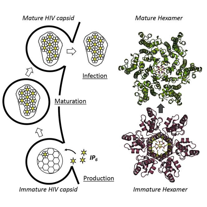
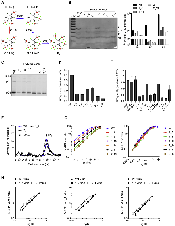
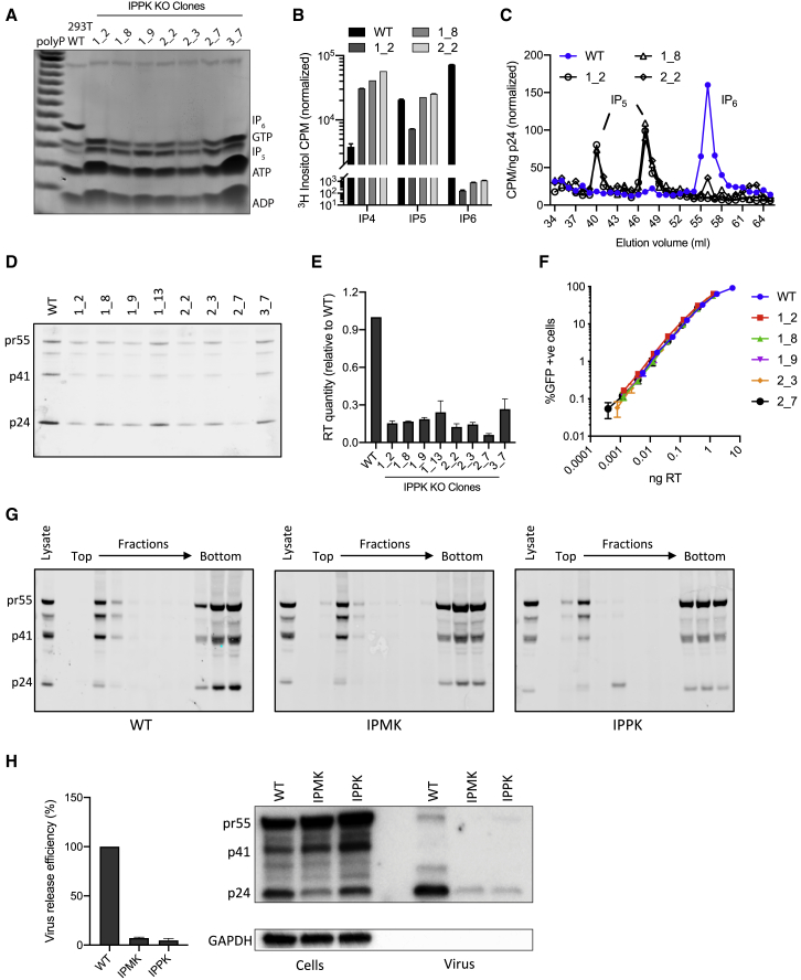
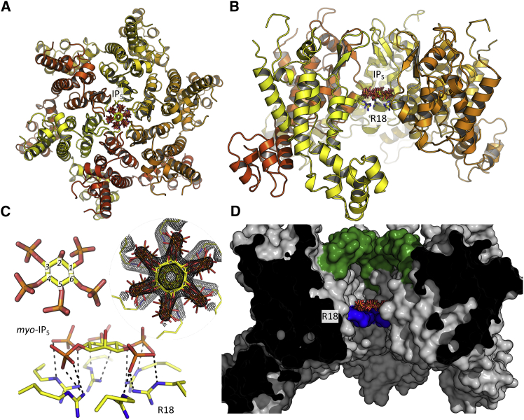
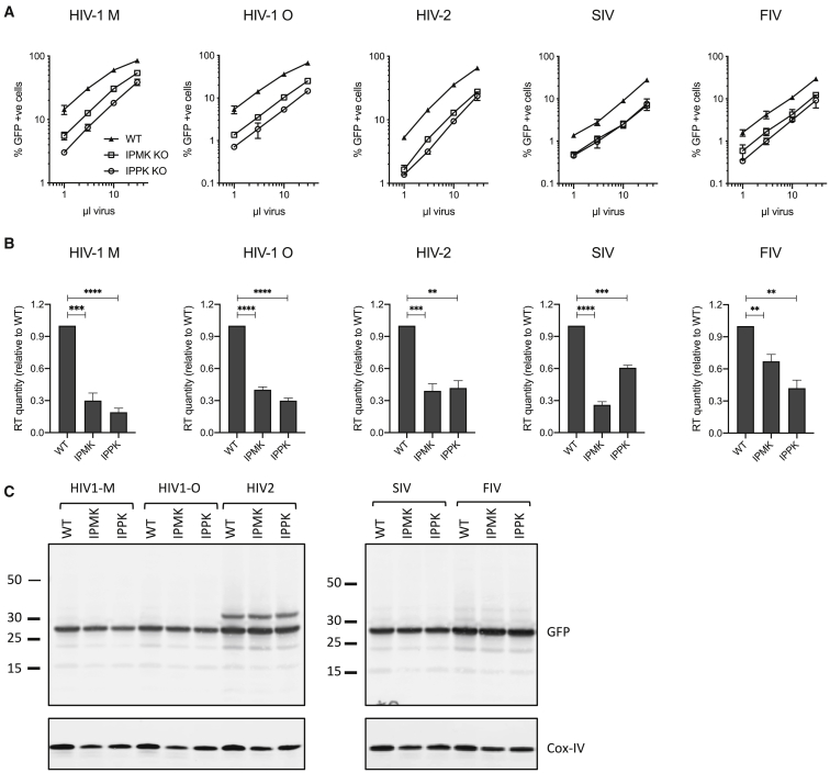
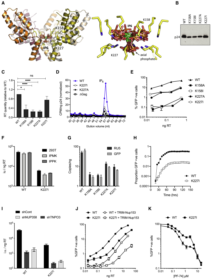
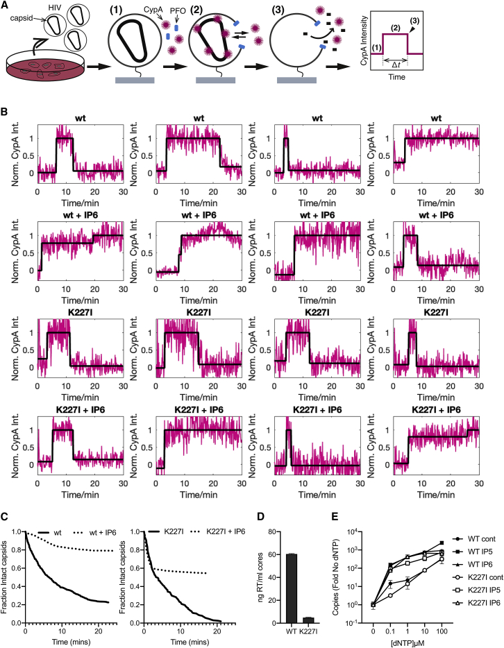
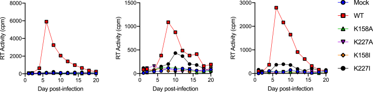
HIV-1 hijacks host proteins to promote infection. Here we show that HIV is also dependent upon the host metabolite inositol hexakisphosphate (IP6) for viral production and primary cell replication. HIV-1 recruits IP6 into virions using two lysine rings in its immature hexamers. Mutation of either ring inhibits IP6 packaging and reduces viral production. Loss of IP6 also results in virions with highly unstable capsids, leading to a profound loss of reverse transcription and cell infection. Replacement of one ring with a hydrophobic isoleucine core restores viral production, but IP6 incorporation and infection remain impaired, consistent with an independent role for IP6 in stable capsid assembly. Genetic knockout of biosynthetic kinases IPMK and IPPK reveals that cellular IP6 availability limits the production of diverse lentiviruses, but in the absence of IP6, HIV-1 packages IP5 without loss of infectivity. Together, these data suggest that IP6 is a critical cofactor for HIV-1 replication.

Keywords: AIDS; HIV; IP6; IPMK; IPPK; capsid; inositol hexakisphosphate; virus.


Conflict of interest statement

The authors declare no competing interests.

Figures

None
Graphical abstract
Figure 1
Figure 1
Depletion of IP6 in IPMK-Knockout Cells Reduces HIV Production but Does Not Affect IP6 Incorporation or Infectivity (A) Biosynthetic pathway of inositol phosphates, illustrating the central role of IPMK and IPPK in IP6 production. (B) Analysis of IP6 levels in IPMK CRISPR/Cas9 knockout clones by TiO2-PAGE and toluidine blue staining of cell extracts (left), [3H]inositol labeling with SAX-HPLC, and scintillation counting of fractions, normalized to background (right). Synthetic polyP was used as ladder for gel orientation. (C) p24 western blot of pelleted virions showing p24 levels in HIV virions produced from IPMK-KO clones. (D) Measurement of virus production through quantification of RT in viral supernatants from IPMK-KO clones. Error bars depict mean ± SD of three independent experiments. Values are represented as fold WT virus for comparison. Reduction compared with WT is statistically significant (p < 0.0003 in all cases). (E) Levels of virus production from viral supernatants collected from parental clones and the same cell lines stably transduced with either EV or IPMK. Error bars depict mean ± SD of two independent experiments. Values normalized to the levels in 293T untransduced cells. (F) Quantification of IP6 packaging in virions produced in wild-type cells and IPMK-KO clones by [3H]inositol labeling, SAX-HPLC, and scintillation counting of fractions then normalized to background. (G) Infectivity of viruses produced in selection of IPMK clones plotted against viral volume input (left) and plotted against quantity of RT in viral supernatants to normalize for differences in production (right). (H) Infectivity of viruses purified from wild-type, 1_7, or 2_1 IPMK-KO producer cells and titrated onto the three different cell lines, normalized to RT levels. Error bars for infection data depict mean ± SD of three replicates from one experiment representative of three independent experiments.
Figure 2
Figure 2
HIV Incorporates IP5 in the Absence of IP6 without Loss of Production or Infectivity (A) TiO2-PAGE and toluidine blue staining of cell extracts showing IP5 and IP6 levels in IPPK CRISPR/Cas9 knockout clones. (B) Inositol phosphate quantification in selected IPPK-KO clones using 3H-inositol labeling and inositol phosphate fractionation by SAX-HPLC. (C) Quantification of IP5 and IP6 packaging in virions produced in wild-type and IPPK-KO cells through [3H]inositol labeling, SAX-HPLC, and scintillation counting of fractions. (D) p24 western blot of pelleted virions showing p24 levels in HIV virions produced from IPPK-KO clones. (E) Measurement of virus production through quantification of RT in viral supernatants from IPMK-KO clones. Error bars depict mean ± SD of three independent experiments. Values are represented as fold WT virus, and reduction compared with WT is statistically significant (p < 0.0012 in all cases). (F) Infectivity of viruses from (E), as a function of viral dose measured by RT levels. Error bars depict mean ± SD of three replicates from one experiment representative of three independent experiments. (G) Membrane flotation analysis of cell lysates from WT, IPMK-KO, and IPPK-KO cells. Western blotting of sucrose gradient fractions for Gag show that similar levels of Gag are associated with the membrane fractions. Gag precursor Pr55Gag (pr55), p41, and mature capsid protein (p24) are indicated. (H) Virus release assays showing levels of Gag in lysates and virions after transduction of WT and KO cells with virus produced in WT cells. Graph shows relative quantification of p24 from two independent experiments and western blots. Representative data from at least two independent experiments are shown, unless otherwise indicated. CPM data are normalized to background.
Figure 3
Figure 3
Structure of the HIV-1 Capsid Hexamer:IP5 Complex (A and B) Secondary structure representation of the complex shown from above (outside-facing, A) and side (within the capsid lattice, B). The six equivalent binding positions for IP5 are shown, together with the location of the R18 ring. (C) The structure of IP5 alone and interacting with the six arginine residues at position 18. Electron density (2Fo − Fc) centered around the ligand is shown as a mesh contoured at 1.4σ. Putative hydrogen bond interactions between IP5 and R18 side chains are shown as black dashes. (D) Molecular surface of the capsid hexamer sliced through the middle to reveal the internal cavity where IP5 is bound. All surface residues are colored gray except R18 (blue), and the β-hairpin (residues 1–12) that forms one end of the cavity (green). IP5, positioned above the R18 ring, is also shown.
Figure 4
Figure 4
The Requirement for IP6 during Viral Production Is Conserved in Diverse Lentiviruses (A) Titers of viral supernatants for lentiviruses produced in WT, IPMK-KO, and IPPK-KO 293T cells. Titers are determined by percentage GFP infection on WT 293T cells. (B) Quantification of viral production as determined by RT levels in viral supernatants. Values shown are mean ± SD from three independent experiments. Statistically fewer virions are produced in kinase-knockout cells (p < 0.002 in all cases). (C) Western blots for GFP expression during viral production show similar protein levels for each cell line. Bottom panel shows loading control Cox-IV.
Figure 5
Figure 5
Mutation of K158 and K227 Rings in Immature Gag Hexamers Affects Viral Production and Infectivity (A) View of five subunits of the immature hexamer (on the basis of PDB: 6BHR) showing the lysine side-chains responsible for coordinating IP6 (blue sphere denotes the ε-amino group). Symmetrically equivalent molecules of IP6 are shown with the carbon rings in green. (B) Western blot of pelleted virions to show p24 levels in HIV wild-type and mutant virions. (C) Quantification of mutant virus production 293T cells as determined by RT levels in viral supernatants. Error bars depict mean ± SD of three independent experiments. Values are expressed as fold change from levels of RT produced in WT virus. The reduction in virus production between WT and K158A, K158I, and K227A is statistically significant (p = 0.0121, p < 0.0001, and p < 0.0001, respectively). (D) Quantification of IP6 packaging in mutants K227A and K227I after normalization for background and input virus (per nanogram p24). Both K227 mutants package similarly reduced levels of IP6 with respect to wild-type virus. Representative data from two independent experiments are shown. (E) Infectivity of lysine mutant viruses normalized to nanogram RT input. Each mutant pair gives similarly reduced levels of infection, with K158A/I largely impaired. Error bars depict mean ± SD of three replicates from one experiment representative of three independent experiments. (F) Infectious titer of WT and K227I mutant viruses produced in WT 293T, IPMK-KO, and IPPK-KO cells. Infectivity is expressed as infection units (IU) per quantity of input virus (nanogram RT). (G) Levels of reverse transcription products strong-stop (RU5) and post-strand-transfer (GFP) 4 h post-infection. Error bars depict mean ± SD of three replicates from one experiment representative of three independent experiments. (H) Infectivity of WT and K227I virus matched for RT input over 150 h to determine whether K227I could recover infectivity over time. Infectivity is measured using an IncuCyte and determined as proportion of cell area that is GFP positive. (I) HeLa cells stably expressing shRNA control or shNUP358 or shTNPO3 were infected with wild-type or K227I virus. Infection is quantified as infection units (IU) per quantity of input virus (nanogram RT). (J) WT or TRIM-Nup153 expressing HeLa cells were infected with wild-type or K227I virus. The percentage infected cells was determined by percentage GFP positive cells for a range of input virus (quantified by nanogram RT). (K) HeLa cells were infected with wild-type or K227I virus in the presence of anti-capsid inhibitor PF-74. Data are normalized to infection in the absence of inhibitor. Error bars depict mean ± SD of three replicates from one experiment representative of at least two independent experiments.
Figure 6
Figure 6
Addition of IP6 Is Able to Stabilize Both WT and K227I Mutant Virions (A) Outline of single-molecule analysis for virus stability. Briefly, viral particles were immobilized, and fluorescence traces were obtained at the locations of individual virions while permeabilizing their membranes with PFO in the presence of fluorescently labeled CypA (1). CypA binds to the capsid, resulting in the appearance of a stable fluorescence signal (2). As the capsid uncoats, the CypA signal disappears (3), and the fluorescence trace returns to background levels. (B) Example traces for individual virions demonstrating the tracking of fluorescence over time. Addition of IP6 to the virions results in a delay in the fluorescence signal reduction signaling uncoating. (C) Capsid survival curves collated from single-virion traces showing that K227I particles have a high fraction of short-lived capsids in the presence and absence of IP6, while the remainder of capsids is strongly stabilized by IP6 addition for both wild-type and K227I particles. (D) Capsid core yields determined by RT quantification (representative of at least two independent experiments). (E) ERT assay using cores isolated from WT and K227I virions, showing a dNTP titration in the absence of IPs or in the presence of either IP5 or IP6. Both WT and K227I cores are competent for ERT and are stabilized by both IP5 and IP6 to the same extent. Data are average of three biological replicas and have been normalized to the copies of RU5 measured in the absence of dNTPs.
Figure 7
Figure 7
Capsid Mutants Prevent Efficient Replication in PBMCs PBMCs from three donors were infected with viruses carrying capsid mutations K158A, K158I, K227A, or K227I for 2 h. Cells were cultured over 20 days and supernatants sampled every other day and assayed for RT activity.

References

    1. Azevedo C., Saiardi A. Extraction and analysis of soluble inositol polyphosphates from yeast. Nat. Protoc. 2006;1:2416–2422. - PubMed
    1. Böcking T., Aguet F., Harrison S.C., Kirchhausen T. Single-molecule analysis of a molecular disassemblase reveals the mechanism of Hsc70-driven clathrin uncoating. Nat. Struct. Mol. Biol. 2011;18:295–301. - PMC - PubMed
    1. Brissault B., Kichler A., Guis C., Leborgne C., Danos O., Cheradame H. Synthesis of linear polyethylenimine derivatives for DNA transfection. Bioconjug. Chem. 2003;14:581–587. - PubMed
    1. Bush D.L., Vogt V.M. In vitro assembly of retroviruses. Annu. Rev. Virol. 2014;1:561–580. - PubMed
    1. Dehairs J., Talebi A., Cherifi Y., Swinnen J.V. CRISP-ID: decoding CRISPR mediated indels by Sanger sequencing. Sci. Rep. 2016;6:28973. - PMC - PubMed

Publication types