

Restoration of the inferomedial orbital strut using a standardized three-dimensional printing implant
- PMID: 31852015
- PMCID: PMC7163695
- DOI: 10.1111/joa.13136
Restoration of the inferomedial orbital strut using a standardized three-dimensional printing implant
Abstract
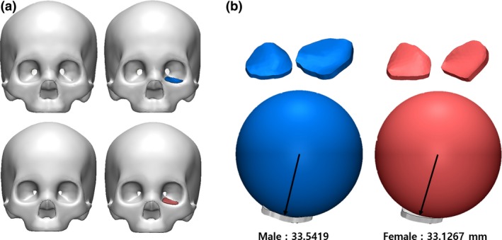
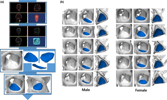
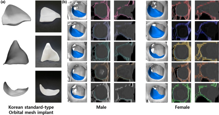
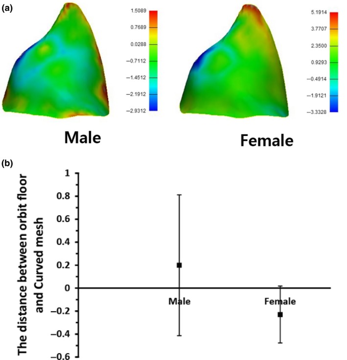
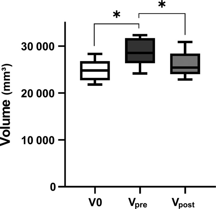
The inferomedial orbital strut (IOS) is the thin bony junction of the orbital medial wall and floor. Its fracture is common and leads to serious complications, including enophthalmos, globe dystopia and diplopia. However, anatomical restoration of the IOS is challenging owing to reduced structural support; sound anatomical background and accurate implants are therefore essential. The aim of the present study was to incorporate data from cadaveric orbit anatomy into three-dimensional (3D) printing technology and to reconstruct the complex orbital fracture elaborately. After averaging the data from computed tomography (CT) images of 100 adult cadavers, the dimensions of the IOS were extracted, and a tangent sphere was created using a computer-aided design program. The curves were compared with the CT data of 10 adult patients from the simulation test. Based on these data, a standardized 3D implant, 1.15 mm thick, was designed using polycaprolactone. The implant was placed in five patients with complex orbital fractures. The radius of the sphere in contact with the orbit, measuring 33.54 mm, was confirmed to be appropriate. A comparison between the normal side volume (V0) and the postoperative volume (Vpost ) showed that they were statistically similar. Furthermore, a comparison between V0 and the preoperative volume (Vpre ), and Vpost compared with Vpre also showed a statistically significant difference (P < 0.05). On follow-up, the preoperative ocular symptoms were resolved. The orbital data obtained from 100 cadavers provided standardized orbital anatomy, and 3D printed implants were created. The implants were anatomically accurate with regard to the orbital cavity and adequately covered the simulation model. The implant also showed satisfactory results when applied clinically in actual patients.
Keywords: 3D printed implants; computer-aided design; inferomedial orbital strut; orbital fracture.
© 2019 Anatomical Society.
Conflict of interest statement
None declared.
Figures
Similar articles
-
Patient-specific puzzle implant preformed with 3D-printed rapid prototype model for combined orbital floor and medial wall fracture.J Plast Reconstr Aesthet Surg. 2018 Apr;71(4):496-503. doi: 10.1016/j.bjps.2017.11.016. Epub 2017 Nov 29. J Plast Reconstr Aesthet Surg. 2018. PMID: 29233510
-
Generation of customized orbital implant templates using 3-dimensional printing for orbital wall reconstruction.Eye (Lond). 2018 Dec;32(12):1864-1870. doi: 10.1038/s41433-018-0193-1. Epub 2018 Aug 28. Eye (Lond). 2018. PMID: 30154573 Free PMC article.
-
Customized Orbital Wall Reconstruction Using Three-Dimensionally Printed Rapid Prototype Model in Patients With Orbital Wall Fracture.J Craniofac Surg. 2016 Nov;27(8):2020-2024. doi: 10.1097/SCS.0000000000003195. J Craniofac Surg. 2016. PMID: 28005746
-
Reconstructing a Traumatic Empty Orbit: Principles, Difficulties of Treatment, and Literature Review.J Oral Maxillofac Surg. 2018 Sep;76(9):1952.e1-1952.e4. doi: 10.1016/j.joms.2018.04.013. Epub 2018 Apr 21. J Oral Maxillofac Surg. 2018. PMID: 29775558 Review.
-
Update on orbital reconstruction.Curr Opin Otolaryngol Head Neck Surg. 2010 Aug;18(4):311-6. doi: 10.1097/MOO.0b013e32833aafd2. Curr Opin Otolaryngol Head Neck Surg. 2010. PMID: 20631536 Review.
Cited by
-
Comparative effectiveness of various orbital decompression techniques in treating thyroid-associated ophthalmopathy: a systematic review and meta-analysis.BMC Ophthalmol. 2024 Dec 18;24(1):526. doi: 10.1186/s12886-024-03749-3. BMC Ophthalmol. 2024. PMID: 39696149 Free PMC article.
-
The Use of Functional Biomaterials in Aesthetic and Functional Restoration in Orbital Surgery.J Funct Biomater. 2024 Jan 29;15(2):33. doi: 10.3390/jfb15020033. J Funct Biomater. 2024. PMID: 38391886 Free PMC article. Review.
-
Towards Precision Ophthalmology: The Role of 3D Printing and Bioprinting in Oculoplastic Surgery, Retinal, Corneal, and Glaucoma Treatment.Biomimetics (Basel). 2024 Feb 27;9(3):145. doi: 10.3390/biomimetics9030145. Biomimetics (Basel). 2024. PMID: 38534830 Free PMC article. Review.
-
Tongue-in-Groove: A Novel Implant Design for a Blow-Out Fracture.J Clin Med. 2024 Mar 19;13(6):1766. doi: 10.3390/jcm13061766. J Clin Med. 2024. PMID: 38541989 Free PMC article.
-
Effect of Pixel Offset Adjustments for XY Plane Dimensional Compensation in Digital Light Processing 3D Printing on the Surface Trueness and Fit of Zirconia Crowns.J Funct Biomater. 2025 Mar 14;16(3):103. doi: 10.3390/jfb16030103. J Funct Biomater. 2025. PMID: 40137382 Free PMC article.
References
-
- Bartoli D, Fadda MT, Battisti A, et al. (2015) Retrospective analysis of 301 patients with orbital floor fracture. J Craniomaxillofac Surg 43, 244–7. - PubMed
-
- Burnstine MA (2003) Clinical recommendations for repair of orbital facial fractures. Curr Opin Ophthalmol 14, 236–40. - PubMed
-
- Cordewener FW, Bos RR, Rozema FR, et al. (1996) Poly(L‐lactide) implants for repair of human orbital floor defects: clinical and magnetic resonance imaging evaluation of long‐term results. J Oral Maxillofac Surg 54(1), 9–13. - PubMed
Publication types
MeSH terms
LinkOut - more resources
Full Text Sources
