

Ectopic thyroid in the gallbladder accompanied with gallbladder adenoma: A case report
- PMID: 31852107
- PMCID: PMC6922594
- DOI: 10.1097/MD.0000000000018293
Ectopic thyroid in the gallbladder accompanied with gallbladder adenoma: A case report
Abstract
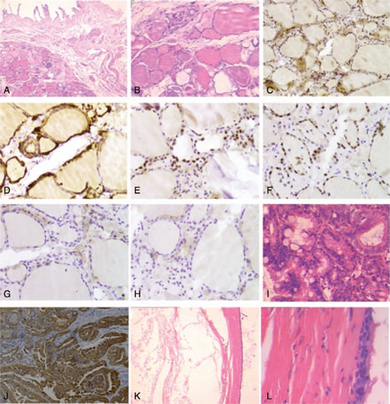
Rationale: Ectopic thyroid is most common in the tongue. Here we reported a rare case of thyroid tissue located in the gallbladder wall, accompanied with adenoma and a cyst lined with pseudostratified ciliated columnar epithelium in the neck region of gallbladder neck.
Patient concerns: A 39-year-old female presented with recurrent upper abdominal pain and radiating back pain.
Diagnoses: Based on ultrasonography, gallbladder polyps and calculous cholecystitis were suspected.
Interventions: The patient was treated by laparoscopic cholecystectomy, and thyroid tissue located in the gallbladder wall was found. Histopathological examination showed no features of papillary thyroid neoplasm.
Outcomes: The patient had no thyroid nodules or suspicious enlarged lymph nodes, and no other symptoms or complications by follow-up for 2.5 years up to September 2019.
Lessons: We should pay attention to the rare location of ectopic thyroid tissue in the gallbladder and rule out primary thyroid malignancy to avoid unnecessary overtreatment.
Conflict of interest statement
The authors report no conflicts of interest.
Figures
References
-
- Noussios G, Anagnostis P, Goulis DG, et al. Ectopic thyroid tissue: anatomical, clinical, and surgical implications of a rare entity. Eur J Endocrinol 2011;165:375–82. - PubMed
-
- Harach HR. Ectopic thyroid tissue adjacent to the gallbladder. Histopathology 1998;32:90–1. - PubMed
-
- Ihtiyar E, Isiksoy S, Algin C, et al. Ectopic thyroid in the gallbladder: report of a case. Surg Today 2003;33:777–80. - PubMed
-
- Venditti M, Hay RW, Kulaga A, et al. Diagnosis of ectopic tissue versus contamination by genetic fingerprinting in a routine surgical pathology specimen. Hum Pathol 2007;38:378–82. - PubMed
-
- Cassol CA, Noria D, Asa SL. Ectopic thyroid tissue within the gall bladder: case report and brief review of the literature. Endocr Pathol 2010;21:263–5. - PubMed
Publication types
MeSH terms
LinkOut - more resources
Full Text Sources
Medical
