. 2020 Jan-Dec;12(1):1685832.
doi: 10.1080/19420862.2019.1685832.

Harnessing MerTK agonism for targeted therapeutics

Affiliations

Harnessing MerTK agonism for targeted therapeutics

Vivekananda Kedage et al. MAbs. 2020 Jan-Dec.

Abstract

Phagocytosis plays important roles both in homeostasis and under pathological conditions. Fcγ receptor-mediated phagocytosis has been exploited as an integral mechanism for antibody-based therapies. Unlike Fcγ receptor-mediated phagocytosis, MerTK-mediated phagocytic clearance is immunologically silent. Here, we describe a bispecific antibody approach to harness MerTK for targeted clearance without inducing proinflammatory cytokine release associated with Fcγ receptor engagement. We generated bispecific antibodies targeting live B cells or amyloid beta aggregates to demonstrate the feasibility and versatility of this new approach.

Keywords: Fcγ receptor; MerTK; Phagocytosis; bispecific antibody; immunologically silent.


Figures
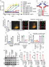
Figure 1.
Figure 1.
Identifying and characterizing MerTK agonist antibodies for engineering anti-CD20/MerTK bispecific antibodies capable of inducing phagocytosis of B cells by macrophages (a) Characterization of lead MerTK agonist antibodies measured by their ability to induce pAKT (Ser-473) in human MerTK+ macrophages using HTRF assay (Mean ± S.D of 2 technical replicates). Gas6-Fc is a positive control for MerTK activation. (b) Schematic depiction of B cell-dependent engagement of MerTK via anti-CD20/MerTK bispecific antibody. (c) Imagestream analysis of phagocytosis of live Raji cells by human primary macrophages in the presence of indicated bispecific antibody. A representation of Imagestream analysis is shown. Quantification is also presented (Mean ± S.D of one replicate each from 3 donors). (d-f) Flow cytometric analysis of phagocytosis of live Raji cells by human primary macrophages in the presence of indicated bispecific antibody, inhibitors and blocking agents (Mean ± S.D of one replicate each from 3 donors) (g) Activation of MerTK (phosphotyrosine) by the bispecific antibody in the presence of target Raji cells. Results are from using macrophages derived from two different human donors. See Supplementary Figure 5 for extended western blot. (h-i) TNF and IL-6 production by human primary macrophages stimulated with indicated plate coated antibodies. (Mean ± S.D of one replicate each from three different donors).
Figure 2.
Figure 2.
Anti-Aβ/MerTK bispecific antibody induces uptake of aggregated Aβ by BMDMs or microglia (a) Antibody dose-dependent uptake of aggregated Aβ by mouse BMDMs (Mean ± S.D of 3 technical replicates). (b) Time course of aggregated Aβ uptake by mouse BMDMs (Mean ± S.D of 3 technical replicates). Aggregated Aβ at 1 μM with antibodies at 10 ug/ml concentration. (c-e) BMDM uptake of aggregated Aβ induced by anti-Aβ/MerTK bispecific antibody is MerTK-dependent, as demonstrated by competition with the parental anti-MerTK antibody (20F5 Rabbit IgG) (c), inhibition by a MerTK SMI (d), or loss of activity in MerTK −/- BMDMs (e) (Mean ± S.D of 3 technical replicates). Aggregated Aβ at 1 μM, and antibodies at 10 ug/ml concentration. (f) TNF production by BMDMs stimulated with 3D6 mIgG2a (FcγR agonist) or 3D6/20F5 mIgG2a-LALAPG (MerTK agonist) (Mean ± S.D of 3 technical replicates). (g-i) Microglial cell uptake of aggregated Aβ induced by anti-Aβ/MerTK bispecific antibody is MerTK-dependent, as demonstrated by competition with the parental anti-MerTK antibody (20F5 Rabbit IgG) (g), inhibition by a MerTK SMI (h), or loss of activity in MerTK −/- microglia (i) (Mean ± S.D of 3 technical replicates). (j) TNF production by microglia stimulated with 3D6 mIgG2a (FcγR agonist) and 3D6/20F5 mIgG2a-LALAPG (MerTK agonist) and 3D6/GP120 mIgG2a-LALAPG (control) (Mean ± S.D of 3 technical replicates).

References

    1. Nimmerjahn F, Ravetch JV.. Fcgamma receptors as regulators of immune responses. Nat Rev Immunol. 2008;8:34–8. - PubMed
    1. Morkuniene R, Zvirbliene A, Dalgediene I, Cizas P, Jankeviciute S, Baliutyte G, Jokubka R, Jankunec M, Valincius G, Borutaite V, et al. Antibodies bound to Abeta oligomers potentiate the neurotoxicity of Abeta by activating microglia. J Neurochem. 2013;126:604–15. doi:10.1111/jnc.2013.126.issue-5. - DOI - PubMed
    1. Bugelski PJ, Achuthanandam R, Capocasale RJ, Treacy G, Bouman-Thio E. Monoclonal antibody-induced cytokine-release syndrome. Expert Rev Clin Immunol. 2009;5:499–521. doi:10.1586/eci.09.31. - DOI - PubMed
    1. Rinne JO, Brooks DJ, Rossor MN, Fox NC, Bullock R, Klunk WE, Mathis CA, Blennow K, Barakos J, Okello AA, et al. 11C-PiB PET assessment of change in fibrillar amyloid-beta load in patients with Alzheimer’s disease treated with bapineuzumab: a phase 2, double-blind, placebo-controlled, ascending-dose study. Lancet Neurol. 2010;9:363–72. doi:10.1016/S1474-4422(10)70043-0. - DOI - PubMed
    1. Salloway S, Sperling R, Gilman S, Fox NC, Blennow K, Raskind M, Sabbagh M, Honig LS, Doody R, van Dyck CH, et al. A phase 2 multiple ascending dose trial of bapineuzumab in mild to moderate Alzheimer disease. Neurology. 2009;73:2061–70. doi:10.1212/WNL.0b013e3181c67808. - DOI - PMC - PubMed

MeSH terms

LinkOut - more resources