. 2020 Feb 14;126(4):439-452.
doi: 10.1161/CIRCRESAHA.119.315767. Epub 2019 Dec 19.

Mitochondrial Deacetylase Sirt3 Reduces Vascular Dysfunction and Hypertension While Sirt3 Depletion in Essential Hypertension Is Linked to Vascular Inflammation and Oxidative Stress

Affiliations

Mitochondrial Deacetylase Sirt3 Reduces Vascular Dysfunction and Hypertension While Sirt3 Depletion in Essential Hypertension Is Linked to Vascular Inflammation and Oxidative Stress

Anna E Dikalova et al. Circ Res. .

Abstract

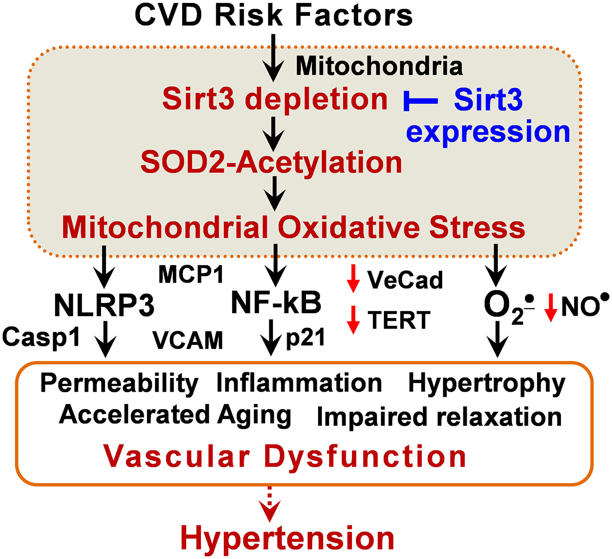
Rationale: Hypertension represents a major risk factor for stroke, myocardial infarction, and heart failure and affects 30% of the adult population. Mitochondrial dysfunction contributes to hypertension, but specific mechanisms are unclear. The mitochondrial deacetylase Sirt3 (Sirtuin 3) is critical in the regulation of metabolic and antioxidant functions which are associated with hypertension, and cardiovascular disease risk factors diminish Sirt3 level.

Objective: We hypothesized that reduced Sirt3 expression contributes to vascular dysfunction in hypertension, but increased Sirt3 protects vascular function and decreases hypertension.

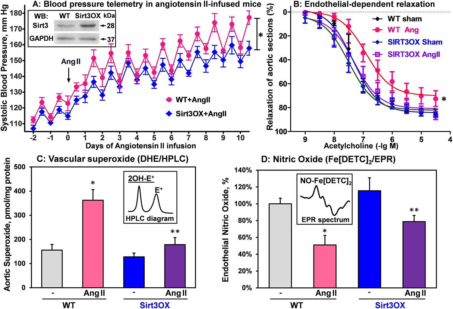
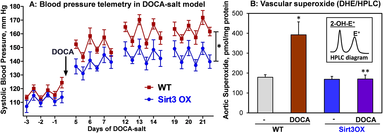
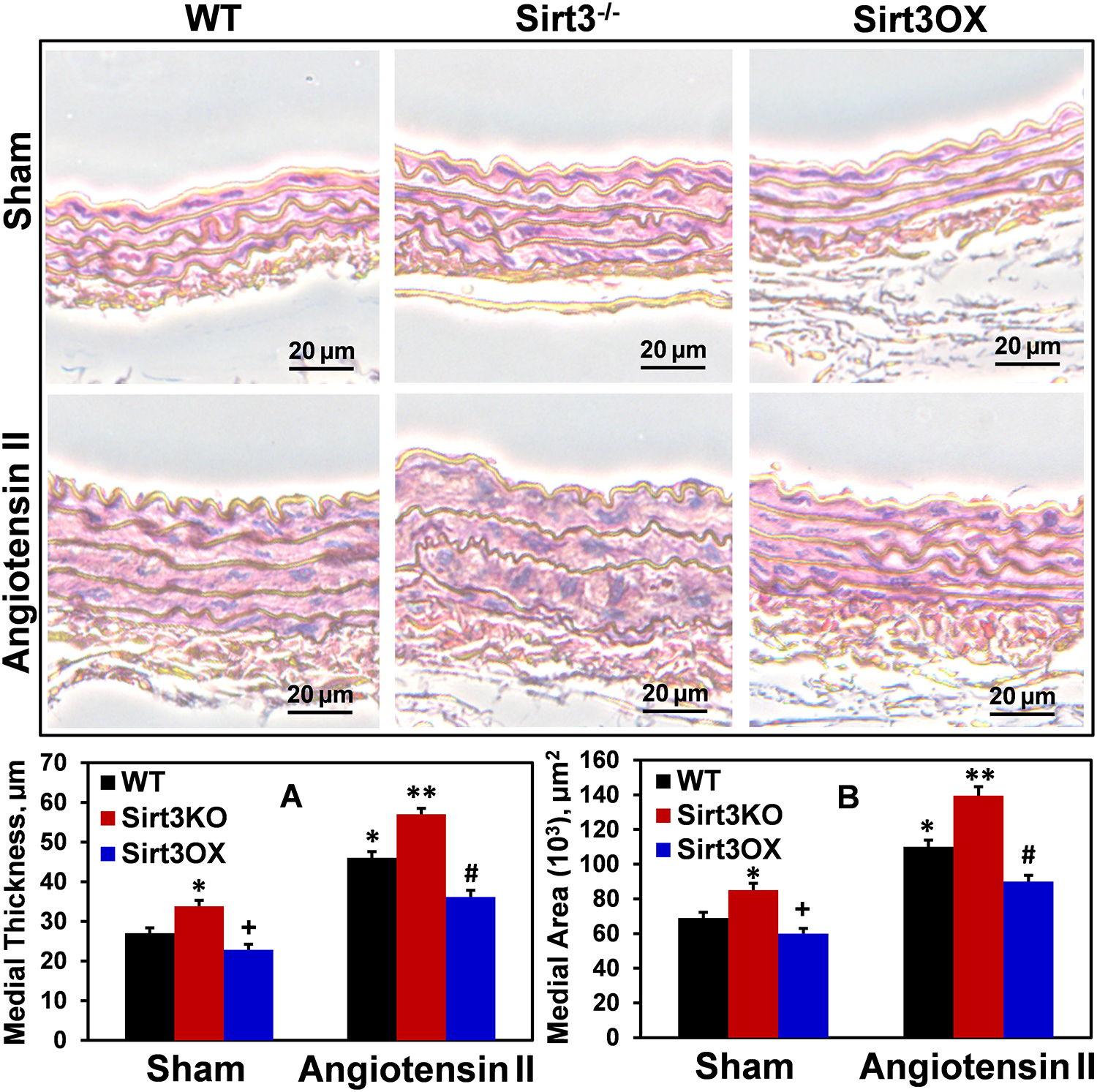
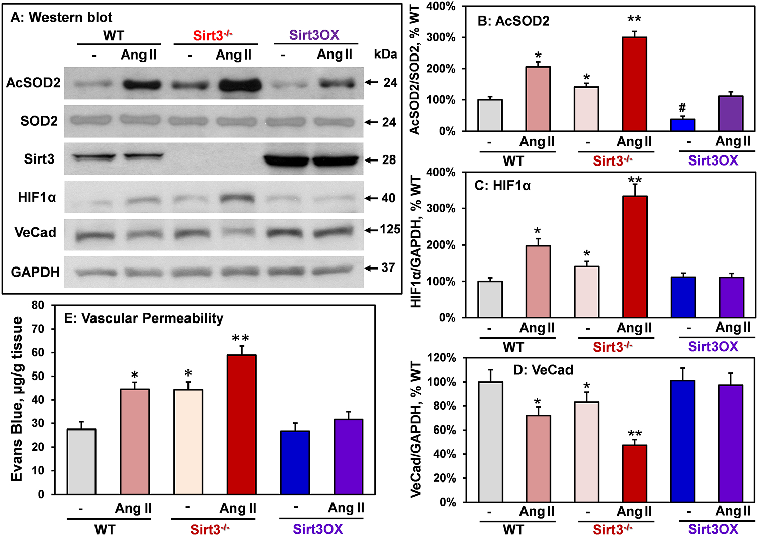
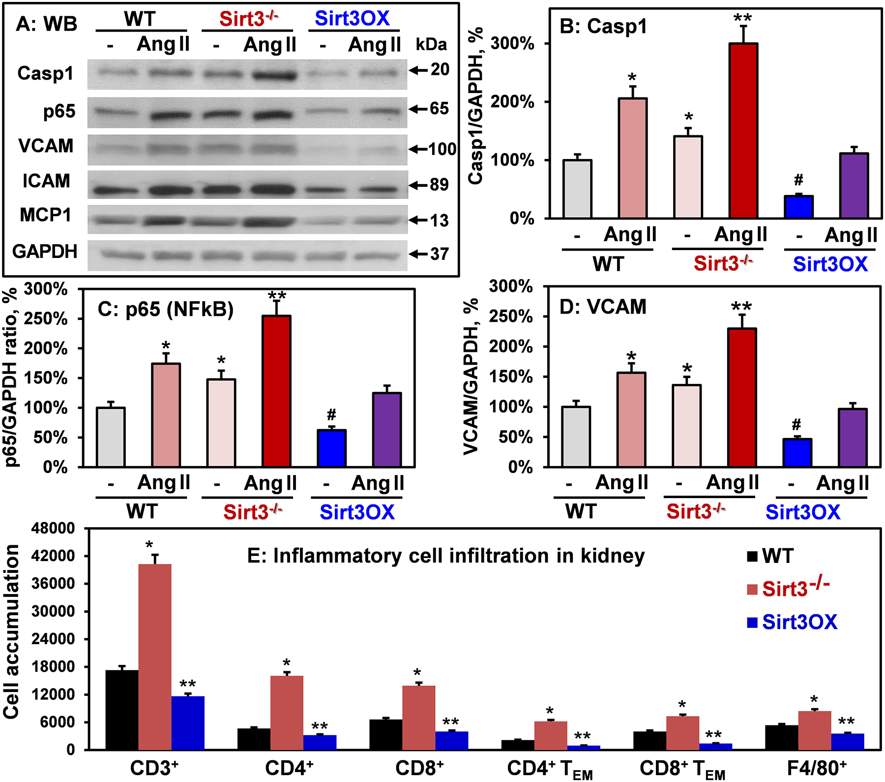
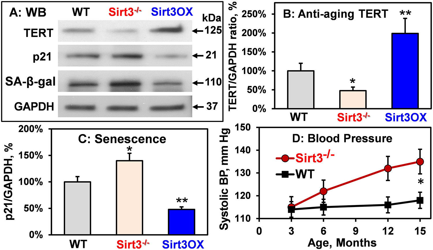
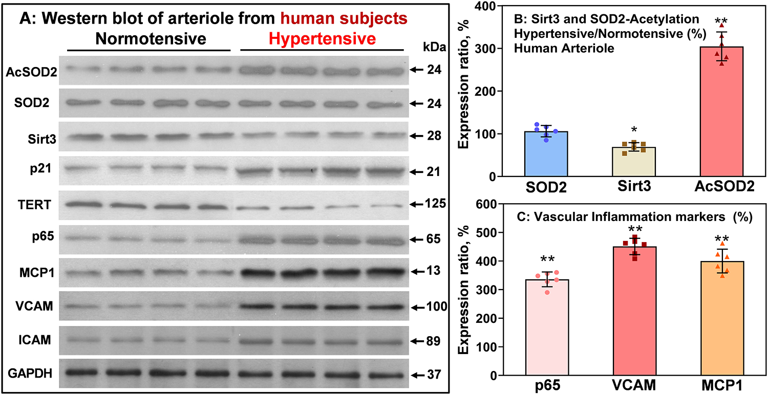
Methods and results: To test the therapeutic potential of targeting Sirt3 expression, we developed new transgenic mice with global Sirt3OX (Sirt3 overexpression), which protects from endothelial dysfunction, vascular oxidative stress, and hypertrophy and attenuates Ang II (angiotensin II) and deoxycorticosterone acetate-salt induced hypertension. Global Sirt3 depletion in Sirt3-/- mice results in oxidative stress due to hyperacetylation of mitochondrial superoxide dismutase (SOD2), increases HIF1α (hypoxia-inducible factor-1), reduces endothelial cadherin, stimulates vascular hypertrophy, increases vascular permeability and vascular inflammation (p65, caspase 1, VCAM [vascular cell adhesion molecule-1], ICAM [intercellular adhesion molecule-1], and MCP1 [monocyte chemoattractant protein 1]), increases inflammatory cell infiltration in the kidney, reduces telomerase expression, and accelerates vascular senescence and age-dependent hypertension; conversely, increased Sirt3 expression in Sirt3OX mice prevents these deleterious effects. The clinical relevance of Sirt3 depletion was confirmed in arterioles from human mediastinal fat in patients with essential hypertension showing a 40% decrease in vascular Sirt3, coupled with Sirt3-dependent 3-fold increases in SOD2 acetylation, NF-κB (nuclear factor kappa-light-chain-enhancer of activated B cells) activity, VCAM, ICAM, and MCP1 levels in hypertensive subjects compared with normotensive subjects.

Conclusions: We suggest that Sirt3 depletion in hypertension promotes endothelial dysfunction, vascular hypertrophy, vascular inflammation, and end-organ damage. Our data support a therapeutic potential of targeting Sirt3 expression in vascular dysfunction and hypertension.

Keywords: Sirtuin 3; acetylation; hypertension; mitochondria; oxidative stress; superoxide dismutase.


Figures

Figure 1:
Figure 1:. Blood pressure, vascular relaxation, aortic superoxide and nitric oxide production in wild-type and Sirt3OX mice in angiotensin II model.
(A) Systolic blood pressure was measured by telemetry before and after Ang II-infusion (0.7 mg/kg/day). Data were analyzed using 2-way ANOVA with repeated measurements. *P=0.02, n=8 in each group. Insert shows representative Western blot of Sirt3 aortas isolated from Sirt3OX mice and wild-type C57Bl/6J littermates. (B) Endothelial-dependent relaxation to acetylcholine. Vasculare relaxation data were analyzed using 2-way ANOVA with repeated measurements. *P=0.01 Sirt3OX+Ang II vs WT+Ang II, n=8 in each group. (C) Aortic superoxide (O2) was measured by DHE-HPLC assay of O2 specific product, 2-hydroxyethidium (2-OH-E+). (D) Endothelial nitric oxide (NO) measured by NO spin trap FeDETC2 and Electron Paramagnetic Resonance (EPR). For (C) and (D), data were analyzed using 2-way ANOVA and Bonferroni post-hoc multiple comparisons. Results are mean ± SEM (n=5). *P<0.01 vs WT, **P<0.01 Sirt3OX+Ang II vs WT+Ang II.
Figure 2:
Figure 2:. Blood pressure and vascular superoxide in wild-type and Sirt3OX mice in DOCA-salt model.
(A) Systolic blood pressure was measured by telemetry before and after DOCA-salt treatment. Results are mean ± SEM. Blood pressure data were analyzed using 2-way ANOVA with repeated measurements. *P=0.03 vs WT+DOCA, n=7 in each group. (B) Aortic superoxide was measured by HPLC analysis of DHE-O2 specific product, 2-hydroxyethidium (2-OH-E+). Results are mean ± SEM (n=5). Data were analyzed using 2-way ANOVA and Bonferroni post-hoc multiple comparisons. *P<0.001 vs WT, **P<0.001 Sirt3OX+DOCA vs WT+DOCA.
Figure 3:
Figure 3:. Vascular hypertrophy in sham and Ang II-infused wild-type, Sirt3−/− and Sirt3OX mice.
Representative images of hematoxylin and eosin-stained aortic cross sections are shown. Aortic thickness (A) and medial area (B) were quantified as described previously. Supplemental Online Figure I displays typical low magnification images with the intact vascular lumen to show the range of vessel wall thickness. Aortic hypertrophy was increased in Sirt3−/− mice, and Sirt3 overexpression reduced vascular hypertrophy in Sirt3OX mice compared to wild-type littermates. Data were analyzed using 2-way ANOVA and Bonferroni post-hoc multiple comparisons. Values are mean ± SEM. (A) *P=0.007 Sirt3−/− Sham vs WT sham, *P=0.00006 WT+Ang II vs WT sham, +P=0.003 vs WT sham, **P=0.0035 vs WT+Ang II, #P=0.0038 vs WT+Ang II (n=5). (B) *P=0.0012 Sirt3−/− Sham vs WT sham, *P=0.00005 WT+Ang II vs WT sham, +P=0.04 vs WT sham, **P=0.001 vs WT+Ang II, #P=0.0007 vs WT+Ang II (n=5).
Figure 4:
Figure 4:. Analysis of SOD2 acetylation, HIF1α, VE-cadherin and vascular permeability in aortas isolated from Sham and Ang II-infused Sirt3−/−, Sirt3OX and wild-type mice.
(A) Typical Western blots of Acetyl-K68-SOD2, total SOD2, HIF1α and VE-cadherin normalized for GAPDH in aortas. (B) SOD2-K68 acetylation, (C) HIF1α expression and (D) VE-cadherin levels normalized by GAPDH compared to Sham wild-type mice (100%). *P<0.05 vs WT sham, #P<0.05 vs WT sham **P<0.01 vs WT+Ang II (n=4). (E) Vascular permeability was measured by accumulation of Evans Blue dye in aortas. Male mice were infused with Ang II (0.7 mg/kg/day) or saline for two weeks prior to Miles assay. Data were analyzed using 2-way ANOVA and Bonferroni post-hoc multiple comparisons. *P<0.01 vs WT, **P<0.01 vs WT+Ang II (n=6).
Figure 5:
Figure 5:. Analysis of inflammasome activation, vascular inflammation and of inflammatory cells infiltration in Sirt3−/−, Sirt3OX and wild-type mice.
(A) Typical Western blots of caspase 1, p65, VCAM, ICAM and MCP1 normalized for GAPDH in aortas. (B) caspase 1, (C) p65 (NFkB subunit) and (D) VCAM levels normalized by GAPDH compared to Sham wild-type mice (100%). Results are mean ± SEM (n=6). *P<0.05 vs WT Sham, **P<0.01 vs WT+Ang II, #P<0.01 vs WT Sham. (E) Accumulation of T cells and macrophages in kidneys of six month old Sirt3−/−, Sirt3OX and wild-type mice measured by flow cytometry as described previously. Online Figure II shows the representative images of flow cytometry and gating strategy. Results are mean ± SEM (n=4). Data were analyzed using 2-way ANOVA and Bonferroni post-hoc multiple comparisons. *P<0.01 vs WT, **P<0.05 vs WT.
Figure 6:
Figure 6:. Western blot analysis of aging and cell senescence markers and age dependent hypertension in Sirt3−/− mice.
(A) Typical Western blots of aortic TERT, p21, and SA-β-gal in 6-month-old wild-type, Sirt3−/− and Sirt3OX mice. (B) TERT and (C) p21 levels normalized by GAPDH compared to Sham wild-type mice (100%). Results are mean ± SEM (n=5). Data were analyzed using 1-way ANOVA and Bonferroni post-hoc multiple comparisons. *P<0.01 vs WT, **P<0.01 vs WT. (D) Systolic blood pressure in Sirt3−/− and WT mice. Data were analyzed with 2-way ANOVA with repeated measurements. *P=0.008 vs WT (n=6).
Figure 7:
Figure 7:. Western blot of SOD2 acetylation, SOD2, Sirt3, inflammation and cell-senescence markers in arterioles isolated from human mediastinal fat in patients with essential hypertension compared with normotensive subjects.
Data were normalized by GAPDH. Results are mean ± SEM. Data were analyzed using 1-way ANOVA and Bonferroni post-hoc multiple comparisons. (A) *P=0.01, *P=0.0001, n=6. (B) **P=0.000001 vs Normotensive, n=6.
Figure 8:
Figure 8:. Proposed role of Sirt3 depletion in vascular dysfunction and hypertension. Sirt3 depletion results in SOD2 hyperacetylation and inactivation leading to mitochondrial oxidative stress.
This in turn inactivates endothelial nitric oxide (NO), induces redox-dependent NF-kB activation and mitochondrial damage, and activates the NLRP3 inflammasome. These pathways disrupt the endothelial barrier, impair vasorelaxation, promote smooth muscle hypertrophy and vascular inflammation, accelerate vascular aging and increase hypertension. These deleterious effects are ameliorated by Sirt3 expression.

Comment in

References

    1. Sacco RL, Benjamin EJ, Broderick JP, Dyken M, Easton JD, Feinberg WM, Goldstein LB, Gorelick PB, Howard G, Kittner SJ, Manolio TA, Whisnant JP, Wolf PA. American heart association prevention conference. Iv. Prevention and rehabilitation of stroke. Risk factors. Stroke. 1997;28:1507–1517 - PubMed
    1. Roth GA, Johnson C, Abajobir A. et al. Global, regional, and national burden of cardiovascular diseases for 10 causes, 1990 to 2015. J Am Coll Cardiol. 2017;70:1–25 - PMC - PubMed
    1. Harrison DG, Marvar PJ, Titze JM. Vascular inflammatory cells in hypertension. Front Physiol. 2012;3:128. - PMC - PubMed
    1. Byrd JB, Zeng C, Tavel HM, Magid DJ, O’Connor PJ, Margolis KL, Selby JV, Ho PM. Combination therapy as initial treatment for newly diagnosed hypertension. Am Heart J. 2011;162:340–346 - PMC - PubMed
    1. Guzik TJ, Touyz RM. Oxidative stress, inflammation, and vascular aging in hypertension. Hypertension. 2017;70:660–667 - PubMed

Publication types

MeSH terms