Task-Free Functional Language Networks: Reproducibility and Clinical Application
- PMID: 31852732
- PMCID: PMC7002153
- DOI: 10.1523/JNEUROSCI.1485-19.2019
Task-Free Functional Language Networks: Reproducibility and Clinical Application
Abstract
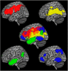
Intrinsic connectivity networks (ICNs) identified through task-free fMRI (tf-fMRI) offer the opportunity to investigate human brain circuits involved in language processes without requiring participants to perform challenging cognitive tasks. In this study, we assessed the ability of tf-fMRI to isolate reproducible networks critical for specific language functions and often damaged in primary progressive aphasia (PPA). First, we performed whole-brain seed-based correlation analyses on tf-fMRI data to identify ICNs anchored in regions known for articulatory, phonological, and semantic processes in healthy male and female controls (HCs). We then evaluated the reproducibility of these ICNs in an independent cohort of HCs, and recapitulated their functional relevance with a post hoc meta-analysis on task-based fMRI. Last, we investigated whether atrophy in these ICNs could inform the differential diagnosis of nonfluent/agrammatic, semantic, and logopenic PPA variants. The identified ICNs included a dorsal articulatory-phonological network involving inferior frontal and supramarginal regions; a ventral semantic network involving anterior middle temporal and angular gyri; a speech perception network involving superior temporal and sensorimotor regions; and a network between posterior inferior temporal and intraparietal regions likely linking visual, phonological, and attentional processes for written language. These ICNs were highly reproducible across independent groups and revealed areas consistent with those emerging from task-based meta-analysis. By comparing ICNs' spatial distribution in HCs with patients' atrophy patterns, we identified ICNs associated with each PPA variant. Our findings demonstrate the potential use of tf-fMRI to investigate the functional status of language networks in patients for whom activation studies can be methodologically challenging.SIGNIFICANCE STATEMENT We showed that a single, short, task-free fMRI acquisition is able to identify four reproducible and relatively segregated intrinsic left-dominant networks associated with articulatory, phonological, semantic, and multimodal orthography-to-phonology processes, in HCs. We also showed that these intrinsic networks relate to syndrome-specific atrophy patterns in primary progressive aphasia. Collectively, our results support the application of task-free fMRI in future research to study functionality of language circuits in patients for whom tasked-based activation studies might be methodologically challenging.
Keywords: functional connectivity; language networks; primary progressive aphasia; reproducibility; resting-state connectivity.
Copyright © 2020 the authors.
Figures


References
-
- Battistella G, Henry M, Gesierich B, Wilson SM, Borghesani V, Shwe W, Miller Z, Deleon J, Miller BL, Jovicich J, Papinutto N, Dronkers NF, Seeley WW, Mandelli ML, Gorno-Tempini ML (2019) Differential intrinsic functional connectivity changes in semantic variant primary progressive aphasia. Neuroimage Clin 22:101797. 10.1016/j.nicl.2019.101797 - DOI - PMC - PubMed
Publication types
MeSH terms
Grants and funding
- P50 AG023501/AG/NIA NIH HHS/United States
- R01 AG038791/AG/NIA NIH HHS/United States
- U01 AG052943/AG/NIA NIH HHS/United States
- F31 DC009145/DC/NIDCD NIH HHS/United States
- R01 NS100440/NS/NINDS NIH HHS/United States
- U54 NS092089/NS/NINDS NIH HHS/United States
- K24 DC015544/DC/NIDCD NIH HHS/United States
- R01 DC016345/DC/NIDCD NIH HHS/United States
- R03 DC013403/DC/NIDCD NIH HHS/United States
- T32 AG023481/AG/NIA NIH HHS/United States
- P30 AG062422/AG/NIA NIH HHS/United States
- P01 AG019724/AG/NIA NIH HHS/United States
- R01 DC016291/DC/NIDCD NIH HHS/United States
- R56 NS050915/NS/NINDS NIH HHS/United States
- R01 NS050915/NS/NINDS NIH HHS/United States
LinkOut - more resources
Full Text Sources
Miscellaneous
