. 2020 Mar 25;58(4):e01503-19.
doi: 10.1128/JCM.01503-19. Print 2020 Mar 25.

Laboratory Analysis of an Outbreak of Candida auris in New York from 2016 to 2018: Impact and Lessons Learned

Affiliations

Laboratory Analysis of an Outbreak of Candida auris in New York from 2016 to 2018: Impact and Lessons Learned

YanChun Zhu et al. J Clin Microbiol. .

Abstract

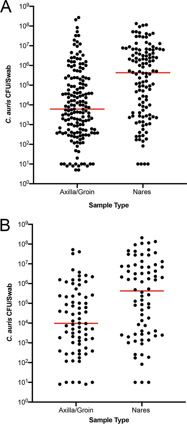
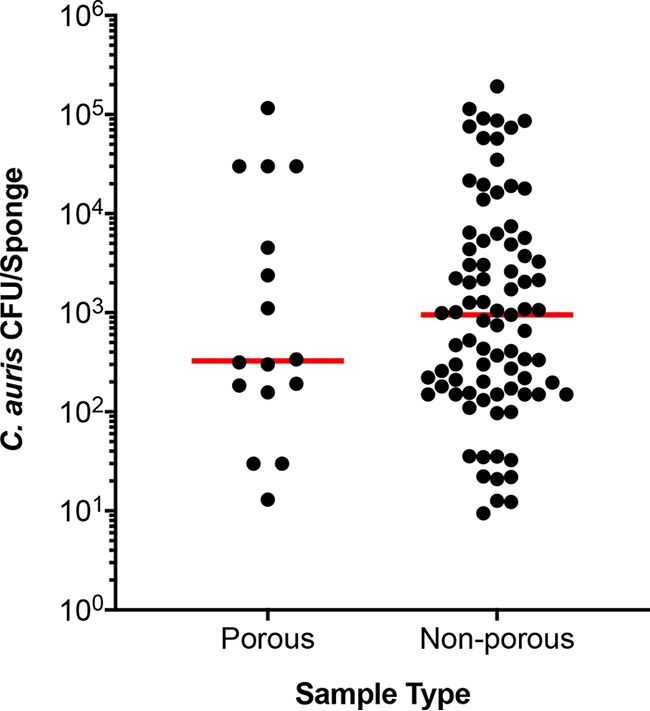
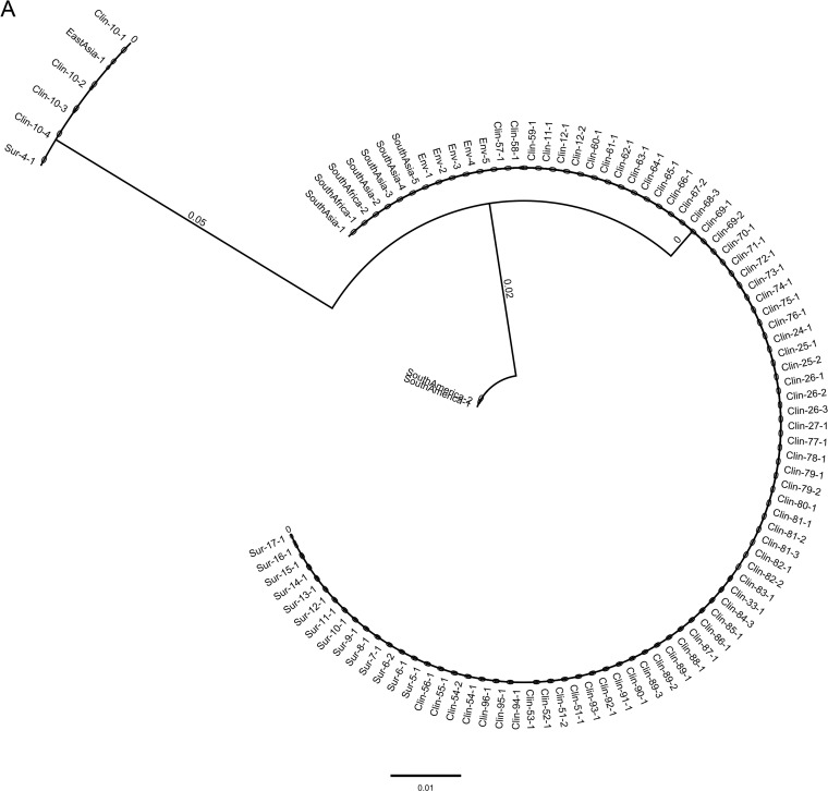
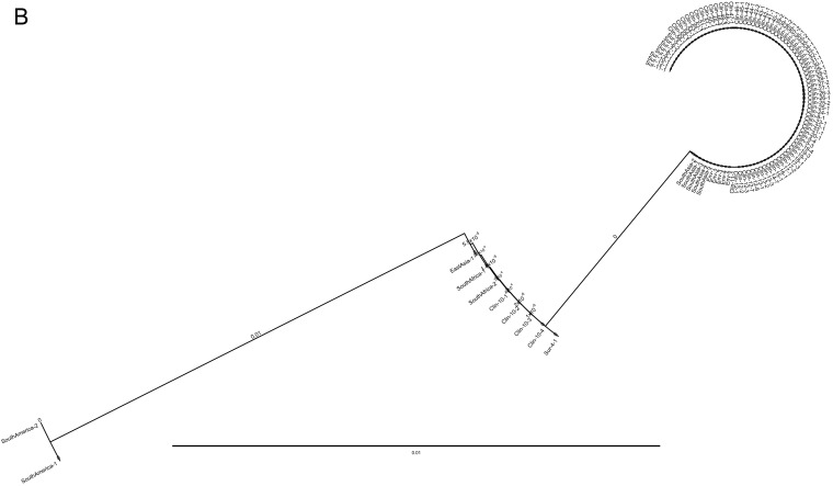
Candida auris is a multidrug-resistant yeast which has emerged in health care facilities worldwide; however, little is known about identification methods, patient colonization, environmental survival, spread, and drug resistance. Colonization on both biotic (patients) and abiotic (health care objects) surfaces, along with travel, appear to be the major factors for the spread of this pathogen across the globe. In this investigation, we present laboratory findings from an ongoing C. auris outbreak in New York (NY) from August 2016 through 2018. A total of 540 clinical isolates, 11,035 patient surveillance specimens, and 3,672 environmental surveillance samples were analyzed. Laboratory methods included matrix-assisted laser desorption ionization-time of flight mass spectrometry (MALDI-TOF MS) for yeast isolate identification, real-time PCR for rapid surveillance sample screening, culture on selective/nonselective media for recovery of C. auris and other yeasts from surveillance samples, antifungal susceptibility testing to determine the C. auris resistance profile, and Sanger sequencing of the internal transcribed spacer (ITS) and D1/D2 regions of the ribosomal gene for C. auris genotyping. Results included (a) identification and confirmation of C. auris in 413 clinical isolates and 931 patient surveillance isolates as well as identification of 277 clinical cases and 350 colonized cases from 151 health care facilities, including 59 hospitals, 92 nursing homes, 1 long-term acute care hospital (LTACH), and 2 hospices, (b) successful utilization of an in-house developed C. auris real-time PCR assay for the rapid screening of patient and environmental surveillance samples, (c) demonstration of relatively heavier colonization of C. auris in nares than in the axilla/groin, and (d) predominance of the South Asia clade I with intrinsic resistance to fluconazole and elevated MIC to voriconazole (81%), amphotericin B (61%), flucytosine (5FC) (3%), and echinocandins (1%). These findings reflect greater regional prevalence and incidence of C. auris and the deployment of better detection tools in an unprecedented outbreak.

Keywords: Candida auris; antifungals; molecular biology; mycology; phylogenetics.


Figures

FIG 1
FIG 1
Colonization of the axilla/groin and nares. Axilla/groin and nares were swabbed and processed for culture. Recovered colonies were counted, and results were expressed as CFU/swab. Each dot represents total CFU/swab/patient, and the horizontal bar within each group represents the median. (A) Unpaired samples from patients colonized with C. auris. (B) Paired samples from patients colonized with C. auris. In both cases, the median C. auris CFU was 2-log higher in nares than in axilla/groin (P < 0.0001).
FIG 2
FIG 2
Colonization of environmental surfaces with C. auris. Environmental surfaces of various health care facilities were sponge swabbed and processed for culture. Recovered colonies were counted, and results were expressed as total CFU/sponge. The environmental surfaces were divided into porous (i.e., linen and carpet) and nonporous (i.e., plastic and metal devices) for data analysis. Each dot represents total CFU recovered/per area swabbed, and the horizontal bar within each group represents the median. The median C. auris CFU was approximately 3-fold higher on nonporous surfaces than on the porous surfaces (P < 0.001).
FIG 3
FIG 3
(A) Phylogenetic analysis of C. auris isolates from New York using ITS and D1/D2 sequences. Nucleotide sequences of 622 isolates of C. auris recovered from clinical patients (Clin), colonized patients (Sur), environmental surfaces (Env), and 10 standard isolates from the CDC AR bank were aligned. The neighbor-joining method was used to construct the phylogenetic tree. The bootstrap scores are based on 2,000 reiterations. The outbreak was predominately the South Asia clade I, with a minor population of the East Asia clade II. Trees representing a few C. auris isolates from each group are shown. (A) Analysis of ITS sequences. Note, ITS could not distinguish South Africa clade III (CDC AR) from South Asia clade I. (B) Analysis of D1/D2 sequences. Note, D1/D2 did not differentiate South Africa clade III from East Asia clade II.
FIG 3
FIG 3
(A) Phylogenetic analysis of C. auris isolates from New York using ITS and D1/D2 sequences. Nucleotide sequences of 622 isolates of C. auris recovered from clinical patients (Clin), colonized patients (Sur), environmental surfaces (Env), and 10 standard isolates from the CDC AR bank were aligned. The neighbor-joining method was used to construct the phylogenetic tree. The bootstrap scores are based on 2,000 reiterations. The outbreak was predominately the South Asia clade I, with a minor population of the East Asia clade II. Trees representing a few C. auris isolates from each group are shown. (A) Analysis of ITS sequences. Note, ITS could not distinguish South Africa clade III (CDC AR) from South Asia clade I. (B) Analysis of D1/D2 sequences. Note, D1/D2 did not differentiate South Africa clade III from East Asia clade II.
FIG 4
FIG 4
Prevalence of Candida species in surveillance samples. (A) The bar diagram represents different Candida species and their frequency of isolation (%) from patient surveillance samples. Candida albicans was the dominant pathogen followed by C. auris, C. glabrata, C. tropicalis, and C. parapsilosis. Additional Candida species, and other yeasts, were minor components. (B) Bar diagram represents different Candida species and their frequency of isolation (%) from environmental surveillance samples. Candida parapsilosis was the dominant pathogen followed by C. auris, C. albicans, C. guilliermondii, C. glabrata, and C. tropicalis. Additional Candida species, molds, and other yeasts were minor components.

Comment in

References

    1. Satoh K, Makimura K, Hasumi Y, Nishiyama Y, Uchida K, Yamaguchi H. 2009. Candida auris sp. nov., a novel ascomycetous yeast isolated from the external ear canal of an inpatient in a Japanese hospital. Microbiol Immunol 53:41–44. doi:10.1111/j.1348-0421.2008.00083.x. - DOI - PubMed
    1. Lee WG, Shin JH, Uh Y, Kang MG, Kim SH, Park KH, Jang HC. 2011. First three reported cases of nosocomial fungemia caused by Candida auris. J Clin Microbiol 49:3139–3142. doi:10.1128/JCM.00319-11. - DOI - PMC - PubMed
    1. Chowdhary A, Sharma C, Duggal S, Agarwal K, Prakash A, Singh PK, Jain S, Kathuria S, Randhawa HS, Hagen F, Meis JF. 2013. New clonal strain of Candida auris, Delhi, India. Emerg Infect Dis 19:1670–1673. doi:10.3201/eid1910.130393. - DOI - PMC - PubMed
    1. Wang X, Bing J, Zheng Q, Zhang F, Liu J, Yue H, Tao L, Du H, Wang Y, Wang H, Huang G. 2018. The first isolate of Candida auris in China: clinical and biological aspects. Emerg Microbes Infect 7:93. doi:10.1038/s41426-018-0095-0. - DOI - PMC - PubMed
    1. Magobo RE, Corcoran C, Seetharam S, Govender NP. 2014. Candida auris-associated candidemia, South Africa. Emerg Infect Dis 20:1250–1251. doi:10.3201/eid2007.131765. - DOI - PMC - PubMed

Publication types

Substances