Review
. 2020 Mar;32(3):547-572.
doi: 10.1105/tpc.19.00158. Epub 2019 Dec 18.

Diatom Molecular Research Comes of Age: Model Species for Studying Phytoplankton Biology and Diversity

Affiliations
Review

Diatom Molecular Research Comes of Age: Model Species for Studying Phytoplankton Biology and Diversity

Angela Falciatore et al. Plant Cell. 2020 Mar.

Abstract

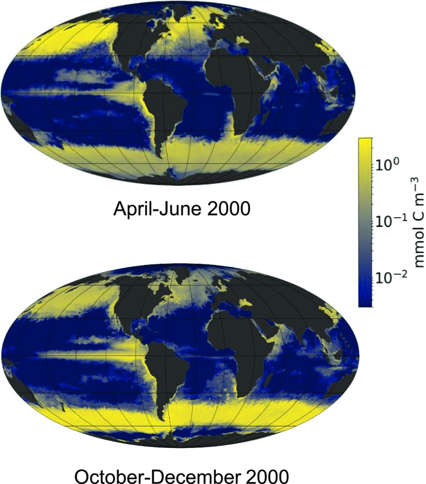
Diatoms are the world's most diverse group of algae, comprising at least 100,000 species. Contributing ∼20% of annual global carbon fixation, they underpin major aquatic food webs and drive global biogeochemical cycles. Over the past two decades, Thalassiosira pseudonana and Phaeodactylum tricornutum have become the most important model systems for diatom molecular research, ranging from cell biology to ecophysiology, due to their rapid growth rates, small genomes, and the cumulative wealth of associated genetic resources. To explore the evolutionary divergence of diatoms, additional model species are emerging, such as Fragilariopsis cylindrus and Pseudo-nitzschia multistriata Here, we describe how functional genomics and reverse genetics have contributed to our understanding of this important class of microalgae in the context of evolution, cell biology, and metabolic adaptations. Our review will also highlight promising areas of investigation into the diversity of these photosynthetic organisms, including the discovery of new molecular pathways governing the life of secondary plastid-bearing organisms in aquatic environments.


Figures

Figure 1.
Figure 1.
Model of the Global Distribution of Diatom Biomass. Model of the global distribution of diatom biomass in the ocean surface layer in April to June (left) and October to December (right) of 2000 (courtesy of Oliver Jahn, Stephanie Dutkiewicz, and Mick Follows). Biomass values, reported in log scale in units of mmol C m−3, were derived from the MIT ecosystem model, which simulates ocean circulation and key biogeochemical processes, e.g., nutrient fluxes, plankton growth, and death occurring in the ocean, at a global scale (Follows et al., 2007; Tréguer et al., 2018). Physiological data were derived from laboratory and field experiments. Because diatom biomass varies between ∼0.4 (P. tricornutum) and ∼7000 (Coscinodiscus wailesii) pmol C per cell, a biomass of 1 mmol C m−3 corresponds to a range of ∼150 cells L−1 to ∼2.5 106 cells L−1 (Marañón et al., 2013).
Figure 2.
Figure 2.
Diatom Characteristics and Morphological Diversity. (A) Scanning electron micrograph of the model centric species T. pseudonana with its characteristic cell wall of silica, viewed from the top. (B) to (I) Light microscopy of the pennate model species P. tricornutum (B). This diatom exists in three interconvertible morphotypes: the fusiform, oval, and triradiate morphotypes, as shown in the figure. Scanning electron micrograph images of (C) Skeletonema tropicum, (D) a valve of a raphid pennate diatom, (E) Shionodiscus oestrupii var venrickiae, and (F) F. cylindrus. Light microscopy of (G) Chaetoceros sp, (H) Fragilariopsis kerguelensis, and (I) Pseudo-nitzschia sp from a natural phytoplankton sample collected at the Long Term Station MareChiara in the Gulf of Naples, Italy. (J) Flipped ice floe in the Southern Ocean with dense population of sea-ice diatoms (e.g., F. cylindrus) at the ice-water interface. Images were kindly provided by Diana Sarno and Marina Montresor (Stazione Zoologica Anton Dohrn, Napoli, Italy) and James A. Raymond (Univeristy of Nevada, United States).
Figure 3.
Figure 3.
Simplified Scheme of the Major Events Leading to the Evolution of Diatoms through Primary and Secondary Endosymbiosis. The initial primary endosymbiosis occurred when a heterotrophic host engulfed a cyanobacterium (represented in blue). Over time, a large proportion of the cyanobacterial genome was transferred to the nucleus of the host, as indicated by the blue arrow. The endosymbiotic process generated the plastids of the Archaeplastida, a major group including Glaucophyta (pale blue), Rhodophyta (pink), and the Viridiplantae (the model green alga C. reinhardtii and plant Arabidopsis are shown). During secondary endosymbiosis, a different heterotrophic cell acquired a red alga and potentially also a green alga. The algal endosymbiont became the plastid (in brown) of the Stramenopila, a group including diatoms, but also other algae such as pelagophytes or the multicellular kelps. Algal nuclear genomes were transferred to the heterotrophic nucleus, as represented by green and red curved arrows, while the algal nucleus and mitochondria were lost. Other bacterial genes in the Stramenopila genome were derived by horizontal gene transfer events from bacterial donors (violet arrow). The figure also shows the approximative dates of diatom evolution and separation between centric and pennate diatoms based on Nakov et al. (2018). Some pennate species further diversified and acquired the capacity to move by secreting mucilage through a longitudinal slit in the cell wall called raphe, hence the division between raphid (motile) and araphid (nonmotile) pennate diatoms. Mya, million years ago.
Figure 4.
Figure 4.
Regulators of Carbon, Nitrogen, and Iron Metabolism Characterized in the Diatom Model Species. (A) Regulators of carbon. (B) Regulators of nitrogen and iron. AOX, alternative oxidase; AQP, aquaporin; CA, carbonic anhydrases (characterized in P. tricornutum in blue, in T. pseudonana in red, in both in purple); CERC, chloroplast/endoplasmic reticulum compartment; CERM, chloroplast endoplasmic reticulum membrane; CP, carbamoyl phosphate; CPSIII, carbamoyl-phosphate synthase; DSP, death-specific protein; FRE, ferrireductase; FTN, ferritin; FTR, Fe(III) permease; iEM, internal plastid envelop membrane; ISIP1 and ISIP2a, iron-starvation-induced proteins; LB, lipid body; M, mitochrondrion; MCO, multicopper oxidase; N, nucleus; NR, nitrate reductase; oEM, outer plastid envelop membrane; P, plastid; PEPC, phosphoenolpyruvate carboxylase; PIS, plastid intermembrane space; PPC, periplastidial compartment; PPM, periplastidial membrane; Px, peroxysome; Pyr, pyrenoid; SCL4, bicarbonate solute carrier 4; T, thylakoids; TCA, tricarboxylic acid; V, vacuole. Question marks indicate still uncharacterized regualtors or uncertain cellular localisation.

References

    1. Ahmed S., et al. (2014). A haploid system of sex determination in the brown alga Ectocarpus sp. Curr. Biol. 24: 1945–1957. - PubMed
    1. Allen A.E., Dupont C.L., Oborník M., Horák A., Nunes-Nesi A., McCrow J.P., Zheng H., Johnson D.A., Hu H., Fernie A.R., Bowler C. (2011). Evolution and metabolic significance of the urea cycle in photosynthetic diatoms. Nature 473: 203–207. - PubMed
    1. Allen A.E., Laroche J., Maheswari U., Lommer M., Schauer N., Lopez P.J., Finazzi G., Fernie A.R., Bowler C. (2008). Whole-cell response of the pennate diatom Phaeodactylum tricornutum to iron starvation. Proc. Natl. Acad. Sci. USA 105: 10438–10443. - PMC - PubMed
    1. Allen J. (2002). Photosynthesis of ATP-electrons, proton pumps, rotors, and poise. Cell 110: 273–276. - PubMed
    1. Amato A., Orsini L. (2015). Rare interspecific breeding in Pseudo-nitzschia (Bacillariophyceae). Phytotaxa 217: 145–154.

Publication types

LinkOut - more resources