. 2019 Dec 18;11(523):eaav5264.
doi: 10.1126/scitranslmed.aav5264.

Human genetics and neuropathology suggest a link between miR-218 and amyotrophic lateral sclerosis pathophysiology

Affiliations

Human genetics and neuropathology suggest a link between miR-218 and amyotrophic lateral sclerosis pathophysiology

Irit Reichenstein et al. Sci Transl Med. .

Abstract

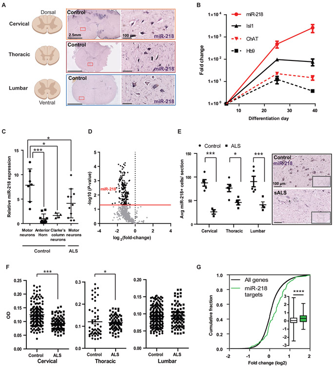
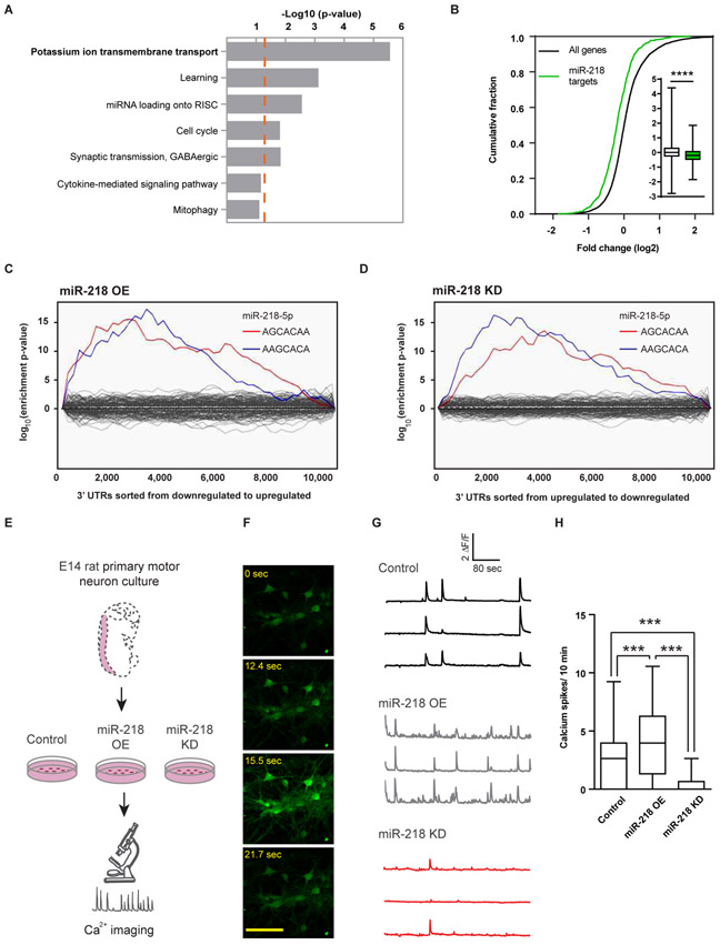
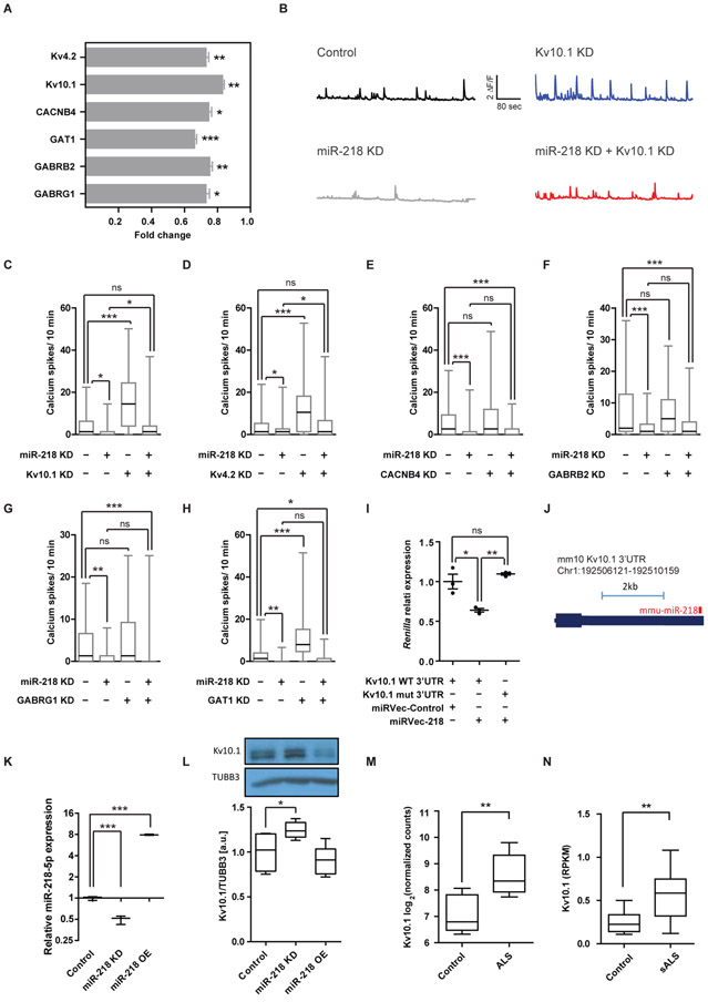
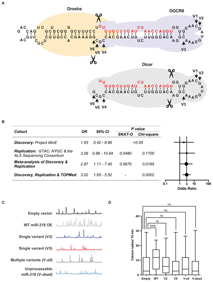
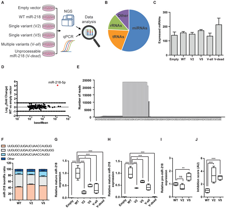
Motor neuron-specific microRNA-218 (miR-218) has recently received attention because of its roles in mouse development. However, miR-218 relevance to human motor neuron disease was not yet explored. Here, we demonstrate by neuropathology that miR-218 is abundant in healthy human motor neurons. However, in amyotrophic lateral sclerosis (ALS) motor neurons, miR-218 is down-regulated and its mRNA targets are reciprocally up-regulated (derepressed). We further identify the potassium channel Kv10.1 as a new miR-218 direct target that controls neuronal activity. In addition, we screened thousands of ALS genomes and identified six rare variants in the human miR-218-2 sequence. miR-218 gene variants fail to regulate neuron activity, suggesting the importance of this small endogenous RNA for neuronal robustness. The underlying mechanisms involve inhibition of miR-218 biogenesis and reduced processing by DICER. Therefore, miR-218 activity in motor neurons may be susceptible to failure in human ALS, suggesting that miR-218 may be a potential therapeutic target in motor neuron disease.


Figures

Fig. 1.
Fig. 1.. miR-218 is expressed in the human spinal motor neurons and is downregulated in human ALS.
(A-F) Three orthogonal miRNA quantification studies in human motor neurons from 20 ALS cases and 14 non-neurodegeneration controls: (A) miR-218 chromogenic in situ hybridization depicting broad expression along the cervical, thoracic and lumbar regions of the adult human spinal cord. (B) qPCR analysis of miR-218, Hb9, Isl1 and ChAT in human iPSCs and differentiated motor neurons. miR-218 normalized to U6 expression. mRNAs normalized to average of HPRT and β–actin expression, presented on a log scale; n=3 independent wells per time point. (C) miR-218 expression in laser-capture micro-dissected human lumbar motor neurons. miR-218 expression in non-neurodegeneration motor neurons (n=7 human spinal cords), relative to surrounding non-motor neuron anterior horn tissue (n=10), to Clarke’s column proprioceptive neurons (n=4), or to ALS motor neurons (n=9 sporadic and 2 familial nervous systems carrying the SOD1 A4V mutation). TaqMan qPCR analysis of miR-218 normalized to the average of RNU48/SNORD48, RNU44/SNORD44 and U6 in the same sample, and to the average miR-218 expression in the anterior horn. One-way ANOVA followed by Newman-Keuls multiple comparison test performed on log-transformed data, Means ± SD. (D) Volcano plot of relative miRNA expression in ALS lumbar ventral horns (n=5), versus non-neurodegeneration controls (n=2; x-axis log2 scale), screened by nanoString nCounter platform. y-axis depicts the differential expression p-values (−log10 scale). Black dots indicate P < 0.05; light gray dots are non-significant. miR-218 is the most downregulated miRNA in ALS nervous systems. Data normalized to the average of five control mRNAs (ACTB, B2M, GAPDH, RPL19, RPLP0). (E) Reduced miR-218+ cell numbers in sALS patient anterior horns (n=4), relative to non-neurodegeneration controls (n=5) and representative miRNA in situ hybridization micrographs. Two way ANOVA followed by Bonferroni’s multiple comparison test, Means ± SEM, and (F) chromogenic miR-218 in situ hybridization signal densitometry in motor neurons at different spinal cord levels (non-neurodegeneration control/ALS cases: Cervical n=151/85 cells; Thoracic n=54/75; Lumbar 189/92). One-tailed Mann-Whitney test, Means ± SEM. (G) Cumulative distribution function (CDF) plot of top 100 predicted miR-218-5p targets (TargetScan (20)), or all expressed mRNAs, in laser capture microdissection-enriched surviving motor neurons from lumbar spinal cords of patients with sALS with rostral onset and caudal progression (n=13) relative to non-neurodegeneration controls (n=6; (21)) and Box-Plot (inset) depicting median, upper and lower quartiles and extreme points. P-value calculated using Kolmogorov–Smirnov test comparing miR-218-5p targets subset distribution to all genes. * P<0.05; *** P<0.001; **** P<0.0001.
Fig. 2.
Fig. 2.. miR-218 controls motor neuron network activity.
(A) Seven most enriched gene ontology terms (22) of predicted miR-218 targets (20). p-value of term enrichment (−log10, dashed orange line indicates P = 0.05). (B) CDF plot of miR-218 predicted targets, relative to all expressed mRNAs, following OE of miR-218 and box-plot (insets), depicting median, upper and lower quartiles and extreme points. P-value calculated using Kolmogorov–Smirnov test comparing miR-218-5p subset distribution to all genes. **** P<0.0001. Binding site enrichment of all known miRNAs, in ~10,000 expressed mRNAs, was tested after (C) miR-218 OE, or (D) miR-218 KD, relative to control virus. Significant enrichment for two miR-218-5p seed-matches (blue, red) and lack of enrichment for any other miRNA (gray) via a Sylamer study (24). (E) Diagram of calcium transient imaging in embryonic rat spinal motor neurons, transduced with lentiviruses encoding control vector, miR-218 OE or a miR-218 KD. Neuron time lapse micrographs (F), representative traces (G) and (H) quantification of spontaneous calcium spike frequencies (ΔF/F >0.5) from Fluo2 HighAff AM study after 12 days in vitro. Recorded from 58 / 76 / 41 control / OE /KD cells, respectively. Box-Plot depicting the median, upper and lower quartiles and extreme points. Kruskal-Wallis test followed by Dunn’s multiple comparison test, *** P<0.001. This experiment was repeated 3 independent times with similar results.
Fig. 3.
Fig. 3.. The potassium channel Kv10.1 acts downstream of miR-218.
(A) qPCR measuring the expression of mRNAs targets, following miR-218 OE (n=15). Data normalized to control virus (n=12) and to average expression of HPRT and β–actin, two technical duplicates, two-sided student’s t-test, Means ± SEM. (B) Representative traces of individual motor neurons and (C-H) quantification of spontaneous calcium spike frequencies (ΔF/F >0.5) of embryonic rat spinal motor neurons, transduced with lentiviruses encoding a control vector or miR-218 KD and further transfected with siRNA for specific target KD or a non-targeting siRNA control (minus sign). ≥55 cells recorded per each experimental condition; N≥2 independent experimental repeats with similar results. Kruskal-Wallis test followed by Dunn’s multiple comparison test. (I) Relative Renilla luminescence upstream of a wild-type Kv10.1 3′UTR or a mutated 3′UTR that is insensitive to miR-218, normalized to co-expressed firefly luciferase and to a negative control miRNA vector. n=3 independent wells per experimental condition. One-way ANOVA followed by Bonferronie’s multiple comparison test. Means ± SEM. (J) miR-218:Kv10.1 3’UTR chimera from an AGO2 CLEAR-CLIP experiment in mouse cortex (29). (K) miR-218 expression (qPCR n=3, normalized to U6) and (L) Kv10.1 protein expression (Western blot n =5), upon miR-218 lentiviral KD or OE, in primary rat motor neurons and a representative blot detected with anti Kv10.1 and anti Tubulin Beta-III (TUBB3) antibodies. Box-Plots depict median, upper / lower quartiles & extreme points, one-way ANOVA followed by Newman-Keuls multiple comparison test. (M) Kv10.1 mRNA expression, as log2 normalized counts, from NGS study of induced ALS motor neurons (n=4 different donors in duplicates) or non-neurodegeneration controls (n=3 different donors in duplicates; (30)). Box-Plots depict median, upper / lower quartiles & extreme points, DESeq analysis. (N) Kv10.1 mRNA expression, as Reads Per Kilobase Million (RPKM) from NGS study of laser capture microdissection-enriched surviving motor neurons from lumbar spinal cords of patients with sALS with rostral onset and caudal progression (n=12) and non-neurodegeneration controls (n=8; (21), GSE76220). Box-Plots depict median, upper / lower quartiles & extreme points, two-sided student’s t-test. * P<0.05; ** P<0.01; *** P<0.001; ns – non-significant.
Fig. 4.
Fig. 4.. Rare genetic miR-218 variants disrupt its ability to regulate neuronal activity.
(A) Diagrams of miR-218-2 pri-miRNA (upper) and the pre-miRNA hairpin (lower), with demarcation of DROSHA, DGCR8 and DICER binding and arrows, revealing variant nucleotides (V1-V6). Guide RNA in red. (B) Table and forest plot depicting odds ratio (OR) estimates with 95% confidence intervals (CI), across study cohorts and p-values, calculated with SKAT-O or Chi-squared test with Yate’s correction. Vertical dotted line denotes OR=3. (C) Representative motor neuron traces and (D) quantification of spontaneous calcium spike frequencies (ΔF/F >0.5) in embryonic rat spinal motor neurons, transduced with lentiviruses encoding WT or mutated human miR-218-2. Number of cells recorded in a single experiment: Control, n=131; WT miR-218-2, n=114; single variant V2, n=137; single variant V5, n=119; multiple variant Vall, n=118; Unprocessable miR-218-2 Vdead, n=111. N=4 independent times with similar results. Kruskal-Wallis test followed by Dunn’s multiple comparison test. *** P<0.001; ns – non-significant.
Fig. 5.
Fig. 5.. Rare genetic variants in miR-218 inhibit biogenesis.
(A) Diagram of experimental design. HEPG2 cells transfected with WT miR-218-2 or miR-218-2 genetic variants and processing of RNA for NGS and qPCR studies. (B) Pie chart of relative representation of different RNA families in NGS data (percentage of reads aligned to miRNA- 56%; tRNA −20%; rRNA – 13%; other RNA types – 11%). (C) The number of expressed miRNAs was comparable across samples. Means ± SEM. (D) MA plot of miRNA expression in HEPG2 cells transfected with wild-type miR-218-2, relative to control vector. Abundance (x-axis; presented on a log scale) against ratio of miRNA in cells overexpressing WT miR-218 vs a control vector (log 2 fold change). (E) Histogram of number of reads-per-base for WT miR-218-2 sequences, aligned over the genomic sequence. (F) Bar graph of miR-218-2-5p isotypes (isomiR-218-2-5p, sequence denoted) in HEPG2 transfected with WT miR-218-2, or V2 / V5 variants. Relative expression of mature miR-218-2 from (G) NGS or (H) TaqMan qPCR studies, normalized to miR-214-3p spike-in mimics. (I) Pre-miR-218-2 expression from NGS. (J) The ratio of pre-miR-218-2 (substrate) to mature miR-218 (product), defined as “inhibition score”. Inhibition score approximates a value of 1 in the WT condition, whereas a value > 1, reflects reduced DICER activity. Control, n=3; WT miR-218-2, n=5; single variant V2, n=4; single variant V5, n=4; multiple variant Vall, n=5; Unprocessable miR-218-2; Vdead, n=3. Box-Plots depict median, upper / lower quartiles & extreme points, One-way ANOVA followed by Bonferroni’s multiple comparison test performed on data (I) or log-transformed data (G, H, J), * P<0.05; ** P<0.01; *** P<0.001; ns – non-significant.

References

    1. Amin ND, Bai G, Klug JR, Bonanomi D, Pankratz MT, Gifford WD, Hinckley CA, Sternfeld MJ, Driscoll SP, Dominguez B, Lee KF, Jin X, Pfaff SL, Loss of motoneuron-specific microRNA-218 causes systemic neuromuscular failure. Science 350, 1525–1529 (2015). - PMC - PubMed
    1. Thiebes KP, Nam H, Cambronne XA, Shen R, Glasgow SM, Cho HH, Kwon JS, Goodman RH, Lee JW, Lee S, Lee SK, miR-218 is essential to establish motor neuron fate as a downstream effector of Isl1-Lhx3. Nat Commun 6, 7718 (2015). - PMC - PubMed
    1. Emde A, Eitan C, Liou LL, Libby RT, Rivkin N, Magen I, Reichenstein I, Oppenheim H, Eilam R, Silvestroni A, Alajajian B, Ben-Dov IZ, Aebischer J, Savidor A, Levin Y, Sons R, Hammond SM, Ravits JM, Moller T, Hornstein E, Dysregulated miRNA biogenesis downstream of cellular stress and ALS-causing mutations: a new mechanism for ALS. The EMBO journal 34, 2633–2651 (2015). - PMC - PubMed
    1. Hoye ML, Koval ED, Wegener AJ, Hyman TS, Yang C, O'Brien DR, Miller RL, Cole T, Schoch KM, Shen T, Kunikata T, Richard JP, Gutmann DH, Maragakis NJ, Kordasiewicz HB, Dougherty JD, Miller TM, MicroRNA Profiling Reveals Marker of Motor Neuron Disease in ALS Models. J Neurosci 37, 5574–5586 (2017). - PMC - PubMed
    1. Hoye ML, Regan MR, Jensen LA, Lake AM, Reddy LV, Vidensky S, Richard JP, Maragakis NJ, Rothstein JD, Dougherty JD, Miller TM, Motor neuron-derived microRNAs cause astrocyte dysfunction in amyotrophic lateral sclerosis. Brain, (2018). - PMC - PubMed

Publication types