. 2020 Jan 7;117(1):317-327.
doi: 10.1073/pnas.1913690116. Epub 2019 Dec 18.

Molecular mechanism of leukocidin GH-integrin CD11b/CD18 recognition and species specificity

Affiliations

Molecular mechanism of leukocidin GH-integrin CD11b/CD18 recognition and species specificity

Nikolina Trstenjak et al. Proc Natl Acad Sci U S A. .

Abstract

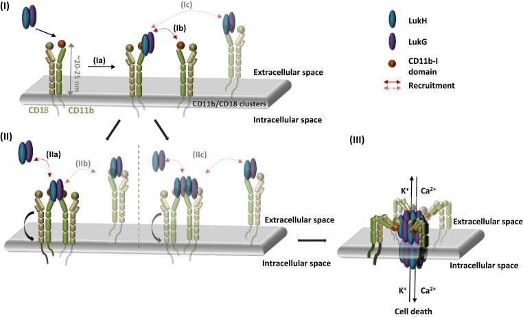
Host-pathogen interactions are central to understanding microbial pathogenesis. The staphylococcal pore-forming cytotoxins hijack important immune molecules but little is known about the underlying molecular mechanisms of cytotoxin-receptor interaction and host specificity. Here we report the structures of a staphylococcal pore-forming cytotoxin, leukocidin GH (LukGH), in complex with its receptor (the α-I domain of complement receptor 3, CD11b-I), both for the human and murine homologs. We observe 2 binding interfaces, on the LukG and the LukH protomers, and show that human CD11b-I induces LukGH oligomerization in solution. LukGH binds murine CD11b-I weakly and is inactive toward murine neutrophils. Using a LukGH variant engineered to bind mouse CD11b-I, we demonstrate that cytolytic activity does not only require binding but also receptor-dependent oligomerization. Our studies provide an unprecedented insight into bicomponent leukocidin-host receptor interaction, enabling the development of antitoxin approaches and improved animal models to explore these approaches.

Keywords: host–pathogen interaction; integrin; leukocidin; pore forming toxins; receptor recognition.


Conflict of interest statement

Competing interest statement: A.B. and H.R. are employees of X4 Pharmaceuticals GmbH, the legal successor of Arsanis Biosciences GmbH, which has developed an antileukocidin-GH antibody.

Figures

Fig. 1.
Fig. 1.
Binding and activity of LukGH wild-type and mutants to CD11b-I and crystal structure of LukGH-CD11b-I. (A) Steady-state analysis of LukGH wild-type binding to moCD11b-I. The steady state Kd is shown in the Inset. (B) Binding of LukGH to hu- or moCD11b-I expressed as response units (mean of 2 to 10 independent experiments ±SEM) and Kd (mean of 2 to 10 independent experiments ±SD). EC50 values of LukGH mutants toward differentiated HL-60 cells or mouse PMNs assessed in a luminescent cell viability assay measuring cellular ATP content (mean of 2 to 8 independent experiments ±SEM). For variants that had limited or no cytotoxicity (could not kill >75% of cells at the highest toxin concentration used), EC50 is not shown. (C) Cytotoxicity of LukGH, LukGHK319A, and LukED toward mouse PMNs assessed in a luminescent cell viability assay measuring cellular ATP content at cytotoxin concentrations of 30 µM, 20 µM, and 100 nM, respectively (mean of 3 independent experiments ±SEM). (D) Front and top view of LukGHK319A–moCD11b-I crystal structure. Dark blue and light green cartoons represent LukH and LukG from dimer 1 and dark green and light blue cartoon represent LukG and LukH from dimer 2, respectively. moCD11b-I is shown as an orange cartoon. Other dimers forming the octamer pore and bound CD11b-I molecules, are shown as a gray cartoon. Red spheres represent bound DMSO molecules from one asymmetric unit (dark red sphere represents DMSO 2). Comparison of moCD11b-I secondary structure (E) and MIDAS residues (F) from LukGHK319A–moCD11b-I structure (orange ribbon) with the active (1IDO, light pink ribbon) and inactive (1JLM, light gray ribbon) form of huCD11b-I. C-terminal α-helix is shown as light pink cartoon (1IDO) and gray cartoon (1JLM). Structures are aligned on moCD11b-I and MIDAS residues in E and F, respectively. The metal ions from the moCD11b-I structure and the inactive form of CD11b-I (1JLM) are shown as orange and gray spheres, respectively.
Fig. 2.
Fig. 2.
Binding epitope of LukGH-CD11b-I. (A) Binding epitopes of LukH–CD11b-I with detailed views of the specific interactions involved in CD11b-I binding in boxes, aligned on LukH. LukG, LukH, and CD11b-I from the LukGHK319A–moCD11b-I structure are shown in green, blue, and orange, respectively. The same protein components from the LukGH–huCD11b-I structure are shown in pale green, pale blue, and pale orange. Hydrogen bonds, salt bridges, the coordinate covalent bonds of Mg2+, as well as some other selected close contacts are shown as dashed lines colored black (for the moCD11b-I complex) or gray (for the huCD11b-I complex). (Left) Conserved interactions; (Right Upper and Lower) nonconserved interactions between the human and the mouse complexes. (B) Binding of LukGH mutants to CD11b-I variants relative to LukGH wild-type (mean of 3 independent experiments ±SEM, except for LukGHD316A with one experiment). Asterisks represent samples where no binding was detected (RU < 0.05 nm). Inset table shows Kd of selected LukGH and CD11b-I variants (mean of 2 to 3 independent experiments ±SD). (C) Binding epitopes of LukG-CD11b-I with a detailed view of the specific interactions involved in CD11b-I binding in the box, aligned on LukH. Color coding as in A.
Fig. 3.
Fig. 3.
Oligomerization of LukGH in solution, binding and activity of LukGH oligomerization variants. (A) Change of LukGH, LukG1H, and LukGHK319A (at 5 mg/mL) plus hu-, rb-, or moCD11b-I (at 2.5 mg/mL) cumulant radius, over time, measured in 25 mM Hepes, pH 7.5, 1 mM MgCl2, 150 mM NaCl (mean of 1 to 2 replicates ±SEM). The dotted lines represent fitting of the data to a one-phase association model with fixed y0 = 5 at x0 = 0 h (GraphPad Prism). (B) Oligomerization rate constant (k) and plateau for LukGH, LukGHK319A, and LukG1H (at 5 mg/mL) plus huCD11bI (2.5 mg/mL) in 25 mM Hepes, pH 7.5, 1 mM MgCl2, 0 to 300 mM NaCl (mean of 1 to 2 replicates ±SEM). Data were fitted as in A giving R2 > 0.93. (C) Oligomerization rate constant (k) of LukGH (4.5 mg/mL) plus increasing amounts of huCD11b-I (2.3 mg/mL) in 25 mM Hepes, pH 7.5, 1 mM MgCl2, 150 mM NaCl (mean of 2 replicates ±SEM). Linear regression fit (GraphPad Prism) is shown in red with equation in Inset. (D) Activity of LukGH mutants toward differentiated HL-60 cells expressed as EC50 and percent cell viability at maximal toxin concentration (100 nM) (mean of 2 independent experiments ±SEM). Red and black line represent EC50 value and percent cell viability of LukGHK319A mutant, respectively. Variants that had limited or no cytotoxicity (could not kill >75% of cells at the highest toxin concentration used) are marked with “#.” (E) Oligomerization rate constant (k) of LukG oligomerization mutants coexpressed with LukHK319A (at 4.5 mg/mL) plus huCD11b-I (2.3 mg/mL) (mean of 2 replicates ±SEM). Data were fitted as in A, in all cases, except for LukGQ31A LukHK319A (#, ambiguous fit), yielding R2 > 0.94. (F) Cumulant radius of LukGH_variant–moCD11b-I complexes (at 4.5 mg/mL for LukGH and 2.3 mg/mL for CD11b-I) and individual LukGH variants at 36 h of incubation in 25 mM Hepes, pH 7.5, 1 mM MgCl2,150 mM NaCl (mean of 1 [circled] or 2 replicates ±SEM). In case the sample shows increased radius at time 36 h, earlier time points are shown (24 and 12 h). Dotted lines represent ±10% change from a 5.5-nm radius. Samples with sum of squares error >10 are marked with “#.”
Fig. 4.
Fig. 4.
SAXS analysis of complex formation. (A) Scattering data as logI(s) vs. s plot compared to the theoretical scattering of the respective models. These comprise the interfaces as retrieved from the crystal structures; χ2 values are indicated. Curves are shifted along the y axis for better visualization. (B) Distance distribution profile of LukGH-Fab (black) and LukGH–Fab–huCD11b-I (red). The Inset shows the expected complex formation as cartoon representation, with the LukG, LukH, huCD11b-I, and Fab subunits in green, blue, orange, and purple cartoons, respectively.
Fig. 5.
Fig. 5.
Interaction of LukGH with Fab of αLukGH-mAb#5.H1H2 and activity in presence of LM2/1 and CBRM1/5. (A) Model of the LukGH–huCD11b-I octamer interacting with the Fab fragment of αLukGH-mAb#5.H1H2 (PDB ID code 5K59). The Fab is shown as purple surface, CD11b-I domain as orange cartoon, LukH1 and LukG1 forming Dimer 1 as dark blue and green cartoons, respectively, and LukH2 from adjacent dimer as light blue cartoon. The other LukG and LukH protomers and CD11b-I are shown in gray. Residues involved in binding of CBRM1/5 are shown as black spheres. (B) Change of the cumulant radius for LukGH plus huCD11b-I and/or αLukGH-mAb#5.H1H2 Fab measured in 25 mM Hepes, pH 7.5, 1 mM MgCl2, 150 mM NaCl at 1 mg/mL (mean of 1 to 2 replicates ±SEM). The red solid line represents fit of the data to a one-phase association model with fixed y0 = 5 at x0 = 0 h (GraphPad Prism). (C and D) Activity of LukGH toward LPS activated human PMNs, in presence and absence of 10 µg/mL LM2/1 (C) and CBRM1/5 (D) antibodies, assessed in a luminescent cell viability assay measuring cellular ATP content at different LukGH concentrations (mean of 3 replicates ±SEM).
Fig. 6.
Fig. 6.
Proposed model of LukGH–CD11b-I interaction and pore formation. (I) Binding of LukGH to CD11b-I via the LukH protomer (Ia) results in recruitment of a second integrin molecule via the LukG protomer (Ib) or alternatively, recruitment of a second integrin molecule with bound LukGH dimer (Ic). (II) After recruitment of the second integrin, via the LukG protomer, further LukGH dimer molecules are bound either as soluble LukGH dimers (IIa) or LukGH dimers bound to integrins (IIb). In the alternative version, 2 LukGH dimers bound to the 2 integrins (IIc) recruit further LukGH dimers in the same way as in IIa and IIb. (III) Bending of the integrin and insertion of the octameric pore containing 2 to 4 bound integrins into the membrane.

References

    1. Tong S. Y. C., Davis J. S., Eichenberger E., Holland T. L., Fowler V. G. Jr, Staphylococcus aureus infections: Epidemiology, pathophysiology, clinical manifestations, and management. Clin. Microbiol. Rev. 28, 603–661 (2015). - PMC - PubMed
    1. Alonzo F. 3rd, Torres V. J., The bicomponent pore-forming leucocidins of Staphylococcus aureus. Microbiol. Mol. Biol. Rev. 78, 199–230 (2014). - PMC - PubMed
    1. Spaan A. N., van Strijp J. A. G., Torres V. J., Leukocidins: Staphylococcal bi-component pore-forming toxins find their receptors. Nat. Rev. Microbiol. 15, 435–447 (2017). - PMC - PubMed
    1. Yamashita D., et al. , Molecular basis of transmembrane beta-barrel formation of staphylococcal pore-forming toxins. Nat. Commun. 5, 4897 (2014). - PubMed
    1. Haapasalo K., et al. , Staphylococcus aureus toxin LukSF dissociates from its membrane receptor target to enable renewed ligand sequestration. FASEB J. 33, 3807–3824 (2019). - PMC - PubMed

Publication types

MeSH terms