. 2020 Jan;19(1):232-240.
doi: 10.3892/etm.2019.8189. Epub 2019 Nov 12.

Protocatechuic acid attenuates anterior cruciate ligament transection-induced osteoarthritis by suppressing osteoclastogenesis

Affiliations

Protocatechuic acid attenuates anterior cruciate ligament transection-induced osteoarthritis by suppressing osteoclastogenesis

Jialin Zhang et al. Exp Ther Med. 2020 Jan.

Abstract

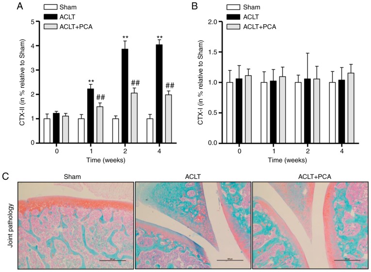
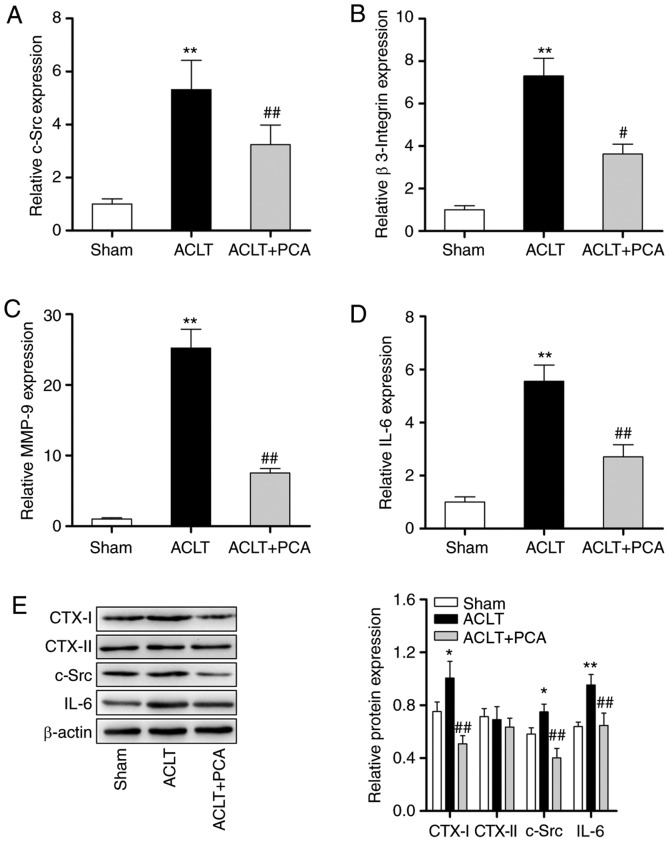
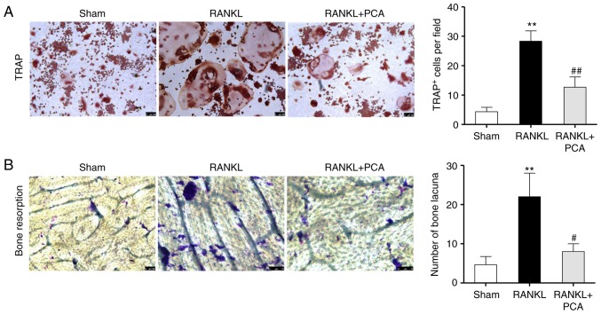
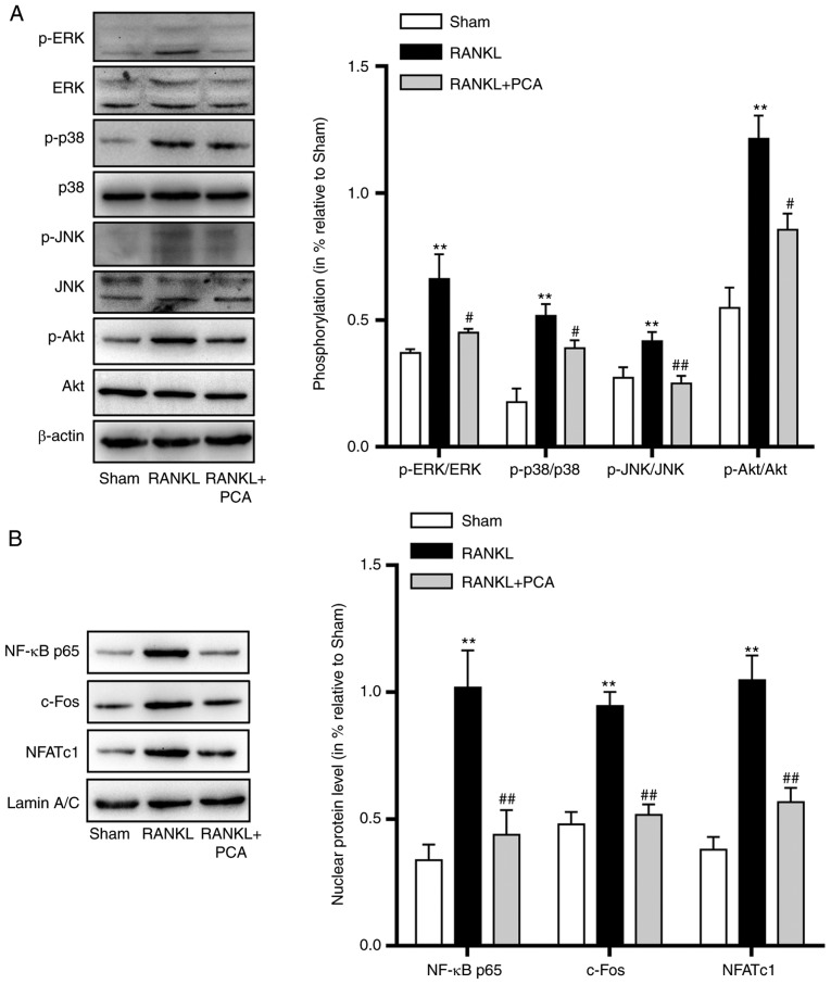
Osteoarthritis (OA) is related to over-proliferation or differentiation of osteoclasts. Although protocatechuic acid (PCA) has been identified to inhibit osteoclast differentiation and stimulate apoptosis in mature osteoclasts, whether it can relieve OA is still unknown. The present study aimed to investigate the effect of PCA on anterior cruciate ligament transection (ACLT)-induced OA and the potential mechanisms of action behind this effect. ACLT was performed on rats, which were then treated with or without PCA. C-terminal telopeptide of type I collagen (CTX-I) and CTX-II were tested in knee joint protein extracts by ELISA. Damage to cartilage was evaluated using Safranin-O/Fast Green staining. Osteoclast-related gene and protein expression was assessed through reverse transcription-quantitative PCR and western blotting. Tartrate-resistant acid phosphatase (TRAP) staining and functional bone resorption pit assays were performed using RAW264.7 murine macrophage cells to determine the effects of PCA on osteoclastic formation and function, respectively, in vitro. Finally, the activity of osteoclastogenesis-related signaling pathways was evaluated by western blotting. Levels of CTX-II were relatively decreased and Safranin-O/fast green staining indicated milder changes in the articular cartilage in the PCA treatment group. PCA downregulated osteoclast specific markers and suppressed receptor activator of nuclear factor-κB ligand-induced formation of TRAP-positive multinucleated cells, bone-resorption and pit formation. Mitogen-activated protein kinase (MAPK) and Akt signaling as well as the downstream factors, were downregulated by PCA. In conclusion, the present study demonstrated that PCA attenuated ACLT-induced OA by suppressing osteoclastogenesis by inhibiting the MAPK, ATK and NF-κB signaling pathways.

Keywords: anterior cruciate ligament transection-induced; osteoarthritis; osteoclastogenesis; protocatechuic acid.


Figures

Figure 1.
Figure 1.
PCA relieves ACLT-induced osteoarthritis. Rats with surgically induced ACLT were treated with or without PCA for 0, 1, 2 and 4 weeks. Levels of (A) CTX–II and (B) CTX–I in the knee joints were tested by ELISA. (C) The degenerative changes in the articular cartilage were examined by Safranin-O/Fast Green staining. Rats undergoing sham surgery were used as a control. The results are shown as the percentage mean ± SEM (n=6). Statistical analysis: ANOVA with Tukey's post-hoc test; NS, **P<0.01 vs. sham; ##P<0.01 vs. ACTL. Scale bars, 500 µm. ACLT, anterior cruciate ligament transection; CTX, C-terminal telopeptide of type I collagen; PCA, protocatechuic acid.
Figure 2.
Figure 2.
PCA decreases the activation of osteoclast in ACLT rats. The relative expression levels of (A) c-Src, (B) β-3 Integrin, (C) MMP-9 and (D) IL-6 in knee tissue samples were evaluated through reverse transcription-quantitative PCR. GAPDH was used as a reference. (E) The protein expressions of CTX–I, CTX–II, c-Src and IL-6 in knee tissue samples were evaluated through western blotting. β-actin was used as a loading control. *P<0.05, **P<0.01 vs. sham; #P<0.05, ##P<0.01 vs. ACTL. ACLT, anterior cruciate ligament transection; c-Src, proto-oncogene tyrosine-protein kinase; MMP, matrix metalloproteinase; PCA, protocatechuic acid.
Figure 3.
Figure 3.
PCA suppresses RANKL-induced osteoclast differentiation and the bone resorbing activity of RAW264.7 cells. (A) The effect of PCA on osteoclast differentiation was evaluated using TRAP staining. The TRAP + multinucleated cells were visualized and counted using an inverted microscope. (B) Pit-forming activity of RANKL-stimulated osteoclasts was analyzed by a microscope. The number of bone lacuna created by osteoclasts was counted. Data are expressed as the mean ± SD (n=4). **P<0.01 vs. sham; #P<0.05, ##P<0.01 vs. RANKL. PCA, protocatechuic acid; RANKL, receptor activator of nuclear factor-κB ligand; TRAP, tartrate-resistance acid phosphatase.
Figure 4.
Figure 4.
PCA negatively regulates the osteoclastogenesis-associated MAPK and Akt signaling pathways and critical nuclear factors. RAW 264.7 cells were treated with PCA for 2 h and cultured with RANKL for the corresponding time. (A) Western blotting was used to analyze the total protein extracts of proteins associated with the MAPKs and Akt signaling pathways. Phosphorylation of MAPKs (ERK, p-38 and JNK) and Akt was quantified. (B) Western blotting was used to analyze the expression of p65, c-Fos and NFATc1. All data represent at least three independent experiments. Values represent the mean ± SEM. **P<0.01 vs. sham; #P<0.05, ##P<0.01 vs. RANKL. MAPK, mitogen-activated protein kinases; NFATc1, nuclear factor of activated T cells 1; p, phosphorylated; PCA, protocatechuic acid; RANKL, receptor activator of nuclear factor-κB ligand.

References

    1. Lluch Girbés E, Nijs J, Torres-Cueco R, López Cubas C. Pain treatment for patients with osteoarthritis and central sensitization. Phys Ther. 2013;93:842–851. doi: 10.2522/ptj.20120253. - DOI - PubMed
    1. Clouet J, Vinatier C, Merceron C, Pot-vaucel M, Maugars Y, Weiss P, Grimandi G, Guicheux J. From osteoarthritis treatments to future regenerative therapies for cartilage. Drug Discov Today. 2009;14:913–925. doi: 10.1016/j.drudis.2009.07.012. - DOI - PubMed
    1. Suri S, Walsh DA. Osteochondral alterations in osteoarthritis. Bone. 2012;51:204–211. doi: 10.1016/j.bone.2011.10.010. - DOI - PubMed
    1. Boyle WJ, Simonet WS, Lacey DL. Osteoclast differentiation and activation. Nature. 2003;423:337–342. doi: 10.1038/nature01658. - DOI - PubMed
    1. Rodan GA, Martin TJ. Therapeutic approaches to bone diseases. Science. 2000;289:1508–1514. doi: 10.1126/science.289.5484.1508. - DOI - PubMed