. 2019 Dec 12;8(6):29.
doi: 10.1167/tvst.8.6.29. eCollection 2019 Nov.

Occurrence of Transmembrane Protein 119 in the Retina is Not Restricted to the Microglia: An Immunohistochemical Study

Affiliations

Occurrence of Transmembrane Protein 119 in the Retina is Not Restricted to the Microglia: An Immunohistochemical Study

Nan Su et al. Transl Vis Sci Technol. .

Abstract

Purpose: Recently, a new marker protein for microglial cells in the brain was postulated, transmembrane protein 119 (TMEM119), raising the hope for a new opportunity to reliably and unambiguously detect microglial cells in histologic sections. It was of interest whether TMEM119 also was a reliable microglial marker in the retina.

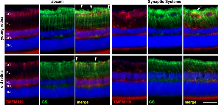
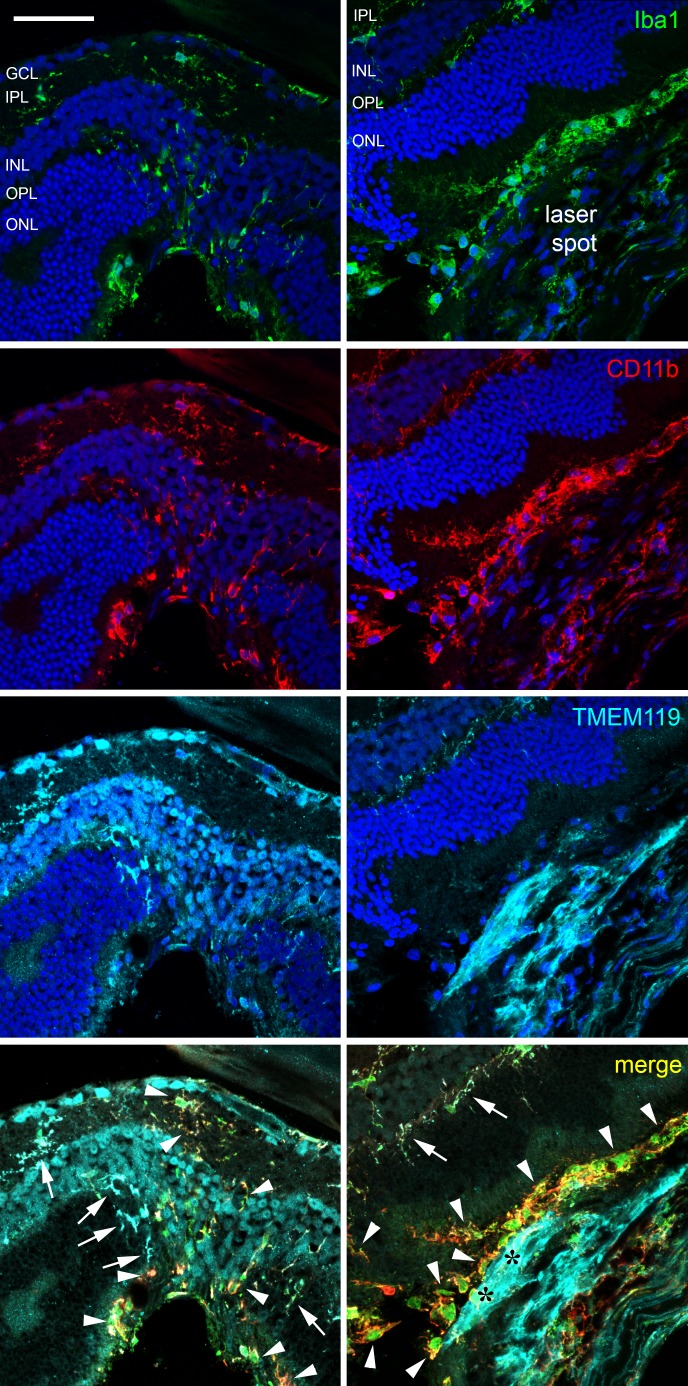
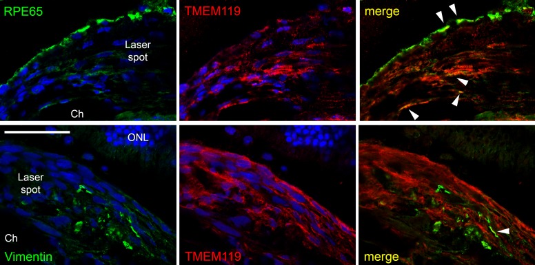
Methods: Anti-TMEM119 antibodies of two providers were used to label microglia in the murine retina, and labeling properties were compared to those of antibodies against Iba1 and CD11b. As an example of a pathologic situation, labeling for TMEM119 was also performed in eyes treated by an argon laser as an experimental model for choroidal neovascularization.

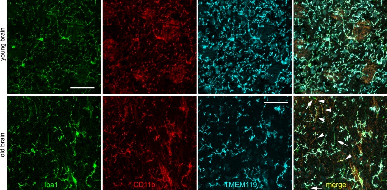
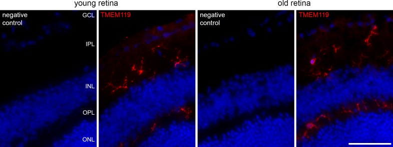
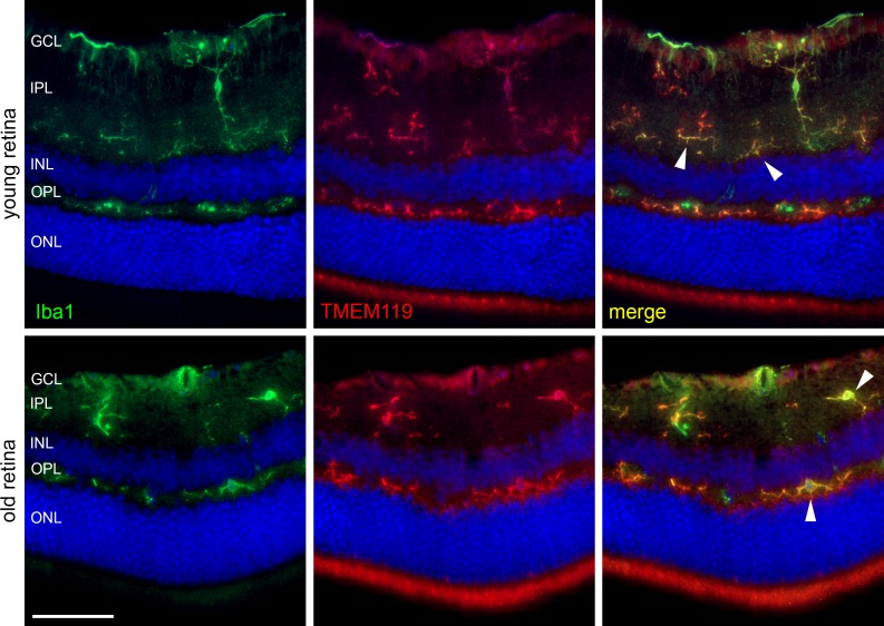
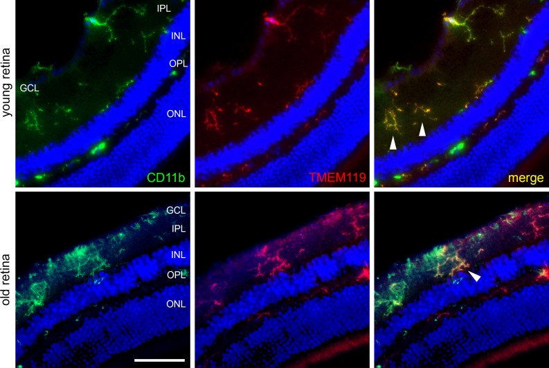
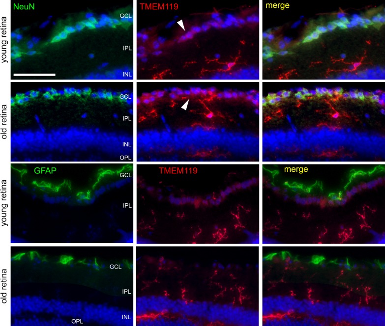
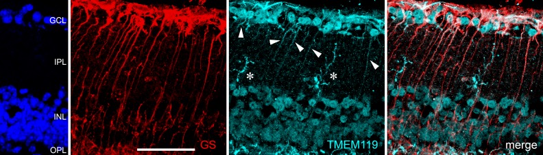
Results: TMEM119 immunoreactivity (IR) was found on microglial cells in the naïve retina. However, specificity and sensitivity of TMEM119 IR varied clearly depending on the source of the antibody, age of the mouse, and location of retinal microglia. After laser treatment, however, microglial cells lost their IR for TMEM119 at the site of the laser spot. Moreover, other cells became positive for TMEM119; for example, Müller cells.

Conclusions: TMEM119 is a useful marker for the microglia in the brain. However, retinal microglia shows variable IR for TMEM119, and the microglia is not the only cell showing TMEM IR. Therefore, TMEM119 appears not to be applicable as a general marker for the retinal microglia in pathologic situations.

Translational relevance: Reliable detection and quantification of microglial cells is of high importance to study disease mechanisms and effects of therapeutic approaches in the retina.

Keywords: TMEM119; immunohistochemistry; microglia; retina.


Figures

Figure 1
Figure 1
Confocal images of immunohistochemical labeling of Iba1 (green), CD11b (red), and TMEM119 (turquoise, antibody by Synaptic Systems) as indicated in brain cryosections obtained from a young (upper row) and old (lower row) mouse. In the merged image of the old mouse, white arrows point to cells that display almost only Iba1 IR, and white arrowheads to cells with Iba1 IR and CD11b IR and almost no TMEM119 IR. Scale bars: 50 μm.
Figure 2
Figure 2
Immunohistochemical labeling of microglial cells in retinal cryosections of a young and old mouse against TMEM119 (red, antibody by Synaptic Systems) as indicated. Corresponding negative controls are included. GCL, ganglion cell layer; IPL, inner plexiform layer; INL, inner nuclear layer; OPL, outer plexiform layer; ONL, outer nuclear layer. Scale bar: 50 μm.
Figure 3
Figure 3
Immunohistochemical staining of a retinal cryosection from a young and old mouse against TMEM119 (red) and GS as indicated. Anti-TMEM119 antibodies were delivered by Abcam (left) and by Synaptic Systems (right). With the anti-TMEM119 antibody by Abcam, a poor labeling of microglial cell was achieved, and a clear IR of other cell populations in the retina; for example, the Müller cells, as demonstrated by GS IR. White arrowheads point to particularly obvious colocalization of TMEM119 IR and GS IR. White arrow points to an overlap of TMEM119 IR and GS IR without real colocalization. Scale bar: 50 μm.
Figure 4
Figure 4
Immunohistochemical labeling of microglial cells in retinal cryosections of a young and old mouse against Iba1 (green) and TMEM119 (red, antibody by Synaptic Systems) as indicated. Arrowheads point to cells that are similarly positive for Iba1 and TMEM119. Scale bar: 50 μm.
Figure 5
Figure 5
Immunohistochemical labeling of microglial cells in retinal cryosections of a young and old mouse against CD11b (green) and TMEM119 (red, antibody by Synaptic Systems) as indicated. Arrowheads point to cells that are similarly positive for CD11b and TMEM119. Scale bar: 50 μm.
Figure 6
Figure 6
Immunohistochemical staining of retinal cryosections to label microglial cells (TMEM119, antibody by Synaptic Systems, red), ganglion cells (NeuN, green), and astrocytes (GFAP, green) in retinas of young and old mice, as indicated. Only inner layers of the retinas are shown. White arrowheads point to TMEM119-positive retinal ganglion cells. Scale bar: 50 μm.
Figure 7
Figure 7
Confocal images of immunohistochemical staining of a retinal cryosection from a young mouse 4 days after laser treatment by labeling of GS (red) and TMEM119 (turquoise, antibody by Synaptic Systems) as indicated. Retinal layers are indicated by DAPI staining on the left margin. Besides microglial cells (white asterisks) and cells in the ganglion cell layer and inner nuclear layer, also the tiny longish Müller cells show TMEM119 IR (white arrowheads). Scale bar: 50 μm.
Figure 8
Figure 8
Confocal images of immunohistochemical staining of a retinal cryosection from a young mouse 4 days after laser treatment against Iba1 (green), CD11b (red), and TMEM119 (turquoise, antibody by Synaptic Systems) as indicated. DAPI staining is shown in the images of single channels and is not shown in the merged images for more clarity. In the left column, part of the inner retina above the laser spot is shown, while the laser spot including the proliferation area is shown in the right column. White arrows point to cells positive for all three microglial markers, whereas white arrowheads point to cells only positive for Iba1 and/or CD11b. Black asterisks indicate the proliferation area where cells are only TMEM+. Scale bar: 50 μm.
Figure 9
Figure 9
Confocal images of double labeling of the proliferation area in the laser spot for TMEM119 (red, antibody by Synaptic Systems) and RPE65 (green, upper row) and vimentin (green, lower row). Colocalization could be found only in very few cases (white arrowheads). ONL, outer nuclear layer; Ch, choroid. Scale bar: 50 μm.

References

    1. Hanisch UK, Kettenmann H. Microglia: active sensor and versatile effector cells in the normal and pathologic brain. Nat Neurosci. 2007;10:1387–1394. - PubMed
    1. Karlstetter M, Ebert S, Langmann T. Microglia in the healthy and degenerating retina: Insights from novel mouse models. Immunobiology. 2010;215:685–691. - PubMed
    1. Madeira MH, Boia R, Santos PF, Ambrósio AF, Santiago AR. Contribution of microglia-mediated neuroinflammation to retinal degenerative diseases. Mediat Inflamm. 2015. 673090. - PMC - PubMed
    1. Li L, Eter N, Heiduschka P. The microglia in healthy and diseased retina. Exp Eye Res. 2015;136:116–130. - PubMed
    1. Jiang ZH, Peng J, Yang HL, et al. Upregulation and biological function of transmembrane protein 119 in osteosarcoma. Exp Mol Med. 2017;49:e329. - PMC - PubMed